Article

Species and Strain Variability among Sarcina Isolates from Diverse Mammalian Hosts

1
Department of Microbiology, Nutrition and Dietetics, Faculty of Agrobiology, Food and Natural Resources, University of Life Sciences Prague, 165 00 Prague, Czech Republic
2
Laboratory of Anaerobic Microbiology, Institute of Animal Physiology and Genetics, Czech Academy of Sciences, 142 20 Prague, Czech Republic
3
Department of Ethology and Companion Animal Science, Faculty of Agrobiology, Food, and Natural Resources, University of Life Sciences Prague, 165 00 Prague, Czech Republic
*
Author to whom correspondence should be addressed.
Animals 2023, 13(9), 1529; https://doi.org/10.3390/ani13091529
Submission received: 2 March 2023 / Revised: 28 April 2023 / Accepted: 29 April 2023 / Published: 3 May 2023
(This article belongs to the Section Mammals)

Abstract

:

Simple Summary

Sporadic but repeated occurrences of Sarcina spp. indicate that these microorganisms with atypical morphology forming packets in the fecal microbiota of animals without health problems may not always be pathogenic and seem to be a common part of the gut microbiota of various mammals. Aside from that, genotyping characterization indicates species and strain variability among Sarcina isolates and the potential presence of two novel taxonomic units originating from dog and elephant hosts.

Abstract

Sarcina spp. has been isolated from the gastrointestinal tracts of diverse mammalian hosts. Their presence is often associated with host health complications, as is evident from many previously published medical case reports. However, only a handful of studies have made proper identification. Most other identifications were solely based on typical Sarcina-like morphology without genotyping. Therefore, the aim of this work was culture detection and the taxonomic classification of Sarcina isolates originating from different mammalian hosts. Sarcina-like colonies were isolated and collected during cultivation analyses of animal fecal samples (n = 197) from primates, dogs, calves of domestic cattle, elephants, and rhinoceroses. The study was carried out on apparently healthy animals kept in zoos or by breeders in the Czech Republic and Slovakia. Selected isolates were identified and compared using 16S rRNA gene sequencing and multi-locus sequence analysis (MLSA; Iles, pheT, pyrG, rplB, rplC, and rpsC). The results indicate the taxonomic variability of Sarcina isolates. S. ventriculi appears to be a common gut microorganism in various captive primates. In contrast, a random occurrence was also recorded in dogs. However, dog isolate N13/4e could represent the next potential novel Sarcina taxonomic unit. Also, a potentially novel Sarcina species was found in elephants, with occurrences in all tested hosts. S. maxima isolates were detected rarely, only in rhinoceroses. Although Sarcina bacteria are often linked to lethal diseases, our results indicate that Sarcina spp. appear to be a common member of the gut microbiota and seem to be an opportunistic pathogen. Further characterization and pathogenic analyses are required.

1. Introduction

The genus Sarcina within the Clostridiaceae family represents morphologically atypical, almost spherical cells forming packets, usually of eight or more units. Two taxa of Sarcina spp. were validly recognized. In 1842, Goodsir et al. first described Sarcina ventriculi in the stomach contents of a human patient with recurrent vomiting. The second recognized valid species with Sarcina-like morphology was Sarcina maxima, isolated from the feces of an elephant in 1969 [1]. Sarcina spp. occur naturally in soil, mud, cereal grains, and in the gastrointestinal tract of animals, as summarized by Lawson & Rainey [2]. Nevertheless, Sarcina findings in the digestive tract of humans and animals are often associated with various pathologies, particularly conditions related to delayed gastric emptying and food movement further into the intestine. Nausea, vomiting, ulcers, and chronic dyspepsia are other related manifestations [3,4,5,6,7]. According to a recently published review [8], more than 100 full-text articles assessing eligibility have been published. Therefore, the number of medical reports on human hosts is alarming. Similarly, in monogastric animals such as horses, dogs, and cats, gastric dilations have also been documented [9,10]. In addition, there are other cases involving gastric problems in farm animals [11,12] or chimpanzees in Sierra Leone [13] with fatal outcomes. However, some publications have described the presence of Sarcina spp. in healthy animals and humans [14,15,16,17].
The pathogenicity of Sarcina bacteria is not entirely clear and is an interesting subject for future investigation. It is striking that, until today, the identification of Sarcina spp. has been mainly based on its morphology and not on a molecular genetic taxonomic approach. This has been documented in several published case reports [8], in which atypical Sarcina-like morphology was used for S. ventriculi identification [7,18,19]. Only a few studies have reported proper taxonomic identification at the species level [20,21,22] compared to the highly studied and characterized clostridia. Sporadic but repeated occurrences of bacterial cells showing Sarcina-like morphology on selective medium for bifidobacteria [15,17] during cultivation analyses of mammalian fecal samples prompted us to target their monitoring. The goal of our study was the culture-dependent screening of Sarcina-like morphology colonies in fecal samples of apparently healthy animals kept in zoos or by breeders in the Czech Republic and Slovakia to get isolates for proper taxonomic identification that would highlight the variability within the genus Sarcina and the need for further research into this dreaded potential pathogen.

2. Materials and Methods

2.1. Ethical Statement, Sampling, and Cultivation Analysis of Fecal Samples

The sampling of animal feces was performed during routine daily procedures at zoos, farms, and breed centers (Czech Republic and Slovakia). Zoological institutions have rigorous standards for animal welfare and are accredited by the European Association of Zoos and Aquaria. All procedures involving animals adhered to the recommendations of the “Guide for the Care and Use of Animals” by the Czech University of Life Sciences Prague and to the legal requirements of the Czech Republic for the ethical treatment of nonhuman primates, as well as in accordance with European Directive 2010/63/EU. Some animal and human isolates were obtained during culture analyses of fecal samples from previously published animal studies [15,17,23].
In total, 197 fecal samples from primates (n = 65), dogs (n = 70), calves of domestic cattle (n = 50), elephants (n = 10), and rhinoceroses (n = 2) were collected in the years 2015–2020 with the original intention of characterizing the cultivable anaerobic bacteria. Fresh fecal samples were collected directly after host defecation. Sampling was done using a sterile spoon from the part that did not touch the ground. Then, fecal samples were placed in an anaerobically prepared medium. Media preparation, sample storage, and cultivation analysis were performed according to Modrackova et al. [17]. The repeated occurrence of the bacterium with Sarcina-like morphology on the modified Wilkins-Chalgren medium directed us to long-term monitoring of Sarcina spp. in tested fecal samples. Yellow-pigmented irregular colonies with a typical Sarcina-like morphology, monitored by phase contrast microscopy, were selected for future identification and characterization. Also, one human isolate of S. maxima 7 from a previous study [24] was included. In total, 58 Sarcina-like morphology isolates of different origins were selected for genotyping.

2.2. Colony Isolation and Identification

Putative Sarcina strains grown in bifidobacterial cultivation media were isolated and consecutively sub-cultivated in WSP broth under anaerobic conditions at 37 °C for 1 d [17]. Bacterial cultures were stored at room temperature (20 °C) and re-inoculated every 3 d. DNA was isolated using PrepMan UltraTM (Applied Biosystems, Waltham, MA, USA) according to the manufacturer’s instructions and stored at −20 °C. DNA samples at a concentration of 10–500 ng were used for polymerase chain reaction (PCR) amplification. Primers fd1 (5′-AGAGTTTGATCCTGGCTCAG-3′) and rP2 (5′-ACGGCTACCTTGTTACGACTT-3′) were used for PCR amplification of the 16S rRNA gene [25]. PCR products were purified using the E.Z.N.A. Cycle Pure Kit (Omega Bio-Tek, Norcross, Georgia, USA) and Sanger sequenced by Eurofins Genomics (Ebersberg, Germany). The obtained sequences were carried out in Chromas Lite 2.5.1 (Technelysium Pty Ltd., Tewantin, Australia) and BioEdit [26,27] using the ClustalW algorithm [28]. Bacteria were preliminarily classified based on a comparative analysis of the EzBioCloud-16S rRNA gene.

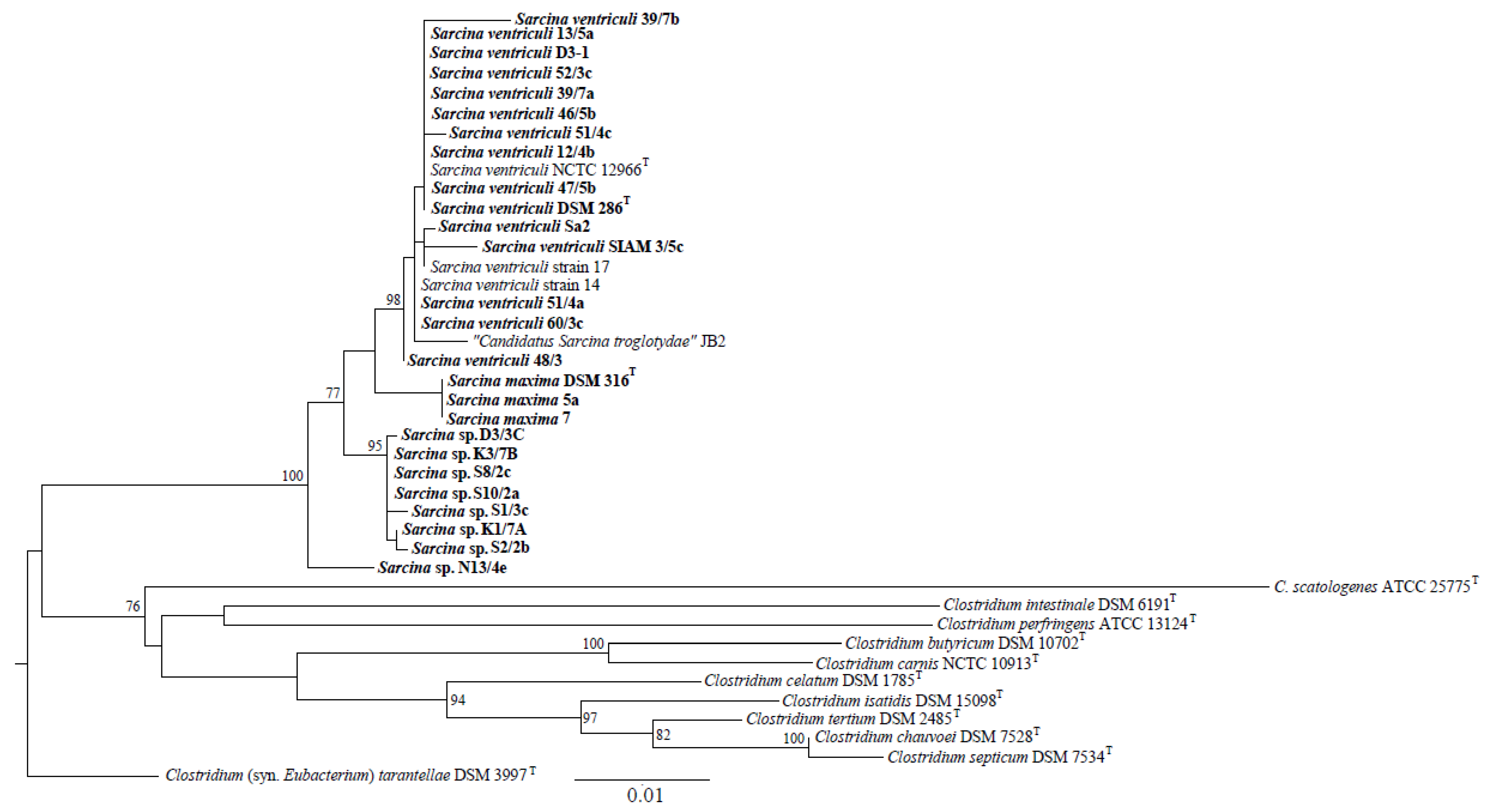
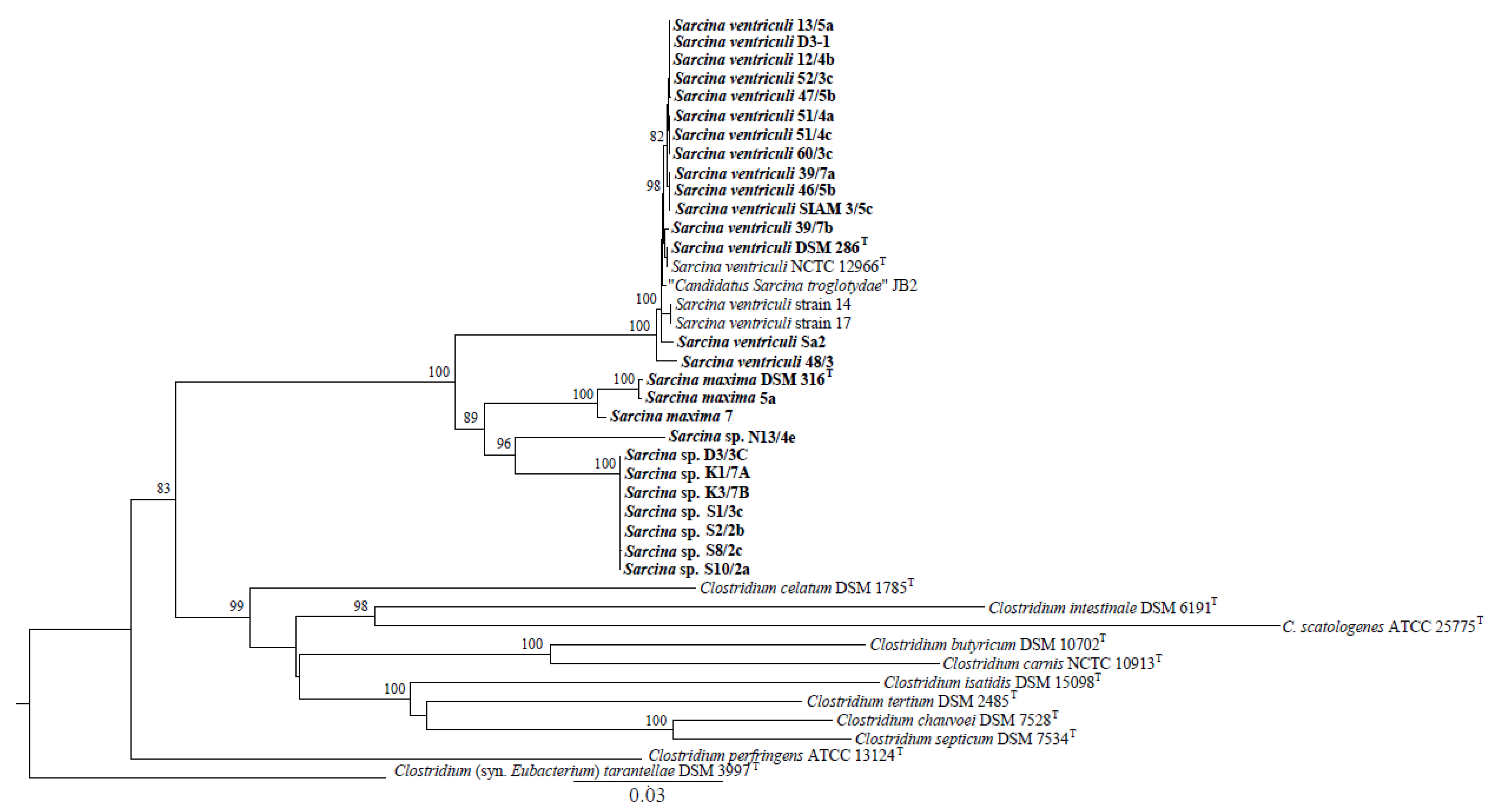
2.3. Phylogenetic Studies

The genomic DNA of 24 selected Sarcina isolates identified based on the 16S rRNA gene sequencing was used for multi-locus sequence analysis (MLSA). The Iles, pheT, pyrG, rplB, rplC, and rpsC operating genes encoding the isoleucyl-tRNA synthetase, phenylalanyl-tRNA synthetase beta subunit, CTP synthase, 50S ribosomal protein L2, 50S ribosomal protein L3, and 30S ribosomal protein S3, respectively, were chosen for MLSA and phylogenetic analyses. Primer pairs flanking variable sections were reconstructed using the sequences of nine bacterial strains retrieved from the complete genomes (Table S1). The consensual sequences obtained in Geneious v7.1.7 software were employed. The obtained sequences were deposited in the NCBI database using the Banklt application (https://www.ncbi.nlm.nih.gov/WebSub/), accessed on 29 October 2022.
Phylogenetic relationships among Sarcina isolates, whose genomes of Sarcina spp. are available in the NCBI database (https://www.ncbi.nlm.nih.gov/nuccore/), accessed on 29 October 2022, and related clostridial taxa based on the 16S rRNA gene phylogeny [2] were reconstructed in the MEGA v5.05 software package using the maximum-likelihood statistical method and a particular multi-locus (ML) model [29]. Gene pairwise identities were calculated using Geneious v7.1.7 software.

2.4. Data Accessibility

The nucleotide sequences of the 16S rRNA gene, Iles, pheT, pyrG, rplB, rplC, and rpsC operating genes are available under the GenBank accession numbers (Table S3).

3. Results

3.1. Occurrence of Sarcina spp. in the Feces of Screened Mammalian Hosts

Due to the recurrent occurrence of Sarcina cells on Wilkins-Chalgren agar supplemented with soya peptone, L-cysteine, Tween 80, acetic acid, and mupirocin, primarily used to isolate bifidobacteria, a total of 197 fresh fecal samples were screened for the presence of Sarcina spp. colonies. Visually distinguishable irregular yellow colonies with Sarcina-like morphology were determined by cultivation in fecal samples of 25/65 primates, 1/70 dogs, 2/50 calves of domestic cattle, 10/10 elephants, and 2/2 rhinoceroses (Table 1 and Table S2). The frequency of Sarcina occurrence was evidently the highest in elephants, where potentially novel Sarcina species were detected even in relatively higher quantities in the Zoo Ustí nad Labem in 107 CFU g−1 of feces than in the Zoo Liberec in 103 CFU g−1 of feces. The presence of cultivable viable Sarcina cells in monkey feces was more common in Old World monkeys, such as guenons and gibbons, which were determined to be ordinarily from 105 to 107 CFU g−1 of fecal samples. The occurrence in dogs, calves of domestic cattle, and humans was random, and Sarcina-like cells were detected at counts of <104 CFU g−1.

3.2. Identification and Taxonomic Classification of Isolated Sarcina Strains

In total, 57 mammalian Sarcina isolates from diverse animal hosts were collected, plus one human isolate and two type strains. Therefore, 60 strains were selected for genotyping (Table 2). Unfortunately, the cultivation of Sarcina spp. is a demanding task. This is not only because of its strictly anaerobic nature but also because of large clusters of Sarcina-like cell arrangements that may harbor some contaminants originating from the complex gut microbiota, which likely decreased the successful identification of a significant number of isolates. Successful 16S rRNA identification was performed for 24 of 60 DNA samples (Table 2), which were also selected for MLSA. Partial gene sequences obtained in the study using specific primers and PCR conditions (Table 3), deposited in the NCBI database with the initial letters OK (for operating genes) and MZ (for 16S rRNA), are listed in Table S3. This table also includes the relevant genes of Sarcina spp. and clostridial taxa available in the NCBI database that were used to reconstruct the phylogenetic relationships.
The 16S rRNA-based phylogenetic tree (Figure 1) grouped Sarcina strains into four clusters, represented by strains of the species S. ventriculi and S. maxima and two other Sarcina sp. groups. The first included seven strains (D3/3C, K3/7B, S8/2c, S10/2a, S1/3c, K1/7A, and S2/2b) and the second strain, Sarcina sp. N13/4e. A very similar topology, including the four Sarcina clusters, was generated based on the concatenated sequences of six operating gene fragments (Figure 2) and amino acid translations (Figure S1).
Members of the Sarcina sp. cluster, including seven strains, have 98.62–99.08% 16S rRNA gene (1302 nucleotides) sequence similarity to type strains of S. maxima and S. ventriculi. Pairwise identities based on the ileS (699 nucleotides in length), pyrG (480), rpsC (444), rplB (516), rplC (360), and pheT (570) gene concatenates were computed in the range of 92.83–94.49%. The strain N13/4e had 98.39–90.0% 16S rRNA gene identity to S. maxima and S. ventriculi type strains and 91.92–94.04% based on the operating gene concatenate. Much higher (>98%) pairwise identities were recorded for other strains clustered with S. maxima and S. ventriculi-type strains.
Phylogenetic analyses suggested that a cluster of seven Sarcina strains (D3/3C, K3/7B, S8/2c, S10/2a, S1/3c, K1/7A, and S2/2b) and Sarcina sp. N13/4e represents a novel Sarcina taxonomic unit. However, sophisticated modern methods, especially those based on whole-genome sequences, must be used to confirm this assumption.

4. Discussion

Sarcina occurrences associated with the mammalian gastrointestinal microbiota were recorded in young ruminants [12,30,31], dogs [9], as well as cats [10], and primates [13,15,17,32,33,34]. Our results indicate that bacteria with Sarcina-like cell morphology originating from fecal samples of mammalian hosts such as primates, dogs, calves of domestic cattle, elephants, and rhinoceroses were clustered into four groups based on 16S rRNA gene sequencing and MLSA.
Animal gut microbiome seems to be significantly modified by dietary changes in the host species and geography. Dynamic microbial communities aid the living and survival of animals in changing environmental conditions, including habitat degradation, captive breeding, and diet. If this host’s gut microbial balance is disturbed and dysbiosis occurs, disease development is presumed [35,36,37,38]. Even though it was possible to isolate Sarcina spp. from various species of primates from different zoos in the Czech Republic and Slovakia. All these isolates were identified as S. ventriculi and grouped into one group. Interestingly, in Zoo Liberec (Czechia), Sarcina-like bacteria from primates (Northern white-cheeked gibbons) and Asian elephant hosts were isolated. However, they were identified as two different species. Based on our results, the taxonomic variability of the analyzed Sarcina strains is more dependent on the animal species than the host location. This confirms the fact that isolates from elephants from two different zoos were also clustered into one group (potentially novel Sarcina sp.). S. ventriculi seems to be a common gut microorganism in captive primates such as guenons (De Brazza’s monkey, Hamlyn’s monkey, Roloway monkey, Lesser spot-nosed monkey, and Vervet monkey) and gibbons (northern white-cheeked gibbon, gibbon siamang, and Yellow-cheeked crested gibbon). S. ventriculi was also detected in the feces of ring-tailed lemurs and golden lion tamarins. Samples of primates belonging to New World monkeys, such as marmosets and other tamarins, were also subjected to culture analysis under the same conditions; however, Sarcina-like cells were detected rarely. Interestingly, S. ventriculi was detected only in one of the eight individuals belonging to the golden lion tamarin species. Similarly, other authors also described S. ventriculi presence in the feces of Old Word monkeys such as guenons and crested gibbons [15,22], macaques [32], wild gorillas [33], and chimpanzees [13,34]. Interestingly, these records indicate that S. ventriculi seems to be common not only for animals kept in captivity but also for primates living in the wild or on nature reserves. The more frequent and successful detection of S. ventriculi may be influenced by the lower representation of bifidobacteria in the microbiota of Old World monkeys compared to New World monkeys [17], which allows the growth of distinguishable Sarcina-like bacterial colonies and their possible isolation and identification. Also, the fecal microbiota of the dog is known for the relatively high occurrence of viable clostridial cells and low or culture-undetectable numbers of bifidobacteria in dog feces which then allow detection of the Clostridiaceae family on bifidobacterial media [23].
Sarcina species have an atypical morphology, with almost spherical cells forming packets, usually of eight or more cells, which is rarely observed in other bacteria. The occurrence of visually distinguishable irregular yellow colonies with Sarcina-like morphology on modified Wilkins-Chalgren agar during the cultivation of primate fecal samples has been previously described [15,17]. Wilkins-Chalgren agar is used to cultivate anaerobic bacteria. Media modification with soya peptone, L-cysteine, Tween 80, acetic acid, and mupirocin is primarily used to isolate Bifidobacterium species [39]. S. ventriculi and S. maxima are anaerobes known for their ability to grow at very low pH values [1,40], and resistance to mupirocin has also been detected [15]. Therefore, the selective factors used in modified Wilkins-Chalgren agar do not limit the cultivation of viable Sarcina species present in the tested fecal samples, and the media used seem to be suitable for their cultivation and detection as well.
A potentially novel Sarcina species represented by seven strains (D3/3C, K3/7B, S8/2c, S10/2a, S1/3c, K1/7A, and S2/2b) isolated from different Asian elephant hosts from the Zoo Liberec and the Zoo Ústí nad Labem was found to be a common commensal of elephants. It is a paradox that S. maxina as a taxon was first described in 1969 in an elephant [1]. However, we identified S. maxima isolates in a human vegetarian host and in both rhinoceroses. Only one Sarcina sp. occurrence was recorded on the modified Wilkins-Chalgren medium out of the 70 analyzed dog fecal samples. Interestingly, this isolate could represent the next novel Sarcina taxonomic unit.
A recently published study [13] dealt with the occurrence of Sarcina bacteria in connection with chimpanzee deaths in Sierra Leone, which were further isolated and characterized. By studying the morphology and growth characteristics of these strains and by completing genome sequencing of an isolate, researchers identified features that distinguish “Candidatus Sarcina troglodytae” from all previously described members of the Sarcina genus. They showed that the bacterium possesses genes encoding biochemical pathways that potentially contribute to enhanced virulence, including an encoded urea degradation biochemical pathway, consistent with the clinical signs observed in chimpanzees. Overall, they concluded that the Sarcina genus probably comprises an overlooked complex of species, ranging from benign commensals to frank pathogens. However, further analyses leading to an understanding of its pathogenicity are often lacking. In addition, most published cases [8] involved the identification of Sarcina isolates only at the genus level based on the morphology and health complications of the host. The threshold value for distinguishing between the two species was 98.65% [41]. Our results were based on 16S rRNA gene sequencing and MLSA (Iles, pheT, pyrG, rplB, rplC, and rpsC genes), indicating species variability across different hosts. In addition to the two known recognized taxa (S. ventriculi and S. maxima), two potentially novel species isolated from dogs and elephants were considered. Based on 16S rRNA gene sequencing and MLSA, there is an assumption of Sarcina variability at the genome level. Remarkably, these analyses clustered the chimpanzee isolates “Candidatus Sarcina troglodytae” [13] into the S. ventriculi taxon together with other primates isolates. Therefore, future complete genome sequencing of our Sarcina isolates can bring new points of interest about their properties.

5. Conclusions

Our screening results indicate the common presence of Sarcina spp. in the gut microbiota of various mammals kept in captivity without obvious health complications. Sarcina spp. occurrence was more significantly affected at the level of animal species than at the level of host location. S. ventriculi appears to be a common part of the primate intestinal microbiota, especially those of Old World monkeys. The presence of potentially novel taxa across detected mammals can bring another view on Sarcina function in the gut microbiome of animals.

Supplementary Materials

The following supporting information can be downloaded at: https://www.mdpi.com/article/10.3390/ani13091529/s1. Figure S1: Maximum likelihood phylogenetic tree revealing relationships among Sarcina strains through the amino acid translation (1023 aa) of ileS, pyrG, rpsC, rplB, rplC, and pheT partial gene sequences, respectively; Table S1: Positions of six genes within the genomes of nine bacterial strains used for primer pair construction Table S2: List of animal hosts cultured to detect Sarcina-like bacteria Table S3: The NCBI accession numbers of genes used for phylogenetic reconstructions and the calculation of pairwise identity.

Author Contributions

Conceptualization, V.N.-B. and P.B.; methodology, V.N.-B. and J.K.; formal analysis, M.M., N.M., E.I. and A.A.; investigation, V.N.-B., M.M. and J.K.; data curation, V.N.-B. and J.K.; writing—original draft, V.N.-B., M.M. and J.K.; writing—review and editing, V.N.-B. and N.M.; funding acquisition, E.V.; supervision, V.N.-B.; project administration, V.N.-B. and E.V. All authors have read and agreed to the published version of the manuscript.

Funding

The research was funded by the European Regional Development Fund-Project “Centre for the investigation of synthesis and transformation of nutritional substances in the food chain in interaction with potentially harmful substances of anthropogenic origin: comprehensive assessment of soil contamination risks for the quality of agricultural products” [No: CZ.02.1.01/0.0/0.0/16_019/0000845].

Institutional Review Board Statement

The sampling of animal feces was performed during routine daily procedures at zoos, farms, and breed centers (Czech Republic and Slovakia). Zoological institutions have rigorous standards for animal welfare and are accredited by the European Association of Zoos and Aquaria. All procedures involving animals adhered to the recommendations of the “Guide for the Care and Use of Animals” by the Czech University of Life Sciences Prague and to the legal requirements of the Czech Republic for the ethical treatment of nonhuman primates, as well as in accordance with European Directive 2010/63/EU.

Informed Consent Statement

Not applicable.

Data Availability Statement

The nucleotide sequences of the 16S rRNA gene, Iles, pheT, pyrG, rplB, rplC, and rpsC operating genes are available under the GenBank accession numbers presented in Table S3, and other data are available on request from the corresponding author.

Acknowledgments

The authors thank the farm in Božkov and zoos in Dvůr Králové, Hodonín, Liberec, Olomouc, Plzeň, Ústí nad Labem, Bojnice, and Bratislava for their kind cooperation.

Conflicts of Interest

The authors declare no conflict of interest.

References

  1. Canale-Parola, E. Genus Sarcina. In Bergey’s Manual of Systematic Bacteriology, 2nd ed.; The Firmicutes; Springer: New York, NY, USA, 2009; Volume 3. [Google Scholar]
  2. Lawson, P.A.; Rainey, F.A. Proposal to restrict the genus Clostridium prazmowski to Clostridium butyricum and related species. Int. J. Syst. Evol. Microbiol. 2016, 66, 1009–1016. [Google Scholar] [CrossRef] [PubMed]
  3. Dumitru, A.; Aliuş, C.; Nica, A.E.; Antoniac, I.; Gheorghiță, D.; Grădinaru, S. Fatal outcome of gastric perforation due to infection with Sarcina spp. A case report. IDCases 2020, 19, e00711. [Google Scholar] [PubMed]
  4. Tintara, S.; Rice, S.; Patel, D. Sarcina Organisms: A Potential Cause of Emphysematous Gastritis in a Patient with Gastroparesis. Am. J. Gastroenterol. 2019, 114, 859. [Google Scholar] [CrossRef] [PubMed]
  5. Singh, K. Emphysematous Gastritis Associated with Sarcina ventriculi. Case Rep. Gastroenterol. 2019, 13, 207–213. [Google Scholar] [CrossRef]
  6. Alvin, M.; Al Jalbout, N. Emphysematous gastritis secondary to Sarcina ventriculi. BMJ Case Rep. 2018, 2018. [Google Scholar] [CrossRef]
  7. de Meij, T.G.; van Wijk, M.P.; Mookhoek, A.; Budding, A.E. Ulcerative gastritis and esophagitis in two children with Sarcina ventriculi infection. Front. Med. 2017, 4, 145. [Google Scholar] [CrossRef]
  8. Tartaglia, D.; Coccolini, F.; Mazzoni, A.; Strambi, S.; Cicuttin, E.; Cremonini, C.; Taddei, G.; Puglisi, A.G.; Ugolini, C.; Di Stefano, I.; et al. Sarcina Ventriculi infection: A rare but fearsome event. A Systematic Review of the Literature. Int. J. Infect. Dis. 2022, 115, 48–61. [Google Scholar] [CrossRef]
  9. Vatn, S.; Gunnes, G.; Nybø, K.; Juul, H.M. Possible involvement of Sarcina ventriculi in canine and equine acute gastric dilatation. Acta Vet. Scand. 2000, 41, 333–337. [Google Scholar] [CrossRef]
  10. Im, J.Y.; Sokol, S.; Duhamel, G.E. Gastric dilatation associated with gastric colonization with Sarcina-like bacteria in a cat with chronic enteritis. J. Am. Anim. Hosp. Assoc. 2017, 53, 321–325. [Google Scholar] [CrossRef]
  11. DeBey, B.M.; Blanchard, P.C.; Durfee, P.T. Abomasal bloat associated with Sarcina-like bacteria in goat kids. J. Am. Vet. Med. Assoc. 1996, 209, 1468. [Google Scholar]
  12. Vatn, S.; Tranulis, M.A.; Hofshagen, M. Sarcina-like bacteria, Clostridium fallax and Clostridium sordellii in lambs with abomasal bloat, haemorrhage and ulcers. J. Comp. Pathol. 2000, 122, 193–200. [Google Scholar] [CrossRef] [PubMed]
  13. Owens, L.A.; Colitti, B.; Hirji, I.; Pizarro, A.; Jaffe, J.E.; Moittié, S.; Bishop-Lilly, K.A.; Estrella, L.A.; Voegtly, L.J.; Kuhn, J.H.; et al. A Sarcina bacterium linked to lethal disease in sanctuary chimpanzees in Sierra Leone. Nat. Commun. 2021, 12, 763. [Google Scholar] [CrossRef] [PubMed]
  14. Haroon Al Rasheed, M.R.; Kim, G.J.; Senseng, C. A Rare Case of Sarcina ventriculi of the Stomach in an Asymptomatic Patient. Int. J. Surg. Pathol. 2016, 24, 142–145. [Google Scholar] [CrossRef]
  15. Makovska, M.; Modrackova, N.; Bolechova, P.; Drnkova, B.; Neuzil-Bunesova, V. Antibiotic susceptibility screening of primate-associated Clostridium ventriculi. Anaerobe 2021, 69, 102347. [Google Scholar] [CrossRef]
  16. Bergey, D.H.G.M.; Whitman, W.B.; Parte, A.C. The actinobacteria. Bergey’s Man. Syst. Bacteriol. 2012, 5, 171–224. [Google Scholar]
  17. Modrackova, N.; Stovicek, A.; Burtscher, J.; Bolechova, P.; Killer, J.; Domig, K.J.; Neuzil-Bunesova, V. The bifidobacterial distribution in the microbiome of captive primates reflects parvorder and feed specialization of the host. Sci. Rep. 2021, 11, 15273. [Google Scholar] [CrossRef]
  18. Marcelino, L.P.; Valentini Junior, D.F.; Machado, S.M.; Schaefer, P.G.; Rivero, R.C.; Osvaldt, A.B. Sarcina ventriculi a rare pathogen. Autops. Case Rep. 2021, 11, e2021337. [Google Scholar] [CrossRef]
  19. Ratuapli, S.; LamHimlin, D.; Heigh, R. Gastroparesis Associated with Sarcina ventriculi Infection of the Stomach. Am. J. Gastroenterol. 2013, 108, S219. [Google Scholar] [CrossRef]
  20. Tuuminen, T.; Suomala, P.; Vuorinen, S. Sarcina ventriculi in blood: The first documented report since 1872. BMC Infect. Dis. 2013, 13, 169. [Google Scholar] [CrossRef]
  21. Bortolotti, P.; Kipnis, E.; Faure, E.; Faure, K.; Wacrenier, A.; Fauquembergue, M.; Penven, M.; Messaadi, S.; Marceau, L.; Dessein, R.; et al. Clostridium ventriculi bacteremia following acute colonic pseudo-obstruction: A case report. Anaerobe 2019, 59, 32–34. [Google Scholar] [CrossRef]
  22. Sauter, J.L.; Nayar, S.K.; Anders, P.D.; D’Amico, M.; Butnor, K.J.; Wilcox, R.L. Co-existence of Sarcina Organisms and Helicobacter pylori Gastritis/Duodenitis in Pediatric Siblings. J. Clin. Anat. Pathol. 2013, 1, 103. [Google Scholar] [CrossRef]
  23. Neuzil-Bunesova, V.; Lugli, G.A.; Modrackova, N.; Makovska, M.; Mrazek, J.; Mekadim, C.; Musilova, S.; Svobodova, I.; Spanek, R.; Ventura, M.; et al. Bifidobacterium canis sp. Nov., a novel member of the bifidobacterium pseudolongum phylogenetic group isolated from faeces of a dog (canis lupus f. familiaris). Int. J. Syst. Evol. Microbiol. 2020, 70, 5040–5047. [Google Scholar] [CrossRef]
  24. Bunešová, V.; Joch, M.; Musilová, S.; Rada, V. Bifidobacteria, Lactobacilli, and Short Chain Fatty Acids of Vegetarians and Omnivores. Sci. Agric. Bohem. 2017, 48, 47–54. [Google Scholar] [CrossRef]
  25. Weisburg, W.G.; Barns, S.M.; Pelletier, D.A.; Lane, D.J. 16S ribosomal DNA amplification for phylogenetic study. J. Bacteriol. 1991, 173, 697–703. [Google Scholar] [CrossRef]
  26. Hall, T.A. BioEdit: A User-Friendly Biological Sequence Alignment Editor and Analysis Program for Windows 95/98/NT; Information Retrieval Ltd.: London, UK, 1999; pp. c1979–c2000. [Google Scholar]
  27. Hall, T.; Biosciences, I.; Carlsbad, C. BioEdit: An important software for molecular biology. GERF Bull. Biosci. 2011, 2, 60–61. [Google Scholar]
  28. Thompson, J.D.; Higgins, D.G.; Gibson, T.J. CLUSTAL W: Improving the sensitivity of progressive multiple sequence alignment through sequence weighting, position-specific gap penalties and weight matrix choice. Nucleic Acids Res. 1994, 22, 4673–4680. [Google Scholar] [CrossRef]
  29. Mekadim, C.; Killer, J.; Pechar, R.; Mrazek, J. Variable regions of the glys, infb and rplb genes usable as novel genetic markers for identification and phylogenetic purposes of genera belonging to the family propionibacteriaceae. Int. J. Syst. Evol. Microbiol. 2018, 68, 2697–2705. [Google Scholar] [CrossRef]
  30. Edwards, G.T.; Woodger, N.G.A.; Barlow, A.M.; Bell, S.J.; Harwood, D.G.; Otter, A.; Wight, A.R. Sarcina-like bacteria associated with bloat in young lambs and calves. Vet. Rec. 2008, 163, 391–393. [Google Scholar] [CrossRef]
  31. Tuzcu, M.; Tuzcu, N.; Akcakavak, G.; Celik, Z. Diagnosis of Sarcina ventriculi-derived haemorrhagic abomasitis in lambs by histopathology and real-time PCR. Acta Vet. Brno 2022, 91, 227–233. [Google Scholar] [CrossRef]
  32. Ushida, K.; Tsuchida, S.; Ogura, Y.; Hayashi, T.; Sawada, A.; Hanya, G. Draft genome sequences of Sarcina ventriculi strains isolated from wild Japanese macaques in Yakushima Island. Genome Announc. 2016, 4, e01694-15. [Google Scholar] [CrossRef]
  33. Frey, J.C.; Rothman, J.M.; Pell, A.N.; Nizeyi, J.B.; Cranfield, M.R.; Angert, E.R. Fecal bacterial diversity in a wild gorilla. Appl. Environ. Microbiol. 2006, 72, 3788–3792. [Google Scholar] [CrossRef] [PubMed]
  34. Moeller, A.H.; Shilts, M.; Li, Y.; Rudicell, R.S.; Lonsdorf, E.V.; Pusey, A.E.; Wilson, M.L.; Hahn, B.H.; Ochman, H. Siv-induced instability of the chimpanzee gut microbiome. Cell Host Microbe 2013, 14, 340–345. [Google Scholar] [CrossRef] [PubMed]
  35. Campbell, T.; Sun, X.; Patel, V.H.; Sanz, C.; Morgan, D.; Dantas, G. The microbiome and resistome of chimpanzees, gorillas, and humans across host lifestyle and geography. ISME J. 2020, 14, 1584–1599. [Google Scholar] [CrossRef] [PubMed]
  36. West, A.G.; Waite, D.W.; Deines, P.; Bourne, D.G.; Digby, A.; McKenzie, V.J.; Taylor, M.W. The microbiome in threatened species conservation. Biol. Conserv. 2019, 229, 85–98. [Google Scholar] [CrossRef]
  37. Wagner Mackenzie, B.; Waite, D.W.; Hoggard, M.; Douglas, R.G.; Taylor, M.W.; Biswas, K. Bacterial community collapse: A meta-analysis of the sinonasal microbiota in chronic rhinosinusitis. Environ. Microbiol. 2017, 19, 381–392. [Google Scholar] [CrossRef] [PubMed]
  38. Carding, S.; Verbeke, K.; Vipond, D.T.; Corfe, B.M.; Owen, L.J. Dysbiosis of the gut microbiota in disease. Microb. Ecol. Health Dis. 2015, 26, 26191. [Google Scholar] [CrossRef]
  39. Rada, V.; Petr, J. A new selective medium for the isolation of glucose non-fermenting bifidobacteria from hen caeca. J. Microbiol. Methods 2000, 43, 127–132. [Google Scholar] [CrossRef]
  40. Goodwin, S.; Zeikus, J.G. Physiological adaptations of anaerobic bacteria to low pH: Metabolic control of proton motive force in Sarcina ventriculi. J. Bacteriol. 1987, 169, 2150–2157. [Google Scholar] [CrossRef]
  41. Kim, M.; Oh, H.-S.; Park, S.-C.; Chun, J. Towards a taxonomic coherence between average nucleotide identity and 16S rRNA gene sequence similarity for species demarcation of prokaryotes. Int. J. Syst. Evol. Microbiol. 2014, 64, 346–351. [Google Scholar] [CrossRef]
Figure 1. Phylogenetic relationships among Sarcina and related Clostridium strains based on 16S rRNA gene sequences (1302 nucleotides in length). The Kimura 2 + G + I best-fit multi-locus (ML) evolutionary model was applied for the reconstruction. The phylogeny was supported by bootstrapping (1000 datasets), while values > 70% are shown at particular nodes. Scale: 0.01 substitutions per nucleotide position. Sarcina isolates whose DNA was used for 16S rRNA sequencing are marked in bold.
Animals 13 01529 g001
Figure 2. Maximum likelihood phylogenetic reconstruction reveals relationships among Sarcina strains based on a concatenate of partial ileS (699 nucleotides in length), pyrG (480), rpsC (444), rplB (516), rplC (360), and pheT (570) gene sequences. The GTR + G + I best-fit multi-locus (ML) model and bootstrap values > 70% (from 1000 replicates) at particular nodes were applied for tree modeling. Bar, 0.03 substitutions per nucleotide position. Sarcina isolates whose DNA was used for multi-locus sequence analysis (MLSA) sequencing are marked in bold.
Animals 13 01529 g002
Table 1. List of animal hosts (n = 197) whose feces were subjected to culture screening for Sarcina spp. (number of positive samples/number of analyzed fecal samples; animal species with detected Sarcina-like isolates are marked in bold).
PRIMATES (n = 65)Moustached tamarin (0/2)Cavalier King Charles spaniel (0/1)
Brown-mantled tamarin (0/1)Northern Talapoin monkey (1/1)Crossbreed dog (0/12)
Campbell’s Mona monkey (1/1)Northern white-cheeked gibbon (3/4)Czechoslovakian wolfdog (0/2)
Common marmoset (1/3)Patas monkey (2/3)Foxterier (0/1)
Cotton-top tamarin (0/1)Putty-nosed monkey (1/1)German Shepherd dog (0/28)
De Brazza’s monkey (1/1)Pygmy marmoset (0/1)Golden retriever (0/2)
Diana monkey (0/1)Red-handed tamarin (0/2)Havanese (0/3)
Emperor tamarin (0/4)Ring-tailed lemur (4/4)Labrador retriever (0/1)
Gibbon siamang (3/3)Roloway monkey (1/1)Not known (0/1)
Goeldi’s marmoset (0/1)Silvery marmoset (0/4)Samoyed (0/1)
Golden lion tamarin (1/8)Yellow-cheeked crested gibbon (3/4)Swiss shepherd (0/2)
Golden-bellied mangabey (0/1)White-faced saki (0/1)Whippet (0/1)
Vervet monkey (1/1)White-headed marmoset (0/2)OTHERS (n = 62)
Hamadryas baboon (0/1)DOGS (n = 70)Asian elephant (10/10)
Hamlyn’s monkey (1/1)American staffordshire terrier (1/1)Eastern black rhinoceros (2/2)
Chimpanzee (0/5)Basenji (0/1)Holstein-Friesian calf (2/50)
Lesser spot-nosed monkey (1/1)Belgian shepherd (0/2)
Lion-tailed macaque (0/1)Border collie (0/11)
Table 2. List of isolates with Sarcina-like morphology, including origin and location of the hosts and species identity based on comparative analysis of the EzBioCloud-16S rRNA gene (selected Sarcina spp. for genotyping are marked in bold).
Isolated fromHost LocationSarcina Isolate16S rRNA IdentificationSimilarity (%)nts
Human (vegetarian)Homo sapiensPrague, CZ7Sarcina maxima99.631356
Northern white-cheeked gibbonNomascus leucogenysZoo Liberec, CZ40/5aNRI
40/5bNRI
59/5aNRI
60/2NRI
60/3aNRI
60/3cSarcina ventriculi99.581419
Gibon siamangSymphalangus syndactylusZoo Olomouc, CZSIAM 3/5bSarcina ventriculi99.331347
SIAM 3/5cSarcina ventriculi99.411362
Yellow-cheeked crested gibbonNomascus gabriellaeZoo Bratislava, SK51/4aSarcina ventriculi99.501414
51/4bNRI
51/4cSarcina ventriculi99.011422
Yellow-cheeked crested gibbonNomascus gabriellaeZoo Olomouc, CZ46/4aNRI
46/4cNRI
46/5bSarcina ventriculi99.641420
46/6NRI
47/4bNRI
47/5bSarcina ventriculi99.501414
De Brazza’s monkeyCercopithecus neglectusZoo Plzeň, CZ39/5aNRI
39/6aNRI
39/7aSarcina ventriculi99.561367
39/7bSarcina ventriculi/Sarcina sp.98.861410
Hamlyn’s monkeyCercopithecus hamlyniZoo Bojnice, SK55/5aNRI
Roloway monkeyCercopithecus rolowayZoo Bojnice, SK56/4aNRI
56/4bNRI
56/5NRI
Lesser spot-nosed monkeyCercopithecus petauristaZoo Bojnice, SK57/3NRI
57/4NRI
57/4aNRI
57/4bNRI
57/4cNRI
57/5NRI
57/5aNRI
57/5bNRI
Vervet monkeyChlorocebus sabaeusZoo Hodonín, CZ52/3aSarcina ventriculi99.501397
52/3bSarcina ventriculi99.561352
52/3cSarcina ventriculi99.581424
Ring-tailed lemurLemur cattaZoo Olomouc, CZ12/4bSarcina ventriculi99.561382
13/5aSarcina ventriculi99.561371
48/3Sarcina ventriculi99.141406
Golden lion tamarinLeontopithecus rosaliaZoo Olomouc, CZD3-1Sarcina ventriculi99.631355
Eastern black rhinoceros Diceros bicornis michaeliSafari park Dvůr Králové, CZ5aSarcina maxima1001403
5bSarcina maxima99.921308
Asian elephantElephas maximusZoo Liberec, CZS1/2aSarcina maxima/Sarcina sp.98.891352
S1/2cNRI
S1/3bNRI
S1/3cSarcina maxima/Sarcina sp.98.741354
S1/3dNRI
S10/2aSarcina maxima/Sarcina sp.98.881346
S2/2bSarcina maxima/Sarcina sp.98.831371
S4/2cNRI
S8/2cSarcina ventriculi/Sarcina sp.98.821356
Asian elephantElephas maximusZoo Ústí nad Labem, CZK1/6ANRI
K1/7ASarcina ventriculi98.951241
K3/7BSarcina maxima98.741356
D1/5ANRI
D3/3CSarcina maxima99.061273
Holstein-Friesian calfBos taurusVražkov, CZSa1NRI
Sa2Sarcina ventriculi99.561370
DogCanis lupus f. familiarisPrague, CZN13/4eSarcina maxima/Sarcina sp.98.901371
Soil DSMZ—type strainDSM 286TSarcina ventriculi99.571392
Elephant DSMZ—type strainDSM 316TSarcina maxima1001349
Footnotes: nts, obtained number of nucleotides for the EzBioCloud-16S rRNA gene comparative analysis; NRI, not reliable identification (probably due to hidden contamination); CZ, Czech Republic; SK, Slovakia.
Table 3. Primer sequences and PCR conditions for amplification of particular operating gene fragments in Sarcina-related bacteria.
GeneForward/Reverse Primers (5′ → 3′)
PCR Conditions
Length of the Amplified/Sequenced Fragment (Nucleotides)
ileSileSSarF: TGGACAACAACTCCGTGG/ileSSarR: TGACCACATTCACATTCCC699
95 °C for 5 min; 30 × (95 °C for 45 s; 56 °C for 40 s; 72 °C for 50 s); 72 °C for 6 min
pheTpheTSarF: TGTAACGGAAAGAGAGCC/pheTSarR: ATCTAAATCAAGCTCTGCC570
95 °C for 5 min; 30 × (95 °C for 45 s; 53 °C for 40 s; 72 °C for 50 s); 72 °C for 6 min
pyrGPyrGSarF: ACAGCAGCATCTTTAGG/PyrGSarR: AACTCTCCTGCTTTTCC480
95 °C for 5 min; 30 × (95 °C for 45 s; 53 °C for 40 s; 72 °C for 50 s); 72 °C for 6 min
rplBrplBSarF: GGTGGTAGAAATGGTCAAGG/rplBSarR: ATCCTCTAACAGTAGGTCTGA516
95 °C for 5 min; 30 × (95 °C for 45 s; 56 °C for 40 s; 72 °C for 50 s); 72 °C for 6 min
rplCrplCSarF: CCAGTAACAGTTGTAGAAGC/rplCSarR: CTTGATGGATCTGATGAAGC360
95 °C for 5 min; 30 × (95 °C for 45 s; 53 °C for 40 s; 72 °C for 50 s); 72 °C for 6 min
rpsCrpsCSarF: CTCACGGACTAAGAGTTGG/rpsCSarR: TTTCTGCACCACCTAATCTACC444
95 °C for 5 min; 30 × (95 °C for 45 s; 56 °C for 40 s; 72 °C for 50 s); 72 °C for 6 min
Disclaimer/Publisher’s Note: The statements, opinions and data contained in all publications are solely those of the individual author(s) and contributor(s) and not of MDPI and/or the editor(s). MDPI and/or the editor(s) disclaim responsibility for any injury to people or property resulting from any ideas, methods, instructions or products referred to in the content.

