
Development of an Acid-Labile Ketal Linked Amphiphilic Block Copolymer Nanoparticles for pH-Triggered Release of Paclitaxel


 and
and
Abstract
:1. Introduction
2. Materials and Methods
2.1. Materials
2.2. Synthesis of MPEO44-b-PCL17 Diblock Copolymer Containing a Ketal Group
2.3. Characterisation Techniques
2.4. Nanoparticle (NP) Preparation
2.5. Drug Loading and Drug Loading Efficiency
2.6. Drug Release Experiments
2.7. Cell Culture and In Vitro Experiments
3. Results and Discussion
4. Conclusions
Supplementary Materials
Author Contributions
Funding
Institutional Review Board Statement
Informed Consent Statement
Data Availability Statement
Conflicts of Interest
References
- Allen, C.; Maysinger, D.; Eisenberg, A. Nano-engineering block copolymer aggregates for drug delivery. Colloids Surf. B Biointerfaces 1999, 16, 3–27. [Google Scholar]
- Atanase, L.I.; Desbrieres, J.; Riess, G. Micellization of synthetic and polysaccharides-based graft copolymers in aqueous media. Prog. Polym. Sci. 2017, 73, 32–60. [Google Scholar] [CrossRef]
- Lee, S.-W.; Yun, M.-H.; Jeong, S.W.; In, C.-H.; Kim, J.-Y.; Seo, M.-H.; Pai, C.-M.; Kim, S.-O. Development of docetaxel-loaded intravenous formulation, Nanoxel-PMTM using polymer-based delivery system. J. Control. Release 2011, 155, 262–271. [Google Scholar] [CrossRef]
- Mikhail, A.S.; Allen, C. Block copolymer micelles for delivery of cancer therapy: Transport at the whole body, tissue and cellular levels. J. Control. Release 2009, 138, 214–223. [Google Scholar] [CrossRef] [PubMed]
- Choucair, A.; Eisenberg, A. Control of amphiphilic block copolymer morphologies using solution conditions. Eur. Phys. J. E 2003, 10, 37–44. [Google Scholar] [CrossRef] [PubMed]
- Atanase, L.; Riess, G. Self-assembly of block and graft copolymers in organic solvents: An overview of recent advances. Polymers 2018, 10, 62. [Google Scholar] [CrossRef] [Green Version]
- Hrubý, M.; Etrych, T.; Kučka, J.; Forsterová, M.; Ulbrich, K. Hydroxybisphosphonate-containing polymeric drug-delivery systems designed for targeting into bone tissue. J. Appl. Polym. Sci. 2006, 101, 3192–3201. [Google Scholar] [CrossRef]
- Xiong, X.B.; Falamarzian, A.; Garg, S.M.; Lavasanifar, A. Engineering of amphiphilic block copolymers for polymeric micellar drug and gene delivery. J. Control. Release 2011, 155, 248–261. [Google Scholar] [CrossRef] [PubMed]
- Li, H.-J.; Wang, H.-X.; Sun, C.-Y.; Du, J.-Z.; Wang, J. Shell-detachable nanoparticles based on a light-responsive amphiphile for enhanced siRNA delivery. RSC Adv. 2014, 4, 1961–1964. [Google Scholar] [CrossRef]
- Bawa, K.K.; Jazani, A.M.; Ye, Z.; Oh, J.K. Synthesis of degradable PLA-based diblock copolymers with dual acid/reduction-cleavable junction. Polymer 2020, 194, 122391. [Google Scholar] [CrossRef]
- Liu, Y.; Wang, W.; Yang, J.; Zhou, C.; Sun, J. pH-sensitive polymeric micelles triggered drug release for extracellular and intracellular drug targeting delivery. Asian J. Pharm. Sci. 2013, 8, 159–167. [Google Scholar] [CrossRef] [Green Version]
- Kedar, U.; Phutane, P.; Shidhaye, S.; Kadam, V. Advances in polymeric micelles for drug delivery and tumor targeting. Nanomed. Nanotechnol. Biol. Med. 2010, 6, 714–729. [Google Scholar] [CrossRef]
- Liu, Y.; Sun, J.; Cao, W.; Yang, J.; Lian, H.; Li, X.; Sun, Y.; Wang, Y.; Wang, S.; He, Z. Dual targeting folate-conjugated hyaluronic acid polymeric micelles for paclitaxel delivery. Int. J. Pharm. 2011, 421, 160–169. [Google Scholar] [CrossRef]
- Imran Ul-haq, M.; Lai, B.F.L.; Kizhakkedathu, J.N. Hybrid polyglycerols with long blood circulation: Synthesis, biocompatibility, and biodistribution. Macromol. Biosci. 2014, 14, 1469–1482. [Google Scholar] [CrossRef]
- Lin, W.; Ma, G.; Ji, F.; Zhang, J.; Wang, L.; Sun, H.; Chen, S. Biocompatible long-circulating star carboxybetaine polymers. J. Mater. Chem. B 2015, 3, 440–448. [Google Scholar] [CrossRef]
- Gibson, M.I.; O’Reilly, R.K. To aggregate, or not to aggregate? considerations in the design and application of polymeric thermally-responsive nanoparticles. Chem. Soc. Rev. 2013, 42, 7204–7213. [Google Scholar] [CrossRef] [PubMed] [Green Version]
- Jeanmaire, D.; Laliturai, J.; Almalik, A.; Carampin, P.; d’Arcy, R.; Lallana, E.; Evans, R.; Winpenny, R.E.P.; Tirelli, N. Chemical specificity in REDOX-responsive materials: The diverse effects of different reactive oxygen species (ROS) on polysulfide nanoparticles. Polym. Chem. 2014, 5, 1393. [Google Scholar] [CrossRef] [Green Version]
- Giacomelli, F.C.; Stepánek, P.; Giacomelli, C.; Schmidt, V.; Jäger, E.; Jäger, A.; Ulbrich, K. pH-triggered block copolymer micelles based on a pH-responsive PDPA (poly[2-(diisopropylamino)ethyl methacrylate]) inner core and a PEO (poly(ethylene oxide)) outer shell as a potential tool for the cancer therapy. Soft Matter 2011, 7, 9316. [Google Scholar] [CrossRef]
- Wang, Y.; Zhou, K.; Huang, G.; Hensley, C.; Huang, X.; Ma, X.; Zhao, T.; Sumer, B.D.; DeBerardinis, R.J.; Gao, J. A nanoparticle-based strategy for the imaging of a broad range of tumours by nonlinear amplification of microenvironment signals. Nat. Mater. 2014, 13, 204–212. [Google Scholar] [CrossRef] [Green Version]
- Yin, H.; Lee, E.S.; Kim, D.; Lee, K.H.; Oh, K.T.; Bae, Y.H. Physicochemical characteristics of pH-sensitive poly(l-Histidine)-b-poly(ethylene glycol)/poly(l-Lactide)-b-poly(ethylene glycol) mixed micelles. J. Control. Release 2008, 126, 130–138. [Google Scholar] [CrossRef] [Green Version]
- Qi, P.; Bu, Y.; Xu, J.; Qin, B.; Luan, S.; Song, S. pH-responsive release of paclitaxel from hydrazone-containing biodegradable micelles. Colloid Polym. Sci. 2017, 295, 1–12. [Google Scholar] [CrossRef]
- Xu, J.; Luan, S.; Qin, B.; Wang, Y.; Wang, K.; Qi, P.; Song, S. Backbone-hydrazone-containing biodegradable copolymeric micelles for anticancer drug delivery. J. Nanoparticle Res. 2016, 18, 316. [Google Scholar] [CrossRef]
- Guan, Y.; Su, Y.; Zhao, L.; Meng, F.; Wang, Q.; Yao, Y.; Luo, J. Biodegradable polyurethane micelles with pH and reduction responsive properties for intracellular drug delivery. Mater. Sci. Eng. C 2017, 75, 1221–1230. [Google Scholar] [CrossRef] [PubMed]
- Pang, X.; Jiang, Y.; Xiao, Q.; Leung, A.W.; Hua, H.; Xu, C. pH-responsive polymer–drug conjugates: Design and progress. J. Control. Release 2016, 222, 116–129. [Google Scholar] [CrossRef] [PubMed]
- Wang, B.; Xu, C.; Xie, J.; Yang, Z.; Sun, S. pH controlled release of chromone from chromone-Fe3O4 nanoparticles. J. Am. Chem. Soc. 2008, 130, 14436–14437. [Google Scholar] [CrossRef] [PubMed] [Green Version]
- Hu, L.; Zhang, P.; Wang, X.; Cheng, X.; Qin, J.; Tang, R. pH-sensitive carboxymethyl chitosan hydrogels via acid-labile ortho ester linkage for potential biomedical applications. Carbohydr. Polym. 2017, 178, 166–179. [Google Scholar] [CrossRef]
- Toncheva, V.; Schacht, E.; Ng, S.Y.; Barr, J.; Heller, J. Use of block copolymers of poly(ortho esters) and poly (ethylene glycol) micellar carriers as potential tumour targeting systems. J. Drug Target. 2003, 11, 345–353. [Google Scholar] [CrossRef]
- Khaja, S.D.; Lee, S.; Murthy, N. Acid-degradable protein delivery vehicles based on metathesis chemistry. Biomacromolecules 2007, 8, 1391–1395. [Google Scholar] [CrossRef]
- Petrova, S.; Jäger, E.; Konefał, R.; Jäger, A.; Venturini, C.G.; Spěváček, J.; Pavlova, E.; Štěpánek, P. Novel poly(ethylene oxide monomethyl ether)-b-poly(ε-caprolactone) diblock copolymers containing a pH-acid labile ketal group as a block linkage. Polym. Chem. 2014, 5, 3884–3893. [Google Scholar] [CrossRef]
- Siepmann, J. Mathematical modeling of bioerodible, polymeric drug delivery systems. Adv. Drug Deliv. Rev. 2001, 48, 229–247. [Google Scholar] [CrossRef]
- Atanase, L.I.; Riess, G. Micellization of pH-stimulable poly(2-vinylpyridine)-b-poly(ethylene oxide) copolymers and their complexation with anionic surfactants. J. Colloid Interface Sci. 2013, 395, 190–197. [Google Scholar] [CrossRef] [PubMed]
- Iurciuc-Tincu, C.-E.; Cretan, M.S.; Purcar, V.; Popa, M.; Daraba, O.M.; Atanase, L.I.; Ochiuz, L. Drug delivery system based on pH-sensitive biocompatible poly(2-vinyl pyridine)-b-poly(ethylene oxide) nanomicelles loaded with curcumin and 5-fluorouracil. Polymers 2020, 12, 1450. [Google Scholar] [CrossRef] [PubMed]
- Daraba, O.M.; Cadinoiu, A.N.; Rata, D.M.; Atanase, L.I.; Vochita, G. Antitumoral drug-loaded biocompatible polymeric nanoparticles obtained by non-aqueous emulsion polymerization. Polymers 2020, 12, 1018. [Google Scholar] [CrossRef] [PubMed]
- Atanase, L.I. Micellar drug delivery systems based on natural biopolymers. Polymers 2021, 13, 477. [Google Scholar] [CrossRef] [PubMed]
- Zhang, Y.; Cao, X.; Liang, T.; Tong, Z. Acid/light dual-responsive biodegradable polymeric nanocarriers for efficient intracellular drug delivery. Polym. Bull. 2019, 76, 1775–1792. [Google Scholar] [CrossRef]
- Rieger, J.; Dubois, P.; Jérôme, R.; Jérôme, C. Controlled synthesis and interface properties of new amphiphilic PCL-g-PEO copolymers. Langmuir 2006, 22, 7471–7479. [Google Scholar] [CrossRef]
- Kumar, N.; Ravikumar, M.N.V.; Domb, A.J. Biodegradable block copolymers. Adv. Drug Deliv. Rev. 2001, 53, 23–44. [Google Scholar] [CrossRef]
- Jakeš, J. Testing of the constrained regularization method of inverting Laplace transform on simulated very wide quasielastic light scattering autocorrelation functions. Czechoslov. J. Phys. 1988, 38, 1305–1316. [Google Scholar] [CrossRef]
- Breßler, I.; Kohlbrecher, J.; Thünemann, A.F. SASfit: A tool for small-angle scattering data analysis using a library of analytical expressions. J. Appl. Crystallogr. 2015, 48, 1587–1598. [Google Scholar] [CrossRef] [Green Version]
- Pedersen, J.S. Form factors of block copolymer micelles with spherical, ellipsoidal and cylindrical cores. J. Appl. Crystallogr. 2000, 33, 637–640. [Google Scholar] [CrossRef]
- Pedersen, J.S.; Gerstenberg, M.C. Scattering form factor of block copolymer micelles. Macromolecules 1996, 29, 1363–1365. [Google Scholar] [CrossRef]
- Trivedi, A.B.; Kitabatake, N.; Doi, E. Toxicity of dimethyl sulfoxide as a solvent in bioassay system with HeLa cells evaluated colorimetrically with 3-(4,5-dimethylthiazol-2-yl)-2,5-diphenyl-tetrazolium bromide. Agric. Biol. Chem. 1990, 54, 2961–2966. [Google Scholar] [CrossRef] [PubMed]
- Jäger, A.; Jäger, E.; Surman, F.; Höcherl, A.; Angelov, B.; Ulbrich, K.; Drechsler, M.; Garamus, V.M.; Rodriguez-Emmenegger, C.; Nallet, F.; et al. Nanoparticles of the poly([N-(2-hydroxypropyl)]methacrylamide)-b-poly[2-(diisopropylamino)ethyl methacrylate] diblock copolymer for pH-triggered release of paclitaxel. Polym. Chem. 2015, 6, 4946–4954. [Google Scholar] [CrossRef] [Green Version]
- Dubikovskaya, E.A.; Thorne, S.H.; Pillow, T.H.; Contag, C.H.; Wender, P.A. Overcoming multidrug resistance of small-molecule therapeutics through conjugation with releasable octaarginine transporters. Proc. Natl. Acad. Sci. USA 2008, 105, 12128–12133. [Google Scholar] [CrossRef] [PubMed] [Green Version]
- Šachl, R.; Uchman, M.; Matějíček, P.; Procházka, K.; Štěpánek, M.; Špírková, M. Preparation and characterization of self-assembled nanoparticles formed by poly(ethylene oxide)-block-poly(ε-caprolactone) copolymers with long poly(ε-caprolactone) blocks in aqueous solutions. Langmuir 2007, 23, 3395–3400. [Google Scholar] [CrossRef] [PubMed]
- Castro, C.E.D.; Mattei, B.; Riske, K.A.; Jäger, E.; Jäger, A.; Stepánek, P.; Giacomelli, F.C. Understanding the structural parameters of biocompatible nanoparticles dictating protein fouling. Langmuir 2014, 30, 9770–9779. [Google Scholar] [CrossRef] [PubMed]
- Rahbari, R.; Sheahan, T.; Modes, V.; Collier, P.; Macfarlane, C.; Badge, R.M. A novel L1 retrotransposon marker for HeLa cell line identification. Biotechniques 2009, 46, 277–284. [Google Scholar] [CrossRef] [PubMed]
- Li, D.; Zhang, Y.; Jin, S.; Guo, J.; Gao, H.; Wang, C. Development of a redox/pH dual stimuli-responsive MSP@P(MAA-Cy) drug delivery system for programmed release of anticancer drugs in tumour cells. J. Mater. Chem. B 2014, 2, 5187–5194. [Google Scholar] [CrossRef] [PubMed]


| Sample | Mn, a (theor.) (g moL−1) | Mn, b (NMR) (g moL−1) | Mn, c (SEC) (g moL−1) | Mw/Mn, d (SEC) |
|---|---|---|---|---|
| MPEO44-b-PCL17 | 4000 | 4200 | 3130 | 1.45 |
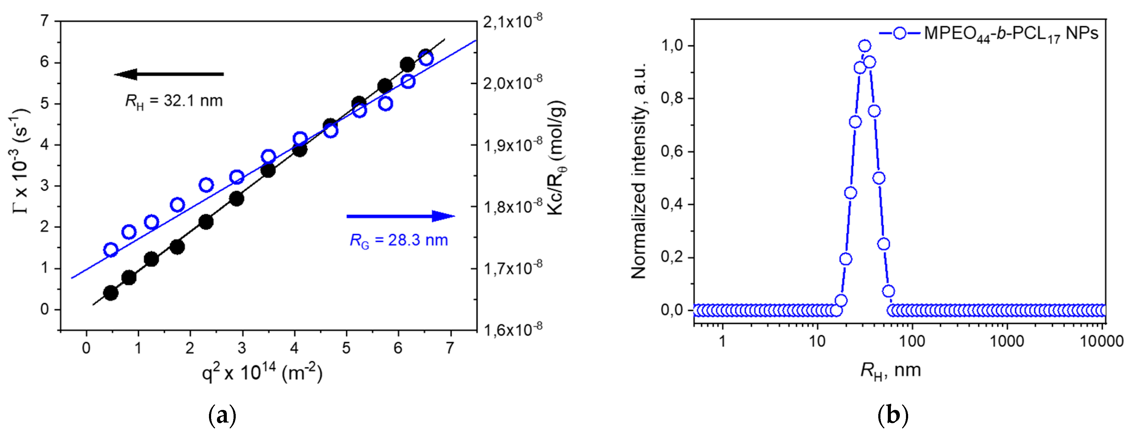
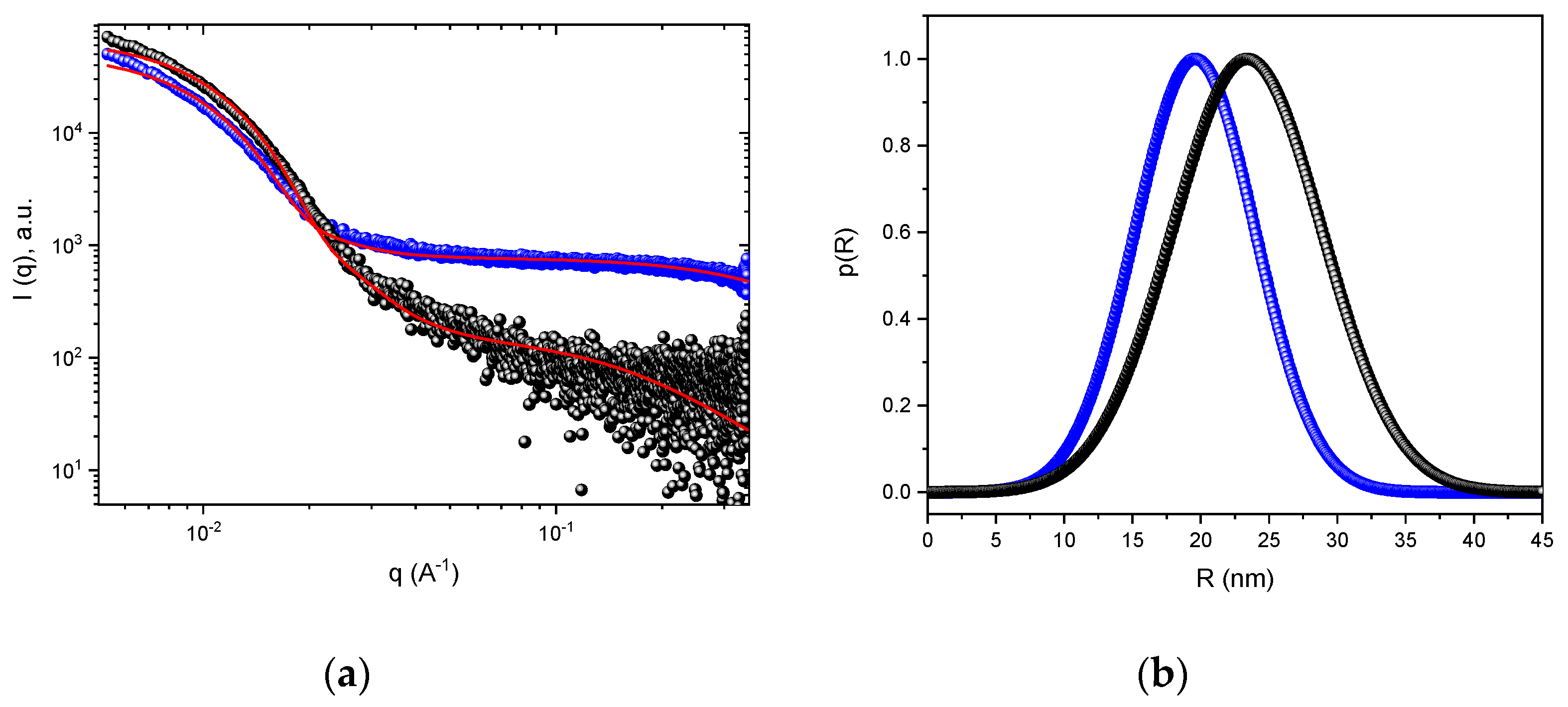
| NPs | RHa | RGb | Rc | Rgshellc | Rggaussc |
|---|---|---|---|---|---|
| MPEO44-b-PCL17 (pH ~7.4) | 32.1 | 28.3 | 23.4 | 1.1 | - |
| MPEO44-b-PCL17 (pH ~5.0) | 15 and 58 | - | 19.5 | 5.6 | 3.0 |
Publisher’s Note: MDPI stays neutral with regard to jurisdictional claims in published maps and institutional affiliations. |
© 2021 by the authors. Licensee MDPI, Basel, Switzerland. This article is an open access article distributed under the terms and conditions of the Creative Commons Attribution (CC BY) license (https://creativecommons.org/licenses/by/4.0/).
Share and Cite
Petrova, S.L.; Jäger, E.; Jäger, A.; Höcherl, A.; Konefał, R.; Zhigunov, A.; Pavlova, E.; Janoušková, O.; Hrubý, M. Development of an Acid-Labile Ketal Linked Amphiphilic Block Copolymer Nanoparticles for pH-Triggered Release of Paclitaxel. Polymers 2021, 13, 1465. https://doi.org/10.3390/polym13091465
Petrova SL, Jäger E, Jäger A, Höcherl A, Konefał R, Zhigunov A, Pavlova E, Janoušková O, Hrubý M. Development of an Acid-Labile Ketal Linked Amphiphilic Block Copolymer Nanoparticles for pH-Triggered Release of Paclitaxel. Polymers. 2021; 13(9):1465. https://doi.org/10.3390/polym13091465
Chicago/Turabian StylePetrova, Svetlana Lukáš, Eliézer Jäger, Alessandro Jäger, Anita Höcherl, Rafał Konefał, Alexander Zhigunov, Ewa Pavlova, Olga Janoušková, and Martin Hrubý. 2021. "Development of an Acid-Labile Ketal Linked Amphiphilic Block Copolymer Nanoparticles for pH-Triggered Release of Paclitaxel" Polymers 13, no. 9: 1465. https://doi.org/10.3390/polym13091465