
Decoration of Ultramicrotome-Cut Polymers with Silver Nanoparticles: Effect of Post-Deposition Laser Treatment


 , , ,
, , , 
Abstract
:1. Introduction
2. Materials and Methods
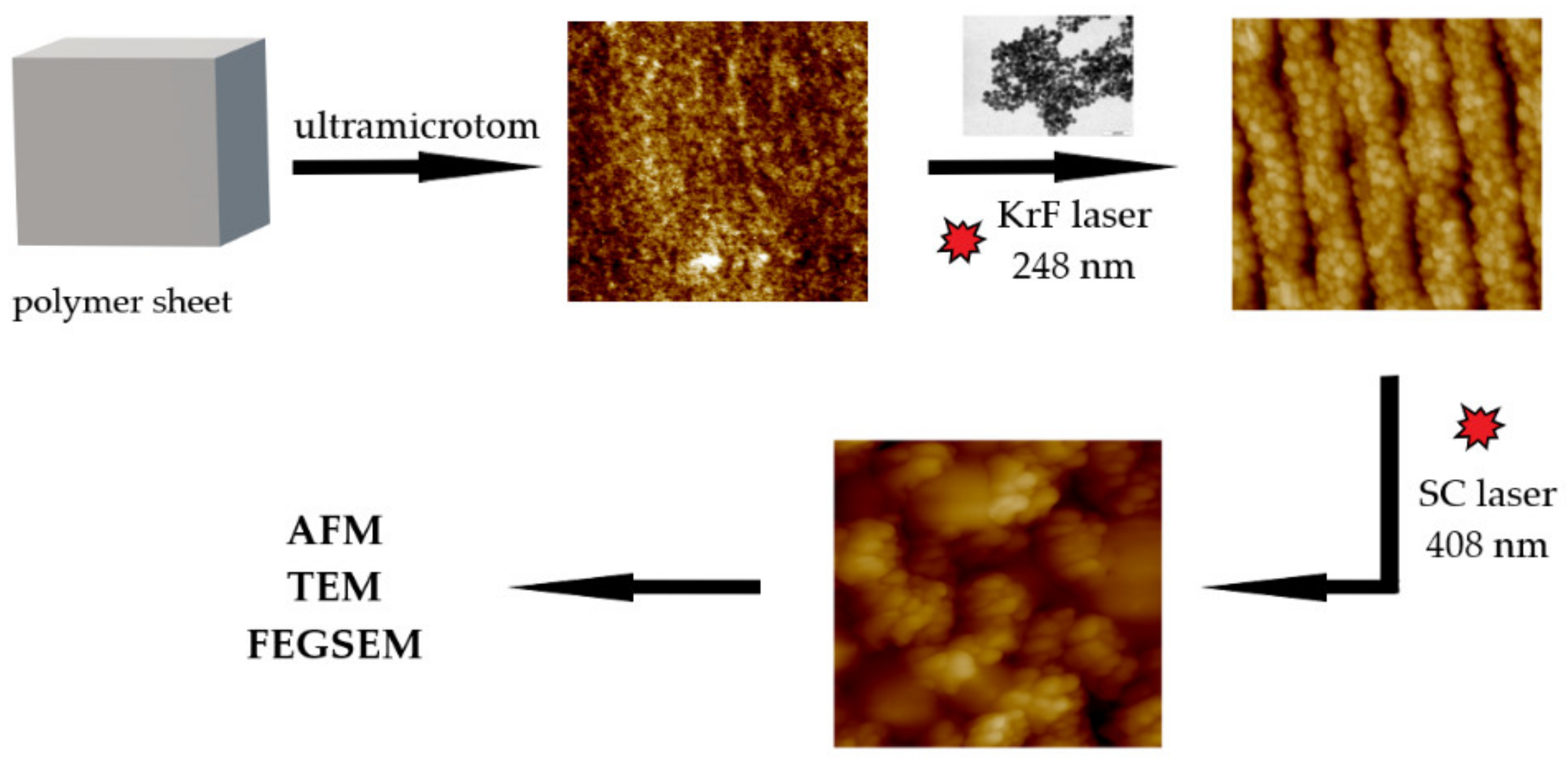
2.1. Materials, Apparatus and Procedures
2.2. Analytical Methods
3. Results and Discussion
4. Conclusions
Author Contributions
Funding
Institutional Review Board Statement
Informed Consent Statement
Data Availability Statement
Acknowledgments
Conflicts of Interest
References
- Kaimlová, M.; Nemogová, I.; Kolářová, K.; Slepička, P.; Švorčík, V.; Siegel, J. Optimization of silver nanowire formation on laser processed PEN: Surface properties and antibacterial effects. Appl. Surf. Sci. 2019, 473, 516–526. [Google Scholar] [CrossRef]
- Pryjmaková, J.; Kaimlová, M.; Vokatá, B.; Hubáček, T.; Slepička, P.; Švorčík, V.; Siegel, J. Bimetallic Nanowires on Laser-Patterned PEN as Promising Biomaterials. Nanomaterials 2021, 11, 2285. [Google Scholar] [CrossRef] [PubMed]
- Pryjmaková, J.; Hryhoruk, M.; Veselý, M.; Slepička, P.; Švorčík, V.; Siegel, J. Engineered Cu-PEN Composites at the Nanoscale: Preparation and Characterisation. Nanomaterials 2022, 12, 1220. [Google Scholar] [CrossRef] [PubMed]
- Polívková, M.; Hubáček, T.; Staszek, M.; Švorčík, V.; Siegel, J. Antimicrobial treatment of polymeric medical devices by silver nanomaterials and related technology. Int. J. Mol. Sci. 2017, 18, 419. [Google Scholar] [CrossRef] [Green Version]
- Wang, J.; Liu, J.; Li, J.; Zhu, J. Characterization of microstructure, chemical, and physical properties of delignified and densified poplar wood. Materials 2021, 14, 5709. [Google Scholar] [CrossRef]
- Do Nascimento Brandão, D.L.; Veiga, A.S.S.; Quaresma, C.C.; Busman, D.V.; de Almeida Lins, A.L.F.; Silveira, F.T.; Campos, M.B.; Percário, S.; Dolabela, M.F. Botanical survey and leishmanicidal activity of grown-love. Res. Soc. Dev. 2020, 9, e3929119983. [Google Scholar] [CrossRef]
- Baden, N. Novel method for high-spatial-resolution chemical analysis of buried polymer-metal interface: Atomic force microscopy-infrared (AFM-IR) spectroscopy with low-angle microtomy. Appl. Spectrosc. 2021, 75, 901–910. [Google Scholar] [CrossRef]
- Al-Aaraji, N.A.-H.; Hashim, A.; Hadi, A.; Abduljalil, H.M. Effect of silicon carbide nanoparticles addition on structural and dielectric characteristics of PVA/CuO nanostructures for electronics devices. Silicon 2022, 14, 4699–4705. [Google Scholar] [CrossRef]
- Sakhno, O.; Yezhov, P.; Hryn, V.; Rudenko, V.; Smirnova, T. Optical and Nonlinear Properties of Photonic Polymer Nanocomposites and Holographic Gratings Modified with Noble Metal Nanoparticles. Polymers 2020, 12, 480. [Google Scholar] [CrossRef] [Green Version]
- Iqbal, S.; Zahoor, C.; Musaddiq, S.; Hussain, M.; Begum, R.; Irfan, A.; Azam, M.; Farooqi, Z.H. Silver nanoparticles stabilized in polymer hydrogels for catalytic degradation of azo dyes. Ecotoxicol. Environ. Saf. 2020, 202, 110924. [Google Scholar] [CrossRef]
- Alshabanah, L.A.; Omran, N.; Elwakil, B.H.; Hamed, M.T.; Abdallah, S.M.; Al-Mutabagani, L.A.; Wang, D.; Liu, Q.; Shehata, N.; Hassanin, A.H. Elastic nanofibrous membranes for medical and personal protection applications: Manufacturing, anti-COVID-19, and anti-colistin resistant bacteria evaluation. Polymers 2021, 13, 3987. [Google Scholar] [CrossRef] [PubMed]
- Diniz, F.R.; Maia, R.C.A.; Rannier Andrade, L.; Andrade, L.N.; Vinicius Chaud, M.; da Silva, C.F.; Corrêa, C.B.; de Albuquerque Junior, R.L.C.; Pereira da Costa, L.; Shin, S.R. Silver nanoparticles-composing alginate/gelatine hydrogel improves wound healing in vivo. Nanomaterials 2020, 10, 390. [Google Scholar] [CrossRef] [PubMed] [Green Version]
- Iida, M.; Goto, T.; Hatakeyama, K.; Ito, T.; Shimizu, Y.; Hakuta, Y.; Terashima, K. Surface modification and Ag nanoparticles support of graphene nanoplates via plasma in liquid. Jpn. J. Appl. Phys. 2020, 59, SHHE08. [Google Scholar] [CrossRef]
- Hupfeld, T.; Wegner, A.; Blanke, M.; Doñate-Buendía, C.; Sharov, V.; Nieskens, S.; Piechotta, M.; Giese, M.; Barcikowski, S.; Gökce, B. Plasmonic seasoning: Giving color to desktop laser 3D printed polymers by highly dispersed nanoparticles. Adv. Opt. Mater. 2020, 8, 2000473. [Google Scholar] [CrossRef]
- Fahmy, A.; Agudo Jácome, L.; Schönhals, A. Effect of silver nanoparticles on the dielectric properties and the homogeneity of plasma poly (acrylic acid) thin films. J. Phys. Chem. C 2020, 124, 22817–22826. [Google Scholar] [CrossRef]
- Dienerowitz, M.; Mazilu, M.; Dholakia, K. Optical manipulation of nanoparticles: A review. J. Nanophotonics 2008, 2, 021875. [Google Scholar] [CrossRef] [Green Version]
- Gao, D.; Shi, R.; Huang, Y.; Gao, L. Fano-enhanced pulling and pushing optical force on active plasmonic nanoparticles. Phys. Rev. A 2017, 96, 043826. [Google Scholar] [CrossRef] [Green Version]
- Lehmuskero, A.; Johansson, P.; Rubinsztein-Dunlop, H.; Tong, L.; Kall, M. Laser trapping of colloidal metal nanoparticles. ACS Nano 2015, 9, 3453–3469. [Google Scholar] [CrossRef]
- Siegel, J.; Kaimlová, M.; Vyhnálková, B.; Trelin, A.; Lyutakov, O.; Slepička, P.; Švorčík, V.; Veselý, M.; Vokatá, B.; Malinský, P. Optomechanical processing of silver colloids: New generation of nanoparticle–polymer composites with bactericidal effect. Int. J. Mol. Sci. 2020, 22, 312. [Google Scholar] [CrossRef]
- Li, H.; Zhang, H.; Luo, W.; Yuan, R.; Zhao, Y.; Huang, J.-A.; Yang, X. Microcontact printing of gold nanoparticle at three-phase interface as flexible substrate for SERS detection of MicroRNA. Anal. Chim. Acta 2022, 1229, 340380. [Google Scholar] [CrossRef]
- Gupta, V.; Sarkar, S.; Aftenieva, O.; Tsuda, T.; Kumar, L.; Schletz, D.; Schultz, J.; Kiriy, A.; Fery, A.; Vogel, N. Nanoimprint Lithography Facilitated Plasmonic-Photonic Coupling for Enhanced Photoconductivity and Photocatalysis. Adv. Funct. Mater. 2021, 31, 2105054. [Google Scholar] [CrossRef]
- Kim, J.Y.; Oh, Y.T.; Lee, S.E.; Park, J.H.; Park, S.; Ko, Y.C.; Hwang, J.P.; Seon, S.W.; Yu, T.S.; Kim, S.H. Collapse-Induced Multimer Formation of Self-Assembled Nanoparticles for Surface Enhanced Raman Scattering. Coatings 2021, 11, 76. [Google Scholar] [CrossRef]
- Angevine, C.E.; Robertson, J.W.; Dass, A.; Reiner, J.E. Laser-based temperature control to study the roles of entropy and enthalpy in polymer-nanopore interactions. Sci. Adv. 2021, 7, eabf5462. [Google Scholar] [CrossRef] [PubMed]
- Huang, J.H.; Cheng, X.Q.; Zhang, Y.; Wang, K.; Liang, H.; Wang, P.; Ma, J.; Shao, L. Polyelectrolyte grafted MOFs enable conjugated membranes for molecular separations in dual solvent systems. Cell Rep. Phys. Sci. 2020, 1, 100034. [Google Scholar] [CrossRef]
- Laucirica, G.; Albesa, A.G.; Toimil-Molares, M.E.; Trautmann, C.; Marmisollé, W.A.; Azzaroni, O. Shape matters: Enhanced osmotic energy harvesting in bullet-shaped nanochannels. Nano Energy 2020, 71, 104612. [Google Scholar] [CrossRef]
- Thomas, A.M.; Peter, J.; Mohan, A.; Nagappan, S.; Selvaraj, M.; Ha, C.-S. Dual stimuli-responsive silver nanoparticles decorated SBA–15 hybrid catalyst for selective oxidation of alcohols under ‘mild’conditions. Microporous Mesoporous Mater. 2021, 311, 110697. [Google Scholar] [CrossRef]
- Karuppaiah, A.; Babu, D.; Selvaraj, D.; Natrajan, T.; Rajan, R.; Gautam, M.; Ranganathan, H.; Siram, K.; Nesamony, J.; Sankar, V. Building and behavior of a pH-stimuli responsive chitosan nanoparticles loaded with folic acid conjugated gemcitabine silver colloids in MDA-MB-453 metastatic breast cancer cell line and pharmacokinetics in rats. Eur. J. Pharm. Sci. 2021, 165, 105938. [Google Scholar] [CrossRef]
- Siegel, J.; Lyutakov, O.; Polívková, M.; Staszek, M.; Hubáček, T.; Švorčík, V. Laser-assisted immobilization of colloid silver nanoparticles on polyethyleneterephthalate. Appl. Surf. Sci. 2017, 420, 661–668. [Google Scholar] [CrossRef]
- Goodfellow Ltd. Cambridge. Available online: https://www.goodfellow.com/de/en-us/displayitemdetails/p/es30-fm-000335/polyethylene-terephthalate-film (accessed on 5 September 2022).
- Goodfellow Ltd. Cambridge. Available online: https://www.goodfellow.com/de/en-us/displayitemdetails/p/ek30-fm-000151/polyetheretherketone-film (accessed on 5 September 2022).
- Kawai, F. Emerging strategies in polyethylene terephthalate hydrolase research for biorecycling. ChemSusChem 2021, 14, 4115–4122. [Google Scholar] [CrossRef]
- Niu, Y.; Zheng, S.; Song, P.; Zhang, X.; Wang, C. Mechanical and thermal properties of PEEK composites by incorporating inorganic particles modified phosphates. Compos. Part B Eng. 2021, 212, 108715. [Google Scholar] [CrossRef]
- Abbott, C.S.; Sperry, M.; Crane, N.B. Relationships between porosity and mechanical properties of polyamide 12 parts produced using the laser sintering and multi-jet fusion powder bed fusion processes. J. Manuf. Process. 2021, 70, 55–66. [Google Scholar] [CrossRef]
- Heitz, J.; Reisinger, B.; Fahrner, M.; Romanin, C.; Siegel, J.; Svorcik, V. Laser-Induced Periodic Surface Structures (LIPSS) on Polymer Surfaces. In Proceedings of the 2012 14th International Conference on Transparent Optical Networks (ICTON), Coventry, UK, 2–5 July 2012; pp. 1–4. [Google Scholar]
- Fazio, E.; Gökce, B.; De Giacomo, A.; Meneghetti, M.; Compagnini, G.; Tommasini, M.; Waag, F.; Lucotti, A.; Zanchi, C.G.; Ossi, P.M. Nanoparticles engineering by pulsed laser ablation in liquids: Concepts and applications. Nanomaterials 2020, 10, 2317. [Google Scholar] [CrossRef] [PubMed]
- Ruiz, S.; Wang, F.; Liu, L.; Lu, Y.; Duan, B.; Korshoj, L.E.; Kielian, T.; Cui, B. Antibacterial properties of silver nanoparticles synthesized via nanosecond pulsed laser ablation in water. J. Laser Appl. 2022, 34, 012031. [Google Scholar] [CrossRef]
- Siegel, J.; Savenkova, T.; Pryjmaková, J.; Slepička, P.; Šlouf, M.; Švorčík, V. Surface Texturing of Polyethylene Terephthalate Induced by Excimer Laser in Silver Nanoparticle Colloids. Materials 2021, 14, 3263. [Google Scholar] [CrossRef] [PubMed]
- Siegel, J.; Grossberger, D.; Pryjmaková, J.; Šlouf, M.; Švorčík, V. Laser-Promoted Immobilization of Ag Nanoparticles: Effect of Surface Morphology of Poly (ethylene terephthalate). Nanomaterials 2022, 12, 792. [Google Scholar] [CrossRef] [PubMed]
- Zhang, J.; Feng, J.; Jia, L.; Xu, R.; Zhao, J.; Zheng, Z.; Zhou, T. Top–Down Direct Preparation of Orange–Yellow Dye Similar to Psittacofulvins from Commercial Polymer by Laser Writing. ACS Appl. Mater. Interfaces 2020, 12, 58339–58348. [Google Scholar] [CrossRef] [PubMed]


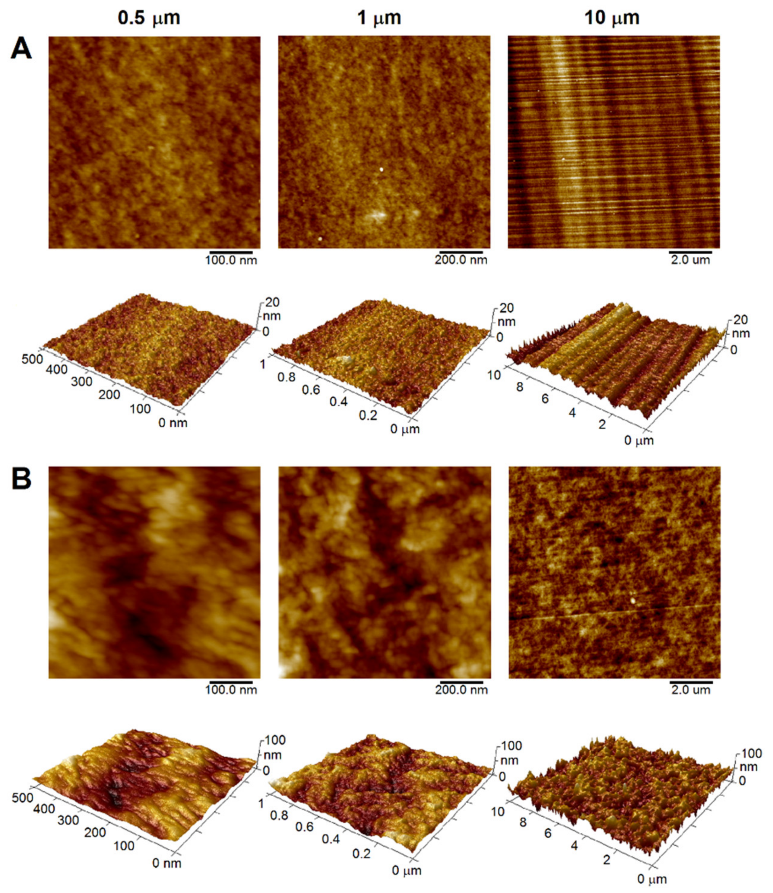
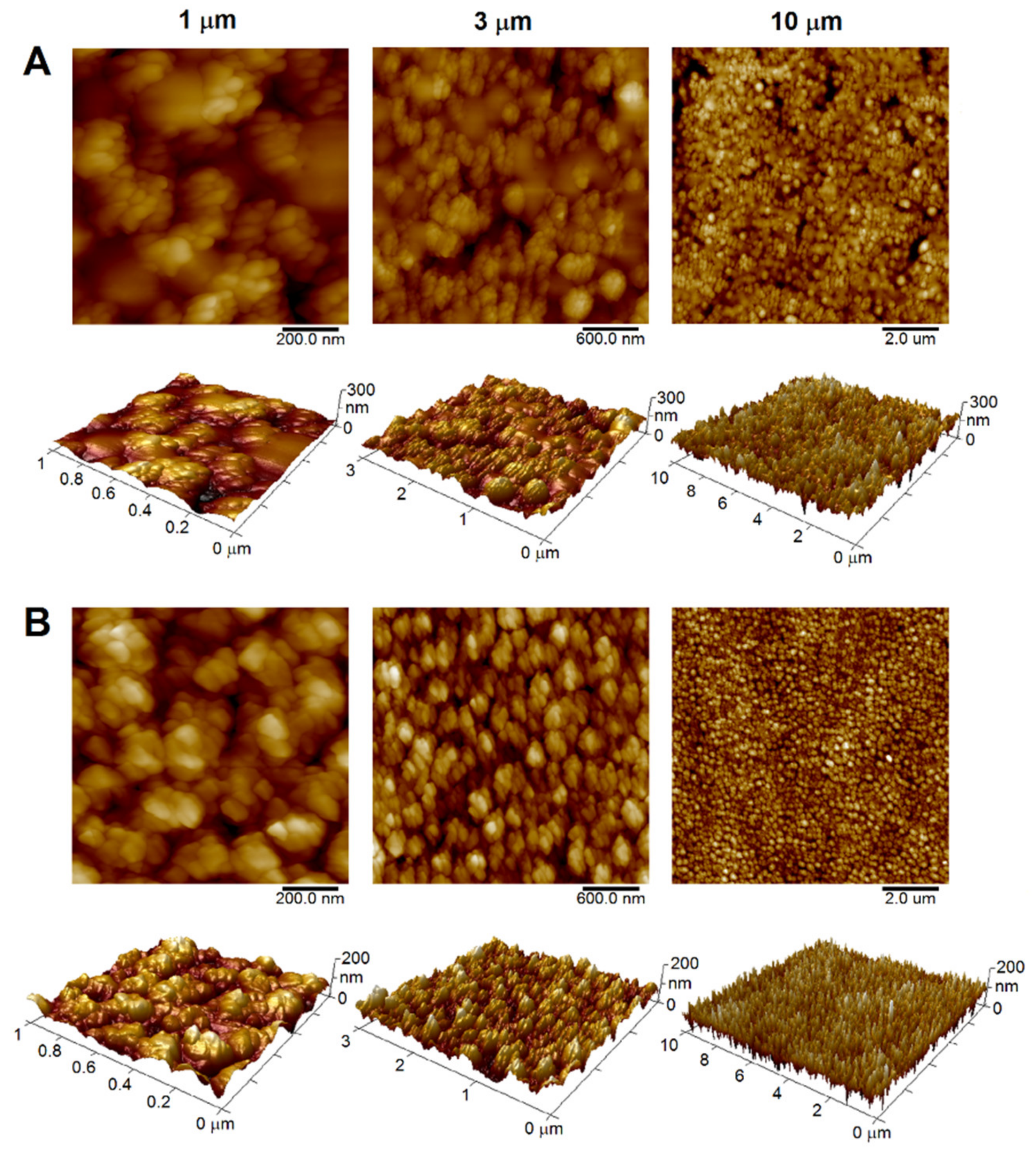
| Sample | Scan Size (µm) | Ra (nm) | SAD (%) |
|---|---|---|---|
| 0.5 | 0.6 | 4.4 | |
| PET (A) | 1 | 0.8 | 2.1 |
| 10 | 1.9 | 0.3 | |
| 0.5 | 4.9 | 9.2 | |
| PEEK (B) | 1 | 4.7 | 5.3 |
| 10 | 8.3 | 2.7 |
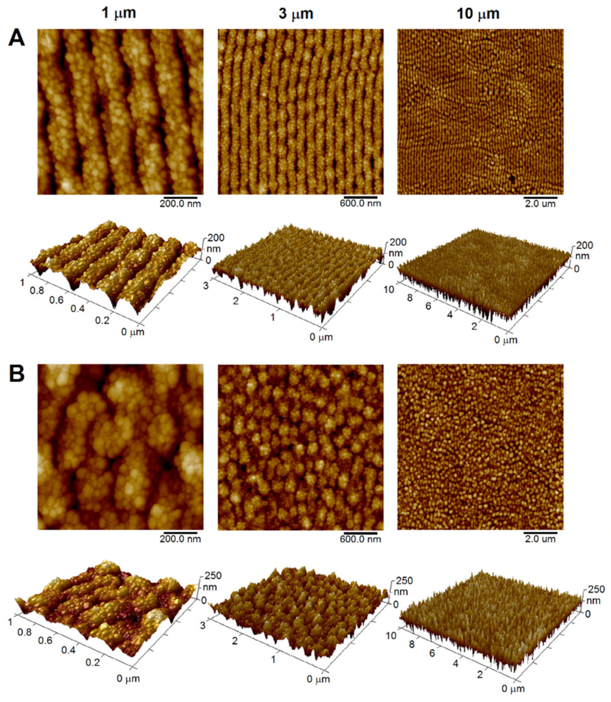
| Sample | Scan Size (µm) | Ra (nm) | SAD (%) |
|---|---|---|---|
| 1 | 20.6 | 89.2 | |
| PET/AgNPs | 3 | 21.8 | 75.2 |
| 10 | 28.1 | 49.2 | |
| 1 | 15.1 | 18.9 | |
| PET/AgNPs + SC center | 3 | 20.0 | 18.0 |
| 10 | 20.6 | 15.2 |
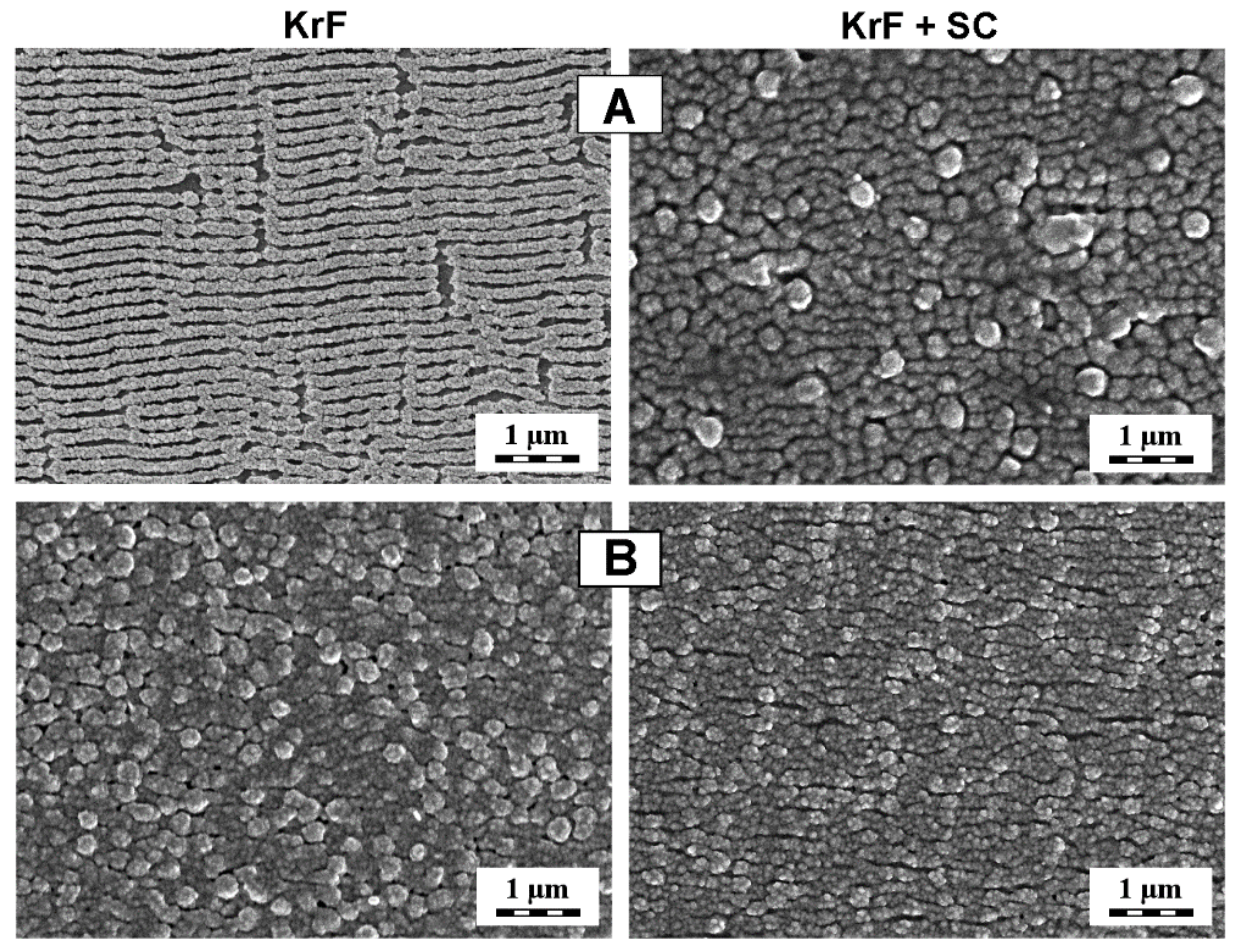
| Sample | Scan Size (µm) | Ra (nm) | SAD (%) |
|---|---|---|---|
| 1 | 21.8 | 67.0 | |
| KrF | 3 | 24.0 | 64.2 |
| 10 | 26.6 | 46.9 | |
| 1 | 18.3 | 42.3 | |
| KrF + SC center | 3 | 19.0 | 25.0 |
| 10 | 20.0 | 23.5 |
Publisher’s Note: MDPI stays neutral with regard to jurisdictional claims in published maps and institutional affiliations. |
© 2022 by the authors. Licensee MDPI, Basel, Switzerland. This article is an open access article distributed under the terms and conditions of the Creative Commons Attribution (CC BY) license (https://creativecommons.org/licenses/by/4.0/).
Share and Cite
Kaimlová, M.; Pryjmaková, J.; Šlouf, M.; Lyutakov, O.; Ceccio, G.; Vacík, J.; Siegel, J. Decoration of Ultramicrotome-Cut Polymers with Silver Nanoparticles: Effect of Post-Deposition Laser Treatment. Materials 2022, 15, 8950. https://doi.org/10.3390/ma15248950
Kaimlová M, Pryjmaková J, Šlouf M, Lyutakov O, Ceccio G, Vacík J, Siegel J. Decoration of Ultramicrotome-Cut Polymers with Silver Nanoparticles: Effect of Post-Deposition Laser Treatment. Materials. 2022; 15(24):8950. https://doi.org/10.3390/ma15248950
Chicago/Turabian StyleKaimlová, Markéta, Jana Pryjmaková, Miroslav Šlouf, Oleksiy Lyutakov, Giovanni Ceccio, Jiří Vacík, and Jakub Siegel. 2022. "Decoration of Ultramicrotome-Cut Polymers with Silver Nanoparticles: Effect of Post-Deposition Laser Treatment" Materials 15, no. 24: 8950. https://doi.org/10.3390/ma15248950