Article

Differential Antioxidant Response to Supplemental UV-B Irradiation and Sunlight in Three Basil Varieties

by
Sonja Milić Komić
1,†,
Bojana Živanović
1,†,
Jelena Dumanović
2,
Predrag Kolarž
3,
Ana Sedlarević Zorić
1,
Filis Morina
4,
Marija Vidović
5 and
Sonja Veljović Jovanović
1,*
1
Institute for Multidisciplinary Research, Department of Life Science, University of Belgrade, Kneza Višeslava 1, 11030 Belgrade, Serbia
2
Department of Analytical Chemistry, Faculty of Chemistry, University of Belgrade, 11158 Belgrade, Serbia
3
Institute of Physics Belgrade, University of Belgrade, 11080 Belgrade, Serbia
4
Biology Center of the Czech Academy of Sciences, Institute of Plant Molecular Biology, Department of Plant Biophysics and Biochemistry, Branišovska 31/1160, 370 05 Ceske Budejovice, Czech Republic
5
Institute of Molecular Genetics and Genetic Engineering, Laboratory for Plant Molecular Biology, University of Belgrade, Vojvode Stepe 444a, 11042 Belgrade, Serbia
*
Author to whom correspondence should be addressed.
These authors contributed equally to this work.
Int. J. Mol. Sci. 2023, 24(20), 15350; https://doi.org/10.3390/ijms242015350
Submission received: 22 September 2023 / Revised: 9 October 2023 / Accepted: 17 October 2023 / Published: 19 October 2023

Abstract

:
Three basil plant varieties (Ocimum basilicum var. Genovese, Ocimum × citriodorum, and Ocimum basilicum var. purpurascens) were grown under moderate light (about 300 µmol photons m−2 s−1) in a glasshouse or growth chamber and then either transferred to an open field (average daily dose: 29.2 kJ m−2 d−1) or additionally exposed to UV-B irradiation in a growth chamber (29.16 kJ m−2 d−1), to reveal the variety-specific and light-specific acclimation responses. Total antioxidant capacity (TAC), phenolic profile, ascorbate content, and class III peroxidase (POD) activity were used to determine the antioxidant status of leaves under all four light regimes. Exposure to high solar irradiation at the open field resulted in an increase in TAC, total hydroxycinnamic acids (HCAs, especially caffeic acid), flavonoids, and epidermal UV-absorbing substances in all three varieties, as well as a two-fold increase in the leaf dry/fresh weight ratio. The supplemental UV-B irradiation induced preferential accumulation of HCAs (rosmarinic acid) over flavonoids, increased TAC and POD activity, but decreased the ascorbate content in the leaves, and inhibited the accumulation of epidermal flavonoids in all basil varieties. Furthermore, characteristic leaf curling and UV-B-induced inhibition of plant growth were observed in all basil varieties, while a pro-oxidant effect of UV-B was indicated with H2O2 accumulation in the leaves and spotty leaf browning. The extent of these morphological changes, and oxidative damage depended on the basil cultivar, implies a genotype-specific tolerance mechanism to high doses of UV-B irradiation.

1. Introduction

Plants are inevitably exposed to seasonal and diurnal variations in light intensity and quality as a result of variations in the ratio of UV-A/UV-B/photosynthetically active radiation (PAR, 400–700 nm) [1,2]. Ultraviolet (UV) irradiation accounts for only a small fraction of total solar radiation, about 6% UV-A (315–400 nm) and 0.5% UV-B (290–315 nm); nevertheless, it can induce a variety of responses in plants. Since the 1980s, the depletion of the stratospheric ozone layer has led to an increase of up to 14% in UV-B radiation reaching the Earth’s surface [3]. The Montreal Protocol and its amendments were remarkably effective in saving the Earth’s stratospheric ozone layer and led to its restoration. Nevertheless, there is a strong link between ozone depletion and climate changes caused by increasing greenhouse gases. Recent studies predict a 3–8% increase in the UV index over the tropics or mid-latitudes by 2100, depending on the greenhouse gas scenario, cloud cover, and aerosol concentration used for modelling [4,5,6]. The predicted decrease in mean cloud cover over the Mediterranean region, due to climate change, could also lead to an increase in the intensity of UV radiation over the Mediterranean region in the near future [7]. The meteorological data for this study were collected in Belgrade (Serbia), situated at the 45th parallel of northern latitude humid subtropical and humid continental climate, exhibiting four distinct seasons and precipitation evenly distributed throughout the year. Malinović-Milićević and co-authors showed that the average daily UV-B dose ranged from 2.062 kJ m−2 (December) to 58.773 kJ m−2 (June) and estimated a 3.7% increase in UV-B radiation intensity per decade in the territory of Vojvodina (northern region of Serbia) [8]. Similar UV-B doses have been measured in Cyprus, where radiation doses vary from 70 kJ m−2 in Larnaca to 55 kJ m−2 in Athalassa, depending on the climate regime of the island [9]. High UV-B radiation can cause DNA damage and/or induce the generation of reactive oxygen species (ROS), which can oxidize proteins and membranes, and inhibit photosynthesis and growth [10]. However, plants rarely show signs of damage even though they are constantly exposed to natural UV-B irradiation [11]. There are numerous studies with conflicting results on the effect of UV-B irradiation on plants, which can be explained with different ratios of UV-B/UV-A/PAR, and unnaturally high UV-B doses applied in the experiments, as well as species/genotype-specific sensitivity to UV-B radiation, different experimental conditions, and previous acclimation periods [12]. Moreover, high PAR, combined with other limiting environmental factors such as water and/or nutrient deficiencies, can overcome the capacity of photosynthetic assimilation and energy dissipation processes and provoke the generation of ROS, resulting in photo-oxidative damage to the photosynthetic apparatus [13,14].
Plants respond to light by developing numerous photoprotective mechanisms against the deleterious effects of excess PAR and UV-B, of which the antioxidant network and redox signaling pathways play an important role in maintaining cellular redox homeostasis [15,16]. The most efficient and rapid photoprotective process of the photosynthetic apparatus is the dissipation of excess excitation energy in photosystem II (PSII) with heat, which is controlled with the trans-thylakoid proton gradient and zeaxanthin formation [17,18]. As part of the acclimation mechanisms to PAR and UV radiation, the enhanced biosynthesis of phenolics can also be considered a photosynthetic energy escape valve, with 20% of the fixed carbon in photosynthesis being directed into the phenylpropanoid pathway [19]. The accumulation of phenolics, particularly flavonoids, is considered a hallmark of the UV-B response in plants [2]. Polyphenols are the largest and most diverse group of secondary metabolites that play a crucial role in the dynamic interactions between plants and the environment. Under full sunlight, plants tend to accumulate UV-absorbing phenolics, especially flavonols, in the epidermal layer of exposed leaves, providing them with an effective protective shield against potentially harmful UV radiation [20]. In addition to their protective role in attenuating UV radiation and PAR, flavonoids and anthocyanidins are potent antioxidants, whose efficiency in ROS scavenging is mainly determined with their structure [21]. Flavonoids and anthocyanidins with ortho-dihydroxyl substitution in the B ring have almost four times higher antioxidant activity than the other polyphenols. Anthocyanins exhibit multiple functional roles in plants that have to cope with a variety of abiotic and biotic stress factors [22,23]. In addition to their absorption in the blue and green regions of the spectrum, they are extremely effective antioxidants that can scavenge ROS in vitro and act as substrates for PODs and participate in H2O2 scavenging [24]. Anthocyanins are commonly induced with high light intensity, low nitrogen content, and low temperatures. On the other hand, both UV-B and high PAR intensities induce phenylpropanoid accumulation in the epidermal layers, such as quercetin and catechin, which also exhibit a strong antioxidant potential through direct interaction with ROS [25].
In contrast to the deleterious effects of high UV-B irradiation, low UV-B levels trigger a UV-B-specific signaling pathway that mediates photomorphogenic responses and the development of the so-called “UV-acclimated phenotype” with shorter petioles and shorter, narrower, and/or thicker leaf blades, as well as leaf curling [26]. The photomorphogenic UV-B signaling pathway is mediated with the UV-B-specific component UV RESISTANCE LOCUS8 (UVR8). Both UVR8 and CONSTITUTIVE PHOTOMORPHOGENESIS 1 (COP1) are required for UV-B-induced expression of the ELONGATED HYPOCOTYL5 (HY5) transcription factor, which plays a central role in regulating genes involved in photomorphogenic UV-B responses [2].
Basil (Ocimum basilicum L.) is a widely used aromatic herbaceous plant in nutrition, as well as in traditional medicine, pharmaceuticals, perfumes, and cosmetics [27,28]. Traditionally, basil was grown in an open field. However, in response to increasing market demand, and unpredictable weather conditions, basil cultivation has strongly shifted to controlled environment agriculture, such as glasshouses, high tunnels, and vertical indoor farms for herb production [29,30]. Light intensity and quality have a significant impact on the basil growth rate, morphology, and anatomical characteristics [31,32,33]. Considering the increased demand for high-quality basil (particularly fresh leaf material), special attention is paid to the cultivation treatment, especially the beneficial effects of low UV radiation on the functional properties, antioxidant value, and content of secondary metabolites [34,35]. In different basil varieties (purple and green), competition in the induction of either flavonoid or anthocyanin metabolic pathways depending on light quality and intensity has been shown [36]. In addition, purple basil varieties have attracted increasing interest in the last decade due to their much higher content of health-promoting substances compared to green varieties, especially when the plants are grown under limited light intensity [37]. A comparison of green and red basil leaves developed at full sunlight showed higher photosynthetic performance and higher stomatal conductance in red leaves than in green ones at a given time point [36]. However, to our knowledge, dynamics of the accumulation of UV screening compounds in basil have not been investigated. Moreover, varieties of O. basilicum with constitutively different contents of anthocyanins and phenolics represent a suitable system to investigate the acclimation response to different UV/PAR ratios. Comparative analyses of green vs. purple leaf response of the same age within the same species to light treatments can reveal the functional role of anthocyanins, which is still debatable as discussed by Landi and co-authors [38]. In addition, lemon basil differs from other varieties not only by its characteristic aroma and high content of biologically active polyphenolic compounds, but also by its particular morphology, characterized by a small stature, early flowering, and narrow leaves [39].
In this study, we aimed to distinguish between UV-B and high PAR effects on their pro-oxidant effects as well as on ascorbate and polyphenol contents, total and epidermal, in the leaves of three commonly consumed basil varieties: “Genovese” (Ocimum basilicum Genovese, GB), “Lemon” (Ocimum × citriodorum, CB), and “Purple” (Ocimum basilicum purpurascens, PB). We compared two green-leaf varieties and one purple-leaf cultivar, to investigate the functional role of anthocyanins in photoprotection (including excessive PAR and UV-B radiation). Based on the physicochemical properties of anthocyanins and other abundant phenylpropanoids in the leaves of three basil varieties, we hypothesized that the purple-leaf cultivar would be more tolerant to high solar radiation, and show fewer symptoms of oxidative stress damage. Furthermore, we aimed to find out whether the varieties differ in their dynamics of acclimation response to different UV/PAR ratios (supplemental UV-B irradiation, and full sun irradiance). To achieve this, the analyses of phenolic profiles, dynamics of accumulation of epidermal UV-absorbing compounds, and redox status were performed.

2. Results

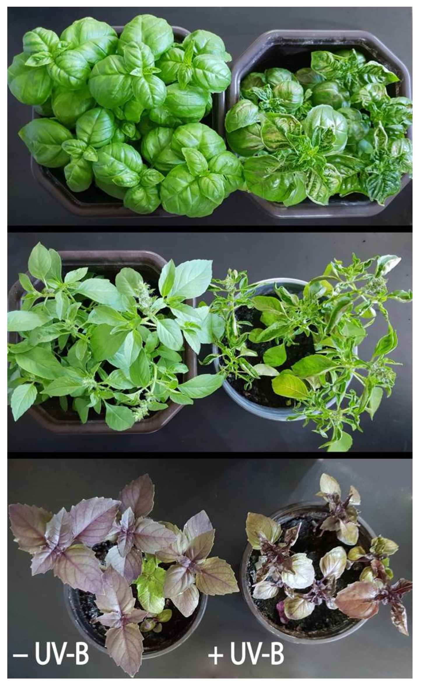
2.1. Effects of Light Regimes on Growth

After 1 month of growth in the glasshouse (GH) with complete attenuation of UV irradiation and 84% of solar PAR, half of the basil plants were transferred to an open field (OF). Upon transferring the basil plants from the glasshouse to full sun for 15 days in the open field (the average daily UV-B dose was 29.20 kJ m−2), all three varieties showed differences in the fresh/dry weight ratio (FW/DW) compared to glasshouse plants (experiment I; Table 1). The FW/DW ratios were lower in plants grown in the open field compared to the glasshouse plants regardless of variety, with the most pronounced difference in Ocimum basilicum var. purpurascens (PB). However, only “lemon” basil (CB), DW, and, to a lesser extent, FW increased in the open field compared to the glasshouse, while growth inhibition (evidenced in FW) was only observed in “Genovese” basil (GB) (Table 1). In addition, 1 week after full sun exposure, leaf thickening was observed in “lemon” basil (CB).
In experiment II, 1-month-old plants were grown in a growth chamber (GC) under controlled conditions (PAR, 300 ± 50 μmol m−2 s−1), and half of the plants of all three varieties were exposed to supplemental UV-B irradiation (1.35 W m−2, i.e., 29.16 kJ m−2) for 6 h per day (UV-B). After only 3 days, the first small brown blotches/spots, characteristic of UV-induced damage, were noticed on the directly exposed leaves. Over the next 5 days, the damaged leaf areas increased in size and became necrotic and the young leaves began to curl, especially in the Genovese variety, which appeared to be the most sensitive variety (Figure 1). Moreover, chlorophyll content was measured with Dualex and no significant changes were observed in both experiments (Supplementary Figure S1, Supplementary Table S1).

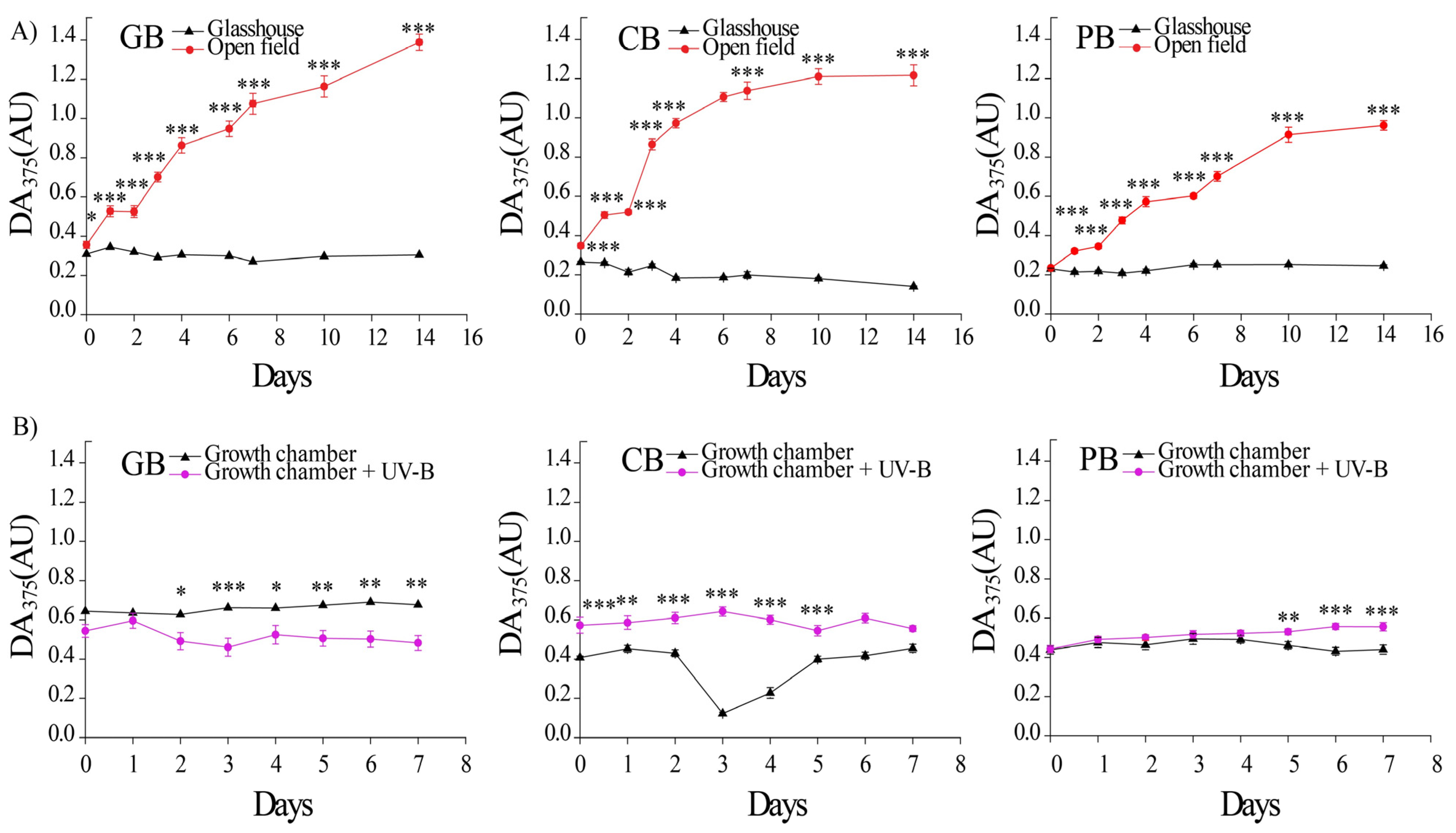
2.2. Light-Induced Accumulation of UV-Absorbing Substances

In the glasshouse, the basal concentration of epidermal flavonoids (EpFlav, DA375) in the leaves of all three basil varieties ranged from 0.2 to 0.4 (AU). Nevertheless, an initial increase in EpFlav accumulation was detected 24 h after transferring the plants to the open field, and it continued to increase in all varieties, but the increase rate was the slowest in Ocimum basilicum var. purpurascens (Figure 2A). On the other hand, EpFlav content in leaves of plants grown continuously in the glasshouse remained constant. After 15 days, the content of epidermal UV-absorbing compounds was seven-fold higher in Ocimum basilicum var. Genovese and Ocimum × citriodorum and five-fold higher in Ocimum basilicum var. purpurascens grown in the open field compared to the respective varieties grown in the glasshouse.
Exposure to supplemental UV-B irradiation at constant PAR, comparable to the intensity of solar UV-B irradiation, resulted in only a slight increase in EpFlav accumulation in all three basil varieties, far less dramatic than in the open field conditions, where the EpFlav content increase was four–nine times higher in comparison to the glasshouse (Figure 2B, significant “light” effects, Supplementary Table S2).

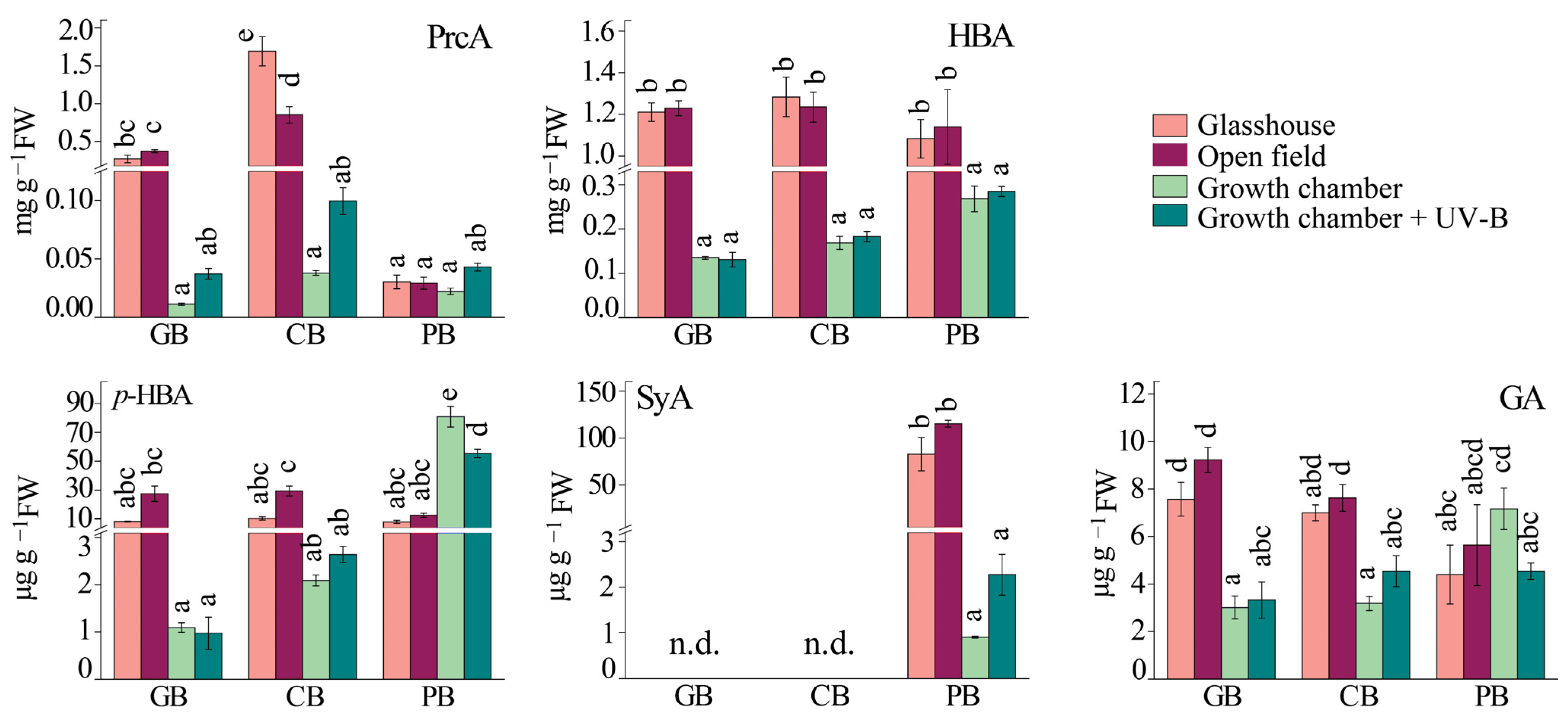
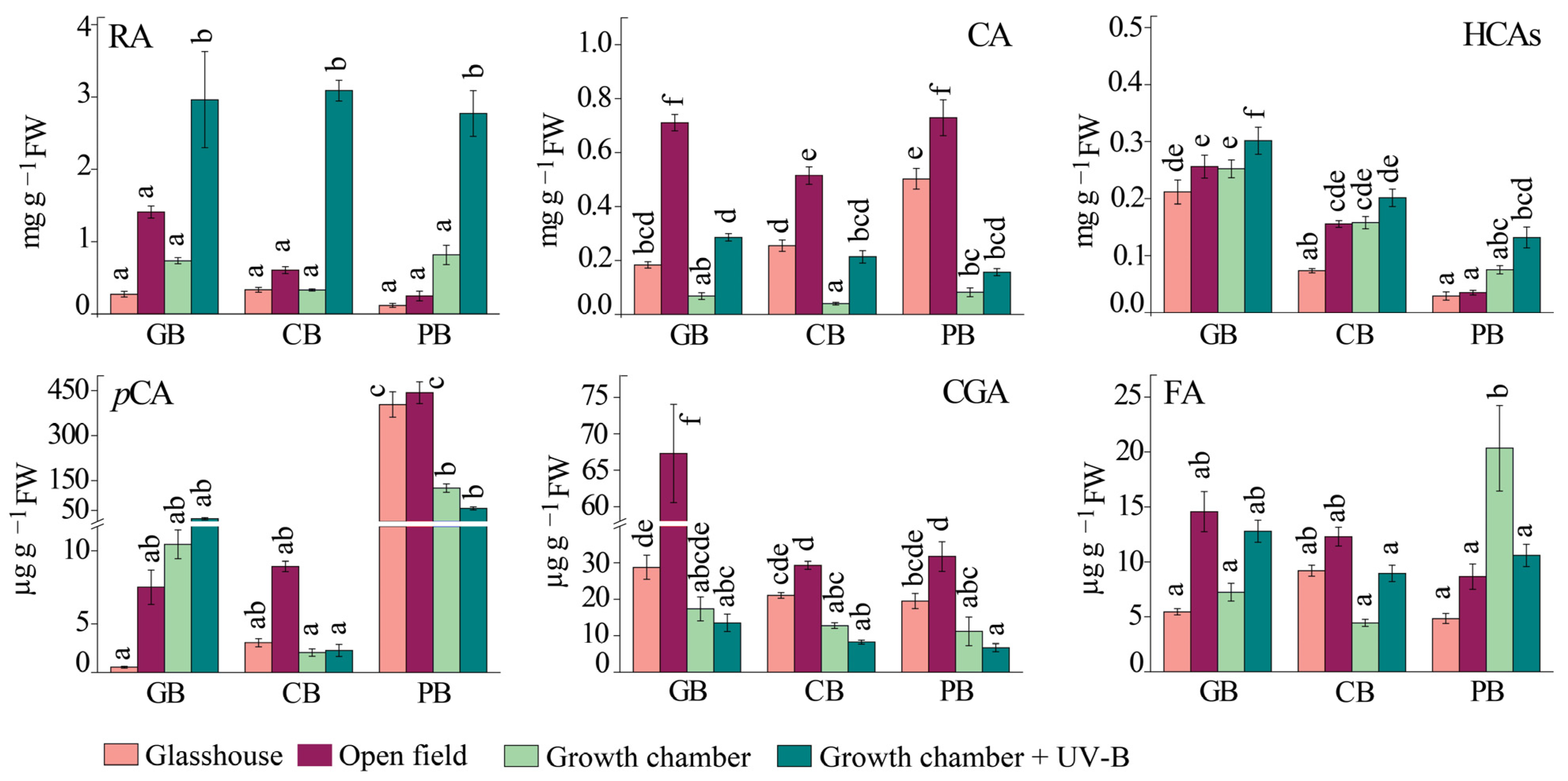
2.3. Phenolic Profile of Basil Leaves under Different Light Regimes

Significantly higher basal levels of syringic acid (SyA), p-coumaric acid (pCA), and cyanidin (Cy) were found in the Ocimum basilicum var. purpurascens variety. Furthermore, anthocyanins were only detected in Ocimum basilicum var. purpurascens. Ocimum basilicum var. Genovese had significantly higher basal levels of hydroxycinnamic acids (HCAs), and the lowest basal levels of quercetin (Q), while Ocimum × citriodorum had the highest basal levels of protocatechuic acid (PrcA).
Generally, the most abundant hydroxybenzoic acids (HBAs) in all three varieties under all light regimes were protocatechuic acid (PrcA) and unidentified HBA, followed by p-HBA, and gallic acid (GA) (Figure 3). The HBA and GA contents were not affected by full sunlight exposure or UV-B supplementation (Figure 3), while significant differences in the levels of PrcA and p-HBA induced with sunlight were observed between the three basil cultivars (Supplementary Table S3, ANOVA, p ≤ 0.05). The accumulation of p-HBA was slightly higher with full sunlight compared to the glasshouse for all three cultivars, while the highest accumulation of this compound was noted in the leaves of Ocimum basilicum var. purpurascens from the growth chamber. The content of syringic acid (SyA) (variety-specific for PB) was significantly higher in the growth chamber and the open field than in the glasshouse and under UV-B (Figure 3).
The dominant hydroxycinnamic acids (HCA) in all three varieties were rosmarinic acid (RA), caffeic acid (CA), and p-coumaric acid (pCA). Sunlight exposure (open field) and UV-B supplementation had the most remarkable effects on the accumulation of RA in all three varieties, but to a greater extent with UV-B supplementation. Nevertheless, the content of RA within the same treatment was relatively similar among basil varieties (Figure 4, Supplementary Table S4, ANOVA, p ≤ 0.05).
Common responses to sunlight (OF) and UV-B supplementation were increased levels of caffeic acid (CA) and other unidentified hydroxycinnamic acids in all three cultivars. While full sunlight resulted in higher accumulation of CA in all three varieties (although the increase was the lowest in purple basil), UV-B supplementation had a much greater impact on total HCA content (the increase with UV-B treatment was from 5 to 9 µmol g−1 FW in Ocimum basilicum var. Genovese, 2.6 to 9.7 µmol g−1 FW in Ocimum × citriodorum, and 0.94 to 9 µmol g−1 FW in Ocimum basilicum var. purpurascens). Opposite were the effects of open field treatment compared to UV-B supplementation that were observed for chlorogenic acid (CGA) accumulation (Supplementary Table S4, Supplementary Figure S2). On the other hand, the content of ferulic acid (FA) was not significantly affected by the different treatments, although an increasing trend was observed in Ocimum basilicum var. Genovese in the open field (Figure 4, Supplementary Table S4, ANOVA, p ≤ 0.05).
Interestingly, the accumulation of flavan-3-ols, catechin (Cat), and epicatechin (ECat) was opposite in Ocimum × citriodorum and Ocimum basilicum var. purpurascens compared to the glasshouse and open field (Figure 5, Supplementary Table S5). ECat levels increased in Ocimum × citriodorum and Ocimum basilicum var. Genovese, whereas Cat levels were higher in Ocimum basilicum var. purpurascens and Cat was not found in Ocimum basilicum var. Genovese. Moreover, UV-B supplementation induced ECat accumulation in all three basil varieties compared with a respective treatment without UV-B (GC).
Furthermore, the induction of quercetin (Q) accumulation was a common response of all three varieties at full sunlight (OF), but to different extents: it increased almost 20-fold in Ocimum basilicum var. Genovese plants compared with the glasshouse plants, and 4-fold and 2-fold in Ocimum × citriodorum and Ocimum basilicum var. purpurascens, respectively (Figure 5, Supplementary Table S5). However, Q content in Ocimum × citriodorum and Ocimum basilicum var. purpurascens did not change significantly after UV-B exposure compared to the growth chamber in any other variety. As expected, the anthocyanin cyanidin (Cy) was only detected in Ocimum basilicum var. purpurascens leaves. Plants grown in the glasshouse and the open field had more than 10 times higher Cy content than plants grown in the growth chamber and under UV-B (Figure 5).

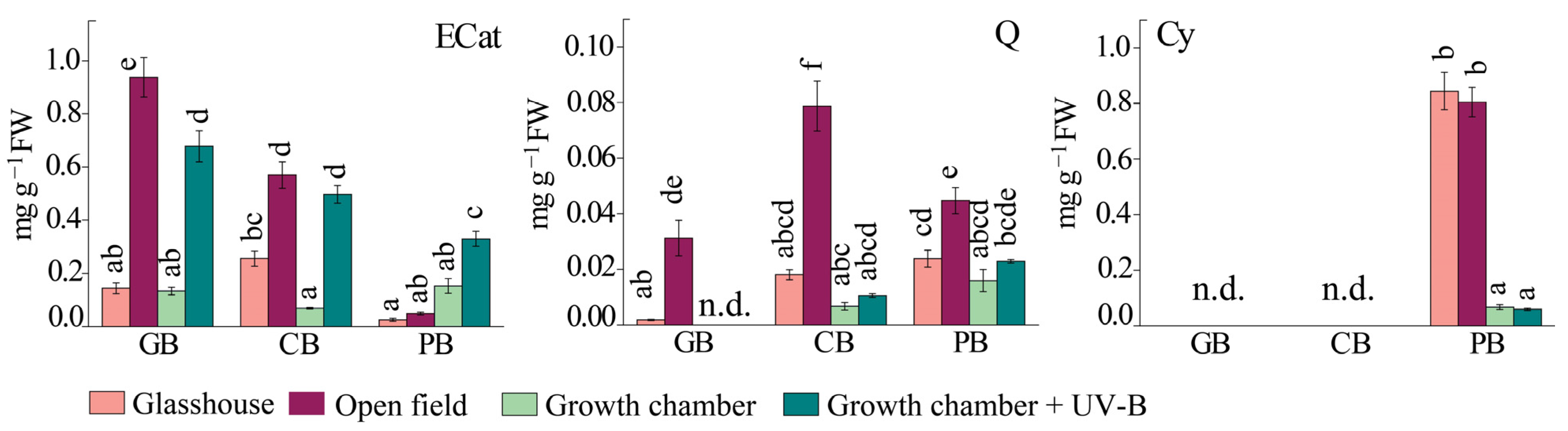
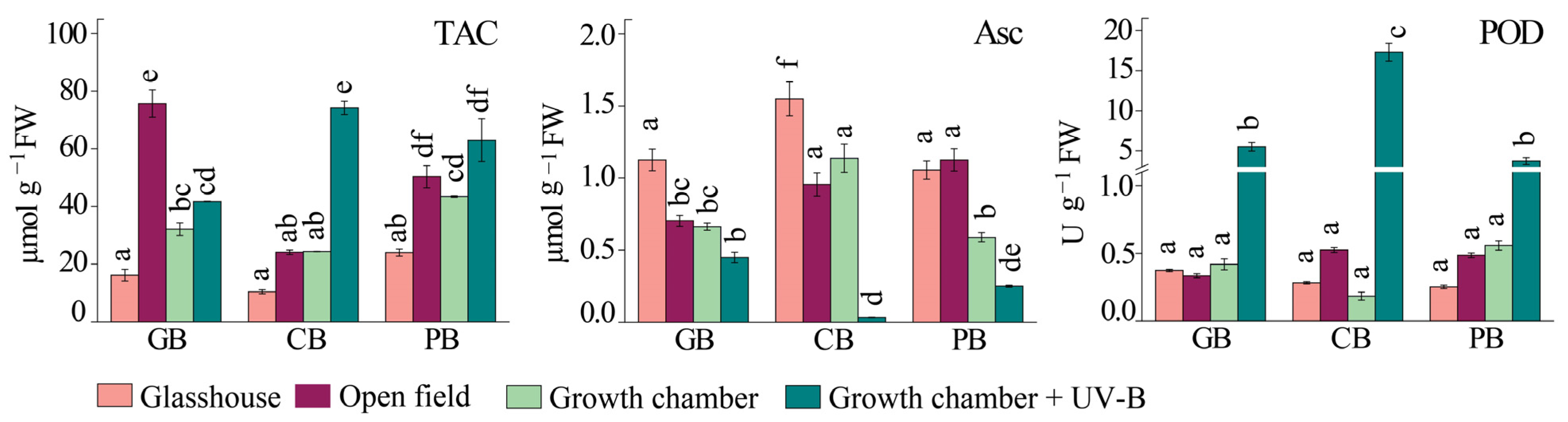
2.4. Antioxidant Response

Total antioxidant capacity (TAC), as determined with the ABTS assay, was highest in the leaves of Ocimum basilicum var. purpurascens plants under glasshouse and growth chamber light conditions (Figure 6). When plants were exposed to full sunlight for 15 days (open field), TAC increased in all three basil varieties compared to plants grown in the glasshouse. The highest increase was measured in Ocimum basilicum var. Genovese (about 4.6-fold higher TAC values compared with the glasshouse), followed by Ocimum × citriodorum and Ocimum basilicum var. purpurascens (about 2-fold higher values) (Figure 6). In contrast, exposure to artificial UV-B irradiation induced only a slight increase in TAC in Ocimum basilicum var. Genovese and Ocimum basilicum var. purpurascens, whereas a three-fold increase was observed in Ocimum × citriodorum (Supplementary Table S6, ANOVA, p ≤ 0.05).
The concentration of reduced ascorbate (Asc) was decreased in Ocimum basilicum var. Genovese and Ocimum × citriodorum plants grown in the open field compared to those grown in the glasshouse. At the same time, no significant changes in reduced ascorbate content were observed in Ocimum basilicum var. purpurascens plants when the plants were exposed to full sunlight. Moreover, the content of reduced ascorbate was significantly decreased in all plants under UV-B treatment in contrast to the control plants in the growth chamber (Figure 6).
No significant changes in the activity of the class III peroxidases (PODs) were observed in the plants grown in the open field compared to plants grown in the glasshouse (Figure 6, Supplementary Table S6). POD activity was similar between basil varieties, as well as between the different treatments. On the other hand, the POD activity drastically increased in response to UV-B supplementation in all three varieties compared to the growth chamber.

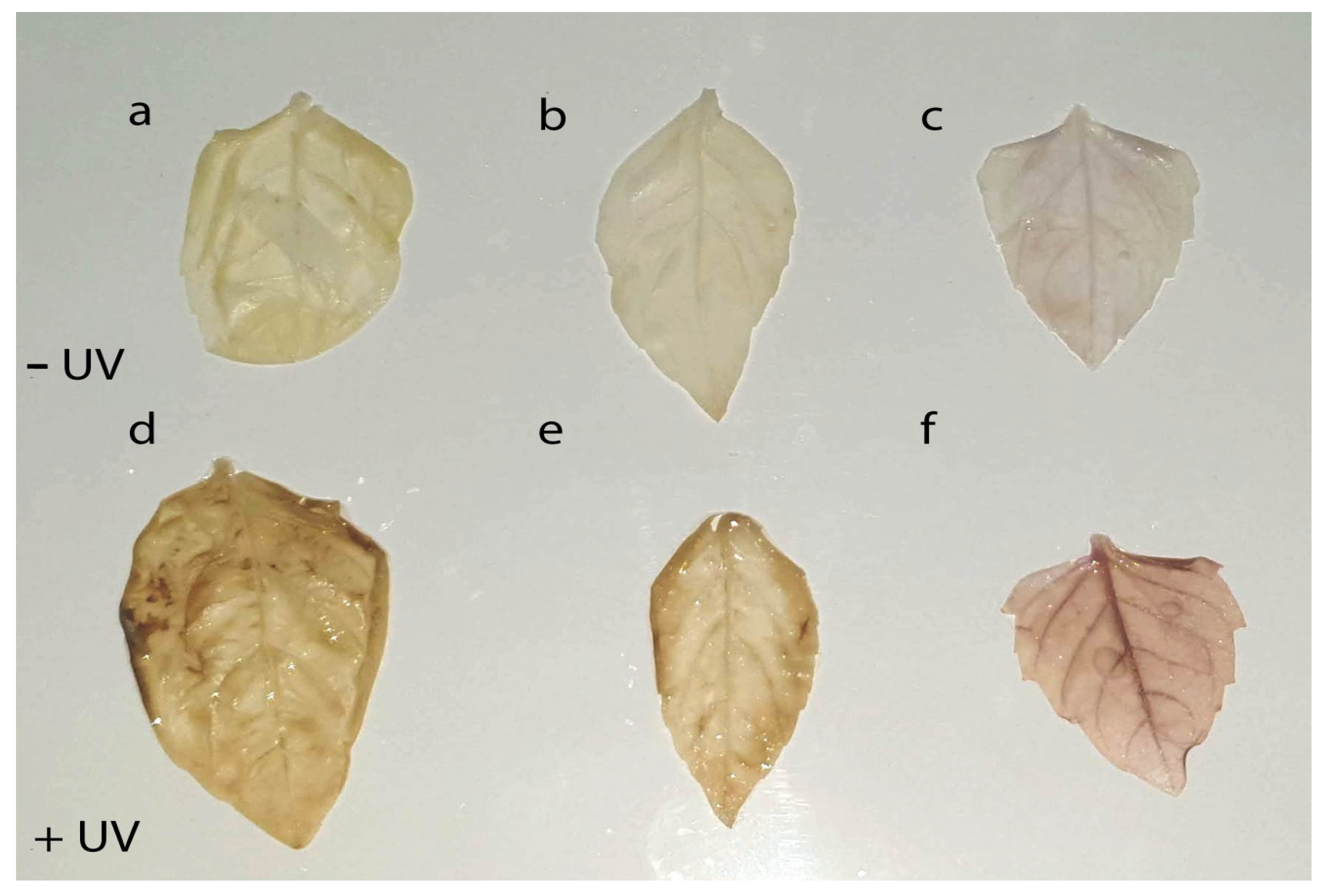
2.5. H2O2 Accumulation in Leaves

Visible signs of damage in response to changing light conditions were observed only in experiment II, following exposure to UV-B irradiation at constant PAR. To elucidate whether UV-B irradiation provoked oxidative injury in leaves, 8 days after the start of treatment, H2O2 accumulation was visualized with the DAB uptake method. Indeed, in all three basil varieties, an increased H2O2 accumulation was observed both in the veins and in mesophyll cells compared to leaves of non-UV-B treated plants (Figure 7).

3. Discussion

3.1. Contrasting Effects of Sunlight and UV-B/Moderate Light

The three varieties of basil used in the study showed specific, as well as general, responses to changing light conditions—full sunlight or UV-B supplementation under moderate PAR. While high sunlight including natural UV-B irradiation developed no visible signs of photo-oxidative damage in any variety, it induced a strong antioxidant response with a significant increase in TAC and total HCAs and both total flavonoid leaf content and epidermal flavonoids indicated ROS signaling in antioxidant defense [40]. In contrast, a supplemental UV-B irradiation (daily dose: 29.16 kJ m−2 d−1, comparable to the intensities of ambient UV-B) combined with moderate background PAR induced oxidative stress within 8 days, which may imply a deficient mechanism of antioxidative defense under the given condition [11]. This was demonstrated with the accumulation of H2O2 and the appearance of brown lesions on the upper surface of mature leaves. Moreover, under supplemental UV-B irradiation, the formation of the UV-absorbing shield failed, which was accompanied by the absence of quercetin accumulation, indicating its important role in UV-B protection and the possible role of UV-A or high PAR in its formation. The results are not in agreement with numerous reports highlighting the central role of UV-B radiation in induction of the epidermal UV shield in a variety of plant species [2,41,42,43]. Interestingly, the high efficiency of UV-B radiation in promoting flavonoid biosynthesis has been demonstrated even at very low UV-B fluence rates [44,45,46]. The failure of UV-B/moderate light to induce epidermal flavonoids’ accumulation could be explained with the lack of interaction with COP1, which can be triggered with an excess of PAR or blue light that upregulates chalcone synthase, a key enzyme for flavonoid biosynthesis, via the COP1/HY5 pathway [44,47]. Preferential accumulation of phenolics under high light over UV was observed in Pelargonium zonale leaves [48]. In addition, supplemental UV-B induced a change in the morphology of developing leaves similar to stress-induced morphogenic responses (SIMRs) commonly induced with high UV-B radiation and ROS accumulation [26,49]. UV-B-induced oxidative stress has been characterized as a non-specific response to ROS generated with high UV-B radiation [50]. The simultaneous drastic increase in PODs’ activity and decrease in reduced Asc content in leaves under supplemental UV-B indicated the predominant pro-oxidative effect of UV-B irradiation (indicated with H2O2 accumulation in the leaf) and the strong antioxidant response of plants [51]. Instead, acclimation to sunlight resulted in leaf thickening, indicating a possible important role of the induced formation of a secondary cell wall, which led to an almost two-fold decrease in the FW/DW biomass ratio, in addition to the known protective formation of an epidermal UV shield of UV-absorbing substances. Similar effects of low UV-B radiation have been reported by other authors [26,52]. However, in addition to the fluence rate of UV-B; the ratio between the UV-B and background PAR intensity, blue light, and UV-A radiation; as well as other climate factors may determine the contradictory UV-B effects on plants, i.e., stress response or regulation [41,43,44]. Therefore, there is a possibility that UV-A radiation could have beneficial effects on plant acclimation with a shifted ratio of UV-B/PAR; however, this remains to be explored. In our study, the stimulation of phenylpropanoid metabolism, but different classes of phenolics, in relation to different UV-B/PAR ratios, suggests the importance of HCAs and flavonoids in antioxidative defense against photo-oxidation.

3.2. The Epidermal UV-Attenuation Capacity in Green vs. Purple Leaves

The most striking difference between the varieties was visible in the dynamics of accumulation of epidermal UV-attenuating compounds upon exposure to the open field conditions. The maximum levels of EpFlav were reached after 11 days, with a delay of 2 to 3 days before the first significant increase. Previously, we observed that the induction period to reach maximal EpFlav levels upon exposure to full sunlight is species-specific and is reached earlier in Solanum lycopersicum, Salvia officinalis, and Eruca sativa compared to basil plants in this study, after 4 days [53].
Similar dynamics in the formation of the protective sun-screening shield have also been reported for other plant species [42,54]. However, the mechanism underlying the delay in EpFlav accumulation has not yet been elucidated. Bidel and co-authors (2015) suggested that the observed kinetics of plant response to a new light regime might depend on the combination of environmental factors, as well as on the previous plant’s history [42]. In their comprehensive study of UV-B-induced dynamics of EpFlav accumulation in Centella aciatica, the authors suggested that the screening effect may contribute to a slower accumulation of flavonoids as a result of negative feedback regulation of flavonol biosynthesis due to exponential attenuation of UV-B radiation through the epidermis [42]. The discrepancy between the high sensitivity and rapid activation of the UVR8 signaling pathway [55,56] on the one hand and the slow attainment of maximal EpFlav on the other hand could be explained with an interplay between UV-B radiation and other specific phenylpropanoid inducers such as UV-A, blue light, or high PAR fluxes in general [57]. The higher basal accumulation of anthocyanins, syringic acid, p-coumaric acid, and caffeic acid in Ocimum basilicum var. purpurascens may be the reason for the slower response to full light exposure. The anthocyanins in red basil leaves are primarily located in the abaxial and adaxial epidermises of basil leaves [58]. Thus, they can preferentially serve as partial absorbers of incident light energy, slowing the generation of ROS with the excess light-exposed chloroplasts. On the other hand, it had the highest chlorophyll concentration in the open field towards the later acclimation phase compared to the other two varieties, as observed previously [36]. Comparative study of full sunlight acclimation of green and purple basil leaves showed that purple leaves have shade avoidance characteristics and that high-light-responsive genes are downregulated [36].

3.3. Accumulation of Asc, HCAs, and Flavonoids Is Affected by the Different UV-B/PAR Ratio

Under both sunlight and supplemental UV-B irradiation, TAC was enhanced in the leaf extract of all three varieties; strikingly, the highest TAC increase occurred in Ocimum × citriodorum under both light regimes. On the other hand, UV-B decreased the level of reduced ascorbate compared with sunlight. Although the leaf constitutive molar content of ascorbate exceeded that of total phenolics by more than 500-fold, the accumulated HCAs and flavonoids contributed more to TAC under stress conditions [21]. Among the HCAs, RA was most abundant in Ocimum basilicum var. purpurascens (74% and 90% of all HCAs under open field conditions, and UV-B, respectively) and least abundant in Ocimum basilicum var. Genovese (64% and 82% of all HCAs at open field, and UV-B, respectively). We measured the antioxidant capacity of each compound present in basil leaves. Their activity decreased in descending order, RA > Cat > ECat > CGA > Q > Cy > FA > CA > Eriodictyol > Asc (Supplementary Table S7), and this is in accordance with Rice-Evans et al. (1997) [21]. Full sunlight exposure (a combination of UV radiation and high PAR intensity) can lead to increased oxidative stress. In our study, it led to a decrease in the ratio of Asc/HCAs (flavonoids), indicating carbon allocation from primary to secondary metabolism, towards the biosynthesis of phenolics and growth inhibition (Supplementary Figure S3).
We propose that induced hydroxycinnamic acids (HCAs), flavonoids, and cyanidin may play a role in H2O2 scavenging via PODs. The resulting phenoxyl radical can be reduced by ascorbate in the so-called phenolics/peroxidase/ascorbate cycle (PPA), which is localized in the apoplast and vacuole, as proposed by Takahama and Oniki (1997) [51]. However, the ratio between HCAs and flavonoids was also changed: in comparison to sunlight (open field including ambient UV-B and UV-A), UV-B radiation supplemented to PAR increased the HCAs/flavonoids ratio in all three basil varieties. The role of HCAs in response mechanisms to excess UV radiation has been underestimated in previous studies [59]. Tattini and co-authors (2004) reported differential spatial accumulation of these two polyphenol groups (flavonoids and HCAs) under excess light and drought [60]. They proposed that flavonoids play a preferential screening role in the epidermis, while HCAs act as important antioxidants in palisade leaf tissue. However, HCAs’ absorbance characteristics imply even greater protection than flavonoids against UV-B-induced damage [61]. Thus, HCAs might be crucial components in the acclimation mechanisms to full solar radiation under field conditions [59,62].

3.4. Differential Photomorphogenic and Antioxidative Response of Three Basil Varieties

The obtained results indicate that UV-B radiation under moderate PAR does not cause significant accumulation of flavonoids in the epidermis, but instead induces morphogenic changes, especially in younger leaves. The most sensitive variety, GB with visible lesions, browning, and vitrification of the upper exposed leaf surface, had the lowest TAC and no epidermal flavonoid accumulation under artificial UV-B irradiation, accompanied by no significant increase in quercetin.
In Ocimum basilicum var. purpurascens and Ocimum × citriodorum, DW increased almost two-fold at OF, which may indicate enhanced content of cell wall compounds (e.g., lignin). Increased lignification in response to UV-B radiation has been observed elsewhere [26,63]. HCAs are structural components of the cell wall and provide mechanical reinforcement of the cell wall through the formation of covalent cross-links with lignin and polysaccharides.
The extent of the antioxidant responses described above depended on the basil variety, in descending order: Ocimum × citriodorum > Ocimum basilicum var. Genovese > Ocimum basilicum var. purpurascens. Furthermore, full sunlight had no effect on both the reduced ascorbate level and POD activity of Ocimum basilicum var. purpurascens, which is consistent with the higher constitutive antioxidative capacity of anthocyanins [64]. A common response in all three basil varieties was the induction of rosmarinic acid (RA) and caffeic acid, under supplementary UV-B irradiation and open field conditions, upon transferring plants, though the induction was smaller in open field conditions. Based on the scavenging capacity of the ABTS radicals, RA was the most potent antioxidant among the phenolics found in basil leaves. Cyanic leaves of Ocimum basilicum var. purpurascens had the lowest content of RA, and the light effect on the biosynthesis of RA was the least pronounced, suggesting that this compound was not the main component of acclimation in this variety. Cyanidin, on the other hand, remained unchanged in the Ocimum basilicum var. purpurascens variety regardless of light conditions. In addition, the slower accumulation of epidermal flavonoids in sunlight in this variety compared to Ocimum × citriodorum and Ocimum basilicum var. Genovese may be attributed to a higher shielding effect of the cyanic leaves [65].
The variety-specific differences not only in the dynamics of epidermal flavonoid induction, but also in the level of reduced ascorbate (reduced in green but not in purple leaves) upon open field acclimation, and in the profile of total phenolics, are important for the selection of the best variety for growers in relation to regional conditions and for the choice of the optimal harvest time to achieve the best functional quality of basil.

4. Materials and Methods

4.1. Plant Material and Growth Conditions

Seeds of Ocimum basilicum var. Genovese (GB), Ocimum × citriodorum (hybrid of Ocimum basilicum and Ocimum americanum, CB), and Ocimum basilicum var. purpurascens (‘Red Rubrum’, PB) were obtained commercially and sown in the substrate Klasman-Potgrond H (Klasmann-Deilmann, Geeste, Germany). Plants were grown in different systems: in a glasshouse (GH), in the open field (OF), and in a growth chamber without UV-B supplementation (GC) and with UV-B supplementation (UV-B).
The first part of the experiments started in spring, when basil was grown in the GH. Two-week-old uniform plants were randomly divided and planted in 8 × 8 cm plastic pots. The seedlings were grown for another 2 weeks in the GH without additional lighting or heating until they reached a height of about 4–5 cm. Then, they were repotted into larger pots and grown for the next 3 weeks until they reached the stage of 4 fully developed leaves and a height of about 15–20 cm.
The basil plants obtained were divided into two groups: the control group, which remained in the GH, and group OF, which was exposed to full sunlight for the rest of the treatment (15 days in total). Radiation conditions (PAR, UV-A, and UV-B) were measured daily in both growing systems. The intensity of PAR was measured with the PAR Quantum Sensor CE (SKP 215 42474; Skye Instruments, Llandrindod Wells, Powys LD1 6DF, UK), whereas intensity of UV-A radiation was measured with the PMA 2100 radiometer (Solar Light Company Inc., Glenside, PA, USA), equipped with UV-A (PMA 2110, 320–400 nm). A UV-B detector for biologically active radiation (Solar Light’s Model 501 Series Biometer-Radiometers), which has continuously measured the UV index over Belgrade since 2009 [66], was used to assess UV-B radiation using the McKenzie and co-authors’ [67] relationship between erythemally weighted UV-B and UV-B280–315 nm. The average daily temperature in the GH and field was 22.0 ± 0.8 °C and 25.2 ± 4.1 °C, respectively, while the relative humidity was 30–40% in the glasshouse and 40–50% in the OF.
For the second part of the experiment, the plants were grown in GC under controlled conditions with a photoperiod of 14/10 h (day/night), a temperature of 26 °C, and a relative humidity of 40–50%. The plants were grown as described for the first part of the experiment. Half of the plantlets were exposed to supplemental UV-B irradiation (1.35 W m−2) for 6 h daily, for 8 days (29.16 kJ m−2 d−1). For UV-B supplementation, a UV lamp with a visible light philter (Carl Roth GmbH, Karlsruhe, Germany; product number: H469.1) and tubes with maximum emission at 312 nm (G15T8E, Sankyo Denki, Tokyo, Japan) were used. Lamp radiation intensity was characterized using the Vilber Lourmat instrument with the CX-312 sensor (312 nm medium-wave UV, bandwidth: 280 to 320 nm). The intensity of irradiation with the UV-B lamp was determined with the distance from the surface of the plant leaves (approx. 0.45 m), while the daily energy of UV-B radiation was chosen to correspond with the previously performed OF measurements.
The content of chlorophyll (Chl), leaf epidermal flavonoids (EpFlav), and the nitrogen balance index (NBI), based on the Chl/flavonoids ratio, were measured daily using the Dualex 4 (FORCE-A, Orsay, France; Cerović et al., 2012) [68]. All measurements were performed on healthy, fully developed, and light-exposed leaves.
Four to five biological replicates of all three genotypes were used for each light treatment (GH, OF, GC, and UV-B). For biochemical measurements, light exposed, fully developed leaves were harvested (three to four leaves per plant, 4–5 plants per treatment). Leaf material was immediately frozen in liquid nitrogen and stored at −80 °C for further analyses. The remaining plants from GH and OF were used to determine fresh weight and dry weight of leaves (n = 4–5). To obtain the dry weight, samples were dried at 70 °C for 48 h.

4.2. Phenolics Determination with HPLC-DAD

Frozen leaves were rapidly homogenized in liquid nitrogen and extracted in methanol containing 0.1% HCl, followed by acid hydrolysis to obtain aglycones. All extracts were purged with nitrogen and stored at −80 °C until a further analysis [43].
The phenolic compounds were identified and quantified with HPLC-PDA (LC-20AB Prominence liquid chromatography, Shimadzu, Kyoto, Japan) with an SPD-M20A diode array Prominence detector using a reversed-phase C18 column (5.0 µm, 250 × 4.6 mm Luna C18 (2); Phenomenex Ltd., Torrance, CA, USA). The elution procedure was 0–5 min, 100% solution B (isocratic step); 5–25 min, 100–80% solution B (linear gradient); 25–35 min, 80–60% solution B (linear gradient); 35–40 min, 60–100% solution B (linear gradient). Chromatograms were recorded at different wavelengths, depending on the characteristic absorption maximum of the selected phenolics: 320 nm for hydroxycinnamic acids and their derivatives, 280 nm for catechins, hydroxybenzoic acids, and their derivatives, and 520 nm for anthocyanins. The individual phenolics were identified by comparing the absorption spectra with authentic standards and quantified with peak area using Shimadzu LC Solution software (LCsolution Version1.25 SP2, Shimadzu, Kyoto, Japan) [43].

4.3. Measurement of Reduced Ascorbate

For the determination of reduced ascorbate content, frozen leaf samples were rapidly homogenized in liquid nitrogen, extracted in 1.5% meta-phosphoric acid containing 1 mM EDTA, and centrifuged for 5 min at 16,000× g and 4 °C, according to Morina et al. (2010) [69]. The reduced form of ascorbate was analyzed with the decrease in absorbance at 265 nm after the addition of one unit of ascorbate oxidase (Sigma Aldrich, Steinheim am Albuch, Germany) in reaction mixtures consisting of 950 µL of a 50 mM sodium phosphate buffer (pH 6.5) containing 2.5 mM EDTA and 50 µL of a plant extract (ε = 14.3 mM−1 cm−1) [69].

4.4. Determination of POD Activity

For soluble class III peroxidases (PODs, EC 1.11.1.7), frozen leaf material was homogenized in liquid nitrogen and extracted in a 100 mM sodium phosphate buffer (pH 6.5) with 2 mM EDTA, 2 mM PMSF (phenylmethanesulfonyl fluoride), and 5% (w/v) insoluble polyvinylpyrrolidine (PVP). The homogenate was centrifuged at 10,000× g for 10 min at 4 °C. The peroxidase activity of POD was measured spectrophotometrically in a reaction mixture of a 100 mM K-phosphate buffer (pH 6.5), 20 mM guaiacol, and aliquots of the plant extract [43]. The reaction was started by adding 5 mM H2O2, and the increase in absorbance at 470 nm was followed. Peroxidase activity was calculated using the extinction coefficient for guaiacol (ε = 26.6 mM−1 cm−1).

4.5. Antioxidant Capacity

The methanol leaf extracts described in Section 4.3. were used for the determination of total antioxidative capacity (TAC). The reaction mixture contained 2 mM 2,2′-azino-bis(3-ethylbenzothiazoline-6-sulfonic acid) (ABTS), 0.015 mM H2O2, and horseradish peroxidase (HRP) in a 50 mM potassium phosphate buffer (pH 7.5) and 20 μL of the extract. The absorbance was measured at 730 nm. The ascorbic acid standard curve was used to determine the relative antioxidant capacity of the samples [70].

4.6. Determination (Visualization) of H2O2 in Basil Leaves

To detect H2O2 accumulation, three sun-exposed, fully developed leaves per plant (three plants per group) were cut from GC and UV-B treatments and incubated with 3,3′-diaminobenzidine (DAB) [71]. Leaves were placed in 1 mg mL−1 (DAB-HCl) in a 100 mM sodium acetate buffer (pH 3.8), infiltrated for 15 s at low vacuum, and incubated for 2 h with shaking. Leaves were cleared in boiling 70% (v/v) ethanol (for 10 min) with 10% glycerol.

4.7. Statistical Analysis

A one-way repeated-measures ANOVA was conducted to determine differences in EpFlav and Chl content during the experiment (within-subject factor) in plants exposed to different treatments (between-subject factors). To test for significant differences in FW, DW, FW/DW, EpFlav, and Chl, content between the different treatments, the Mann–Whitney U-test was used, and the significance threshold was set at 0.05. Two-way ANOVA was used to show the effects of variety type, treatment, and their interaction on the content of phenolic compounds, TAC, reduced ascorbate content, and POD activity. The homogeneity of variance was checked with Levene’s test. The experimental data were analyzed using the software package Statistica 8.0.

5. Conclusions

In this study, we demonstrated a contrasting effect of UV-B radiation on the leaves of three basil varieties depending on the quality and intensity of background light. Different UV-B/UV-A/PAR ratios affected (1) epidermal UV shielding capacity; (2) Asc/HCAs/flavonoids ratio; and (3) antioxidant response in three basil varieties, which may imply the variety-specific tolerance to high PAR and UV-B irradiation. We have shown here that full sunlight overcomes the limitations of supplemental artificial UV-B radiation in combination with moderate PAR to induce the formation of a UV-absorbing shield in the basil leaf based on the accumulation of flavonoids, particularly quercetin. Under full sunlight, the potential risk of UV-B-induced photo-oxidative stress in the leaves of three basil varieties was suppressed. While the response to UV-B-induced photo-oxidation differed among varieties in terms of magnitude and defense strategy, the response to full sunlight was more general, similar among all selected varieties and involved the increase in epidermal flavonoid accumulation. The most striking difference in the responses of the three basil varieties to UV-B is the POD induction and the possible involvement of the PPA cycle, which is greatest in CB and lowest in PB with constitutively high cyanidin content. Thus, the results of our study provided further evidence for the effects of epidermal anthocyanins and flavonoids, as potent antioxidants and philters of PAR and UV-B radiation on the morphological and antioxidant protection responsible for the different acclimation mechanisms to high solar irradiance and UV-B irradiation in red and green leaves. We have shown that purple-leaf basil directs lower assimilated carbon flux to other non-red phenylpropanoids (quercetin, epicatechin) compared to green basil varieties at full sunlight. This indicates competition between flavonol and anthocyanin biosynthesis, possibly leading to two defense strategies in green and purple varieties. We suggest that the role of the philter and sink for reduced carbon in the purple-leaf variety is more responsible for the specific tolerance mechanism in this variety, implying a lower excitation pressure in its leaves. On the other hand, hydroxycinnamates and PODs were found to be the main protective mechanism against UV-B, particularly in the green cultivar CB.

Supplementary Materials

The supporting information can be downloaded at: https://www.mdpi.com/article/10.3390/ijms242015350/s1.

Author Contributions

Conceptualization, S.V.J.; methodology, S.M.K., B.Ž., J.D., M.V., F.M. and P.K.; validation, P.K., S.M.K., M.V. and F.M.; formal analysis, S.M.K., B.Ž., J.D., M.V. and A.S.Z.; investigation, S.M.K., A.S.Z., B.Ž., J.D., M.V., P.K. and F.M.; resources, S.V.J.; data curation, S.V.J. and S.M.K.; writing—original draft preparation, S.V.J., B.Ž., M.V., S.M.K. and A.S.Z.; writing—review and editing, S.V.J., M.V. and F.M.; visualization, B.Ž., A.S.Z. and J.D.; supervision, S.V.J., S.M.K. and M.V.; project administration, S.V.J.; funding acquisition, S.V.J. All authors have read and agreed to the published version of the manuscript.

Funding

This research was funded by the Ministry of Science, Technological Development and Innovation, the Republic of Serbia (Contract Nos.: 451-03-47/2023-011200053; 451-03-47/2023-01/200042), by the Centre for Green Technologies, University of Belgrade, and by Science Fund of the Republic of Serbia, Green Program of Cooperation between Science and Industry, grant no. 5661. The views expressed herein are those of the authors and do not represent the opinion of the Science Fund. FM was funded with grant “KOROLID”, CZ.02.1.01/0.0/0.0/15_003/0000336 (MEYS, CZ with co-funding from EU). The APC was funded by the Ministry of Science, Technological Development and Innovation, and by the Centre for Green Technologies (Contract No. 451-03-47/2023-011200053, and by the Institute of Physics Belgrade).

Institutional Review Board Statement

Not applicable.

Informed Consent Statement

Not applicable.

Data Availability Statement

Not applicable.

Conflicts of Interest

The authors declare no conflict of interest.

References

  1. Favory, J.J.; Stec, A.; Gruber, H.; Rizzini, L.; Oravecz, A.; Funk, M.; Albert, A.; Cloix, C.; Jenkins, G.I.; Oakeley, E.J.; et al. Interaction of COP1 and UVR8 regulates UV-B-induced photomorphogenesis and stress acclimation in Arabidopsis. EMBO J. 2009, 28, 591–601. [Google Scholar] [CrossRef]
  2. Vidović, M.; Morina, F.; Veljović Jovanović, S. Stimulation of various phenolics in plants under ambient UV-B radiation. In UV-B Radiation: From Environmental Stressor to Regulator of Plant Growth; Singh, V.P., Singh, S., Prasad, S.M., Parihar, P., Eds.; Wiley-Blackwell: Hoboken, NJ, USA, 2017; pp. 9–56. [Google Scholar]
  3. Kataria, S.; Jajoo, A.; Guruprasad, K.N. Impact of increasing Ultraviolet-B (UV-B) radiation on photosynthetic processes. J. Photochem. Photobiol. B Biol. 2014, 137, 55–66. [Google Scholar] [CrossRef]
  4. Barnes, P.W.; Robson, T.M.; Zepp, R.G.; Bornman, J.F.; Jansen, M.A.K.; Ossola, R.; Wang, Q.W.; Robinson, S.A.; Foereid, B.; Klekociuk, A.R.; et al. Interactive effects of changes in UV radiation and climate on terrestrial ecosystems, biogeochemical cycles, and feedbacks to the climate system. Photochem. Photobiol. Sci. 2023, 22, 1049–1091. [Google Scholar] [CrossRef]
  5. Bernhard, G.H.; Bais, A.F.; Aucamp, P.J.; Klekociuk, A.R.; Liley, J.B.; McKenzie, R.L. Stratospheric ozone, UV radiation, and climate interactions. Photochem. Photobiol. Sci. 2023, 22, 937–989. [Google Scholar] [CrossRef] [PubMed]
  6. Lamy, K.; Portafaix, T.; Josse, B.; Brogniez, C.; Godin-Beekmann, S.; Bencherif, H.; Revell, L.; Akiyoshi, H.; Bekki, S.; Hegglin, M.I.; et al. Clear-sky ultraviolet radiation modelling using output from the Chemistry Climate Model Initiative. Atmos. Chem. Phys. 2019, 19, 10087–10110. [Google Scholar] [CrossRef]
  7. Sanchez-Lorenzo, A.; Enriquez-Alonso, A.; Calbó, J.; González, J.A.; Wild, M.; Folini, D.; Norris, J.R.; Vicente-Serrano, S.M. Fewer clouds in the Mediterranean: Consistency of observations and climate simulations. Sci. Rep. 2017, 7, 41475. [Google Scholar] [CrossRef] [PubMed]
  8. Malinovic-Milicevic, S.; Mihailovic, D.T.; Lalic, B.; Dreskovic, N. Thermal environment and UV-B radiation indices in the Vojvodina region, Serbia. Clim. Res. 2013, 57, 111–121. [Google Scholar] [CrossRef]
  9. Pashiardis, S.; Kalogirou, S.A.; Pelengaris, A. Statistical Analysis and Inter-Comparison of Solar UVB and Global Radiation for Athalassa and Larnaca, Cyprus. SM J. Biometr. Biostat. 2017, 2, 1006. [Google Scholar]
  10. Podolec, R.; Demarsy, E.; Ulm, R. Perception and signaling of ultraviolet-B radiation in plants. Annu. Rev. Plant Biol. 2021, 72, 793–822. [Google Scholar] [CrossRef]
  11. Hideg, É.; Jansen, M.A.; Strid, Å. UV-B exposure, ROS, and stress: Inseparable companions or loosely linked associates? Trends Plant Sci. 2013, 18, 107–115. [Google Scholar] [CrossRef]
  12. Yadav, A.; Singh, D.; Lingwan, M.; Yadukrishnan, P.; Masakapalli, S.K.; Datta, S. Light signaling and UV-B-mediated plant growth regulation. J. Integr. Plant Biol. 2020, 62, 1270–1292. [Google Scholar] [CrossRef]
  13. Asada, K. Production and scavenging of reactive oxygen species in chloroplasts and their functions. Plant Physiol. 2006, 141, 391–396. [Google Scholar] [CrossRef] [PubMed]
  14. Fischer, B.B.; Hideg, É.; Krieger-Liszkay, A. Production, detection, and signaling of singlet oxygen in photosynthetic organisms. Antioxid. Redox Signal. 2013, 18, 2145–2162. [Google Scholar] [CrossRef] [PubMed]
  15. Foyer, C.H.; Shigeoka, S. Understanding oxidative stress and antioxidant functions to enhance photosynthesis. Plant Physiol. 2011, 155, 93–100. [Google Scholar] [CrossRef] [PubMed]
  16. Vidović, M.; Morina, F.; Prokić, L.; Milić Komić, S.; Živanović, B.; Veljović Jovanović, S. Antioxidative response in variegated Pelargonium zonale leaves and generation of extracellular H2O2 in (peri) vascular tissue induced by sunlight and paraquat. J. Plant Physiol. 2016, 206, 25–39. [Google Scholar] [CrossRef]
  17. Krause, G.H. The high-energy state of the thylakoid system as indicated by chlorophyll fluorescence and chloroplast shrinkage. Biochim. Biophys. Acta Bioenerg. 1973, 292, 715–728. [Google Scholar] [CrossRef] [PubMed]
  18. Briantais, J.M.; Vernotte, C.; Picaud, M.; Krause, G.H. Chlorophyll fluorescence as a probe for the determination of the photo-induced proton gradient in isolated chloroplasts. Biochim. Biophys. Acta Bioenerg. 1980, 591, 198–202. [Google Scholar] [CrossRef]
  19. Hernández, I.; Van Breusegem, F. Opinion on the possible role of flavonoids as energy escape valves: Novel tools for nature’s Swiss army knife? Plant Sci. 2010, 179, 297–301. [Google Scholar] [CrossRef]
  20. Golovatskaya, I.F.; Laptev, N.I. Effect of UV-B radiation on plants growth, active constituents, and productivity. In Plants and Their Interaction to Environmental Pollution; Elsevier: Amsterdam, The Netherlands, 2023; pp. 25–60. [Google Scholar]
  21. Rice-Evans, C.; Miller, N.; Paganga, G. Antioxidant properties of phenolic compounds. Trends Plant Sci. 1997, 2, 152–159. [Google Scholar] [CrossRef]
  22. Neill, S.O.; Gould, K.S. Anthocyanins in leaves: Light attenuators or antioxidants? Funct. Plant Biol. 2003, 30, 865–873. [Google Scholar] [CrossRef]
  23. Gould, K.S. Nature’s Swiss army knife: The diverse protective roles of anthocyanins in leaves. J. Biotechnol. Biomed. 2004, 2004, 314. [Google Scholar] [CrossRef] [PubMed]
  24. Ferreres, F.; Figueiredo, R.; Bettencourt, S.; Carqueijeiro, I.; Oliveira, J.; Gil-Izquierdo, A.; Pereira, D.M.; Valentão, P.; Andrade, P.B.; Duarte, P.; et al. Identification of phenolic compounds in isolated vacuoles of the medicinal plant Catharanthus roseus and their interaction with vacuolar class III peroxidase: An H2O2 affair? J. Exp. Bot. 2011, 62, 2841–2854. [Google Scholar] [CrossRef] [PubMed]
  25. Agati, G.; Matteini, P.; Goti, A.; Tattini, M. Chloroplast-located flavonoids can scavenge singlet oxygen. New Phytol. 2007, 174, 77–89. [Google Scholar] [CrossRef]
  26. Jansen, M.A.; Coffey, A.M.; Prinsen, E. UV-B induced morphogenesis: Four players or a quartet? Plant Signal. Behav. 2012, 7, 1185–1187. [Google Scholar] [CrossRef]
  27. Castronuovo, D.; Russo, D.; Libonati, R.; Faraone, I.; Candido, V.; Picuno, P.; Andrade, P.; Milella, L. Influence of shading treatment on yield, morphological traits and phenolic profile of sweet basil (Ocimum basilicum L.). Sci. Hortic. 2019, 254, 91–98. [Google Scholar] [CrossRef]
  28. Aldarkazali, M.; Rihan, H.Z.; Carne, D.; Fuller, M.P. The growth and development of sweet basil (Ocimum basilicum) and bush basil (Ocimum minimum) grown under three light regimes in a controlled environment. Agronomy 2019, 9, 743. [Google Scholar] [CrossRef]
  29. Dou, H.; Niu, G.; Gu, M.; Masabni, J.G. Effects of light quality on growth and phytonutrient accumulation of herbs under controlled Environments. Horticulturae 2017, 3, 36. [Google Scholar] [CrossRef]
  30. Sipos, L.; Balázs, L.; Székely, G.; Jung, A.; Sárosi, S.; Radácsi, P.; Csambalik, L. Optimization of basil (Ocimum basilicum L.) production in LED light environments—A review. Sci. Hortic. 2021, 289, 110486. [Google Scholar] [CrossRef]
  31. Taulavuori, K.; Pyysalo, A.; Taulavuori, E.; Julkunen-Tiitto, R. Responses of phenolic acid and flavonoid synthesis to blue and blue-violet light depends on plant species. Environ. Exp. Bot. 2018, 150, 183–187. [Google Scholar] [CrossRef]
  32. Stagnari, F.; Di Mattia, C.; Galieni, A.; Santarelli, V.; D’Egidio, S.; Pagnani, G.; Pisante, M. Light quantity and quality supplies sharply affect growth, morphological, physiological and quality traits of basil. Ind. Crops Prod. 2018, 122, 277–289. [Google Scholar] [CrossRef]
  33. Pennisi, G.; Pistillo, A.; Orsini, F.; Cellini, A.; Spinelli, F.; Nicola, S.; Fernandez, J.A.; Crepaldi, A.; Gianquinto, G.; Marcelis, L.F. Optimal light intensity for sustainable water and energy use in indoor cultivation of lettuce and basil under red and blue LEDs. Sci. Hortic. 2020, 272, 109508. [Google Scholar] [CrossRef]
  34. Sakalauskaite, J.; Viškelis, P.; Duchovskis, P.; Dambrauskiene, E.; Sakalauskiene, S.; Samuoliene, G.; Brazaityte, A. Supplementary UV-B irradiation effects on basil (Ocimum basilicum L.) growth and phytochemical properties. J. Food Agric. Environ. 2012, 10, 342–346. [Google Scholar]
  35. Semenova, N.A.; Smirnov, A.A.; Ivanitskikh, A.S.; Izmailov, A.Y.; Dorokhov, A.S.; Proshkin, Y.A.; Yanykin, D.V.; Sarimov, R.R.; Gudkov, S.V.; Chilingaryan, N.O. Impact of Ultraviolet Radiation on the Pigment Content and Essential Oil Accumulation in Sweet Basil (Ocimum basilicum L.). Appl. Sci. 2022, 12, 7190. [Google Scholar] [CrossRef]
  36. Tattini, M.; Sebastiani, F.; Brunetti, C.; Fini, A.; Torre, S.; Gori, A.; Centritto, M.; Ferrini, F.; Landi, M.; Guidi, L. Dissecting molecular and physiological response mechanisms to high solar radiation in cyanic and acyanic leaves: A case study on red and green basil. J. Exp. Bot. 2017, 68, 2425–2437. [Google Scholar] [CrossRef]
  37. Torre, S.; Tattini, M.; Brunetti, C.; Guidi, L.; Gori, A.; Marzano, C.; Landi, M.; Sebastiani, F. De novo assembly and comparative transcriptome analyses of red and green morphs of sweet basil grown in full sunlight. PLoS ONE 2016, 11, e0160370. [Google Scholar] [CrossRef] [PubMed]
  38. Landi, M.; Agati, G.; Fini, A.; Guidi, L.; Sebastiani, F.; Tattini, M. Unveiling the shade nature of cyanic leaves: A view from the “blue absorbing side” of anthocyanins. Plant Cell Environ. 2021, 44, 1119–1129. [Google Scholar] [CrossRef] [PubMed]
  39. Jakovljević, D.; Topuzović, M.; Stanković, M. Nutrient limitation as a tool for the induction of secondary metabolites with antioxidant activity in basil cultivars. Ind. Crops Prod. 2019, 138, 111462. [Google Scholar] [CrossRef]
  40. Foyer, C.H.; Noctor, G. Redox homeostasis and antioxidant signaling: A metabolic interface between stress perception and physiological responses. Plant Cell 2005, 17, 1866–1875. [Google Scholar] [CrossRef] [PubMed]
  41. Caldwell, M.M.; Bornman, J.F.; Ballaré, C.L.; Flint, S.D.; Kulandaivelu, G. Terrestrial ecosystems, increased solar ultraviolet radiation, and interactions with other climate change factors. Photochem. Photobiol. Sci. 2007, 6, 252–266. [Google Scholar] [CrossRef]
  42. Bidel, L.P.; Chomicki, G.; Bonini, F.; Mondolot, L.; Soulé, J.; Coumans, M.; La Fisca, P.; Baissac, Y.; Petit, V.; Loiseau, A.; et al. Dynamics of flavonol accumulation in leaf tissues under different UV-B regimes in Centella asiatica (Apiaceae). Planta 2015, 242, 545–559. [Google Scholar] [CrossRef]
  43. Vidović, M.; Morina, F.; Milić, S.; Zechmann, B.; Albert, A.; Winkler, J.B.; Veljovıć Jovanovıć, S. Ultraviolet-B component of sunlight stimulates photosynthesis and flavonoid accumulation in variegated Plectranthus coleoides leaves depending on background light. Plant Cell Environ. 2015, 38, 968–979. [Google Scholar] [CrossRef]
  44. Jenkins, G.I.; Long, J.C.; Wade, H.K.; Shenton, M.R.; Bibikova, T.N. UV and blue light signalling: Pathways regulating chalcone synthase gene expression in Arabidopsis. New Phytol. 2001, 151, 121–131. [Google Scholar] [CrossRef]
  45. Jenkins, G.I. Signal transduction in responses to UV-B radiation. Annu. Rev. Plant Biol. 2009, 60, 407–431. [Google Scholar] [CrossRef]
  46. Ulm, R.; Baumann, A.; Oravecz, A.; Máté, Z.; Ádám, É.; Oakeley, E.J.; Schäfer, E.; Nagy, F. Genome-wide analysis of gene expression reveals function of the bZIP transcription factor HY5 in the UV-B response of Arabidopsis. Proc. Natl. Acad. Sci. USA 2004, 101, 1397–1402. [Google Scholar] [CrossRef]
  47. Brown, B.A.; Cloix, C.; Jiang, G.H.; Kaiserli, E.; Herzyk, P.; Kliebenstein, D.J.; Jenkins, G.I. A UV-B-specific signaling component orchestrates plant UV protection. Proc. Natl. Acad. Sci. USA 2005, 102, 18225–18230. [Google Scholar] [CrossRef]
  48. Vidović, M.; Morina, F.; Milić, S.; Albert, A.; Zechmann, B.; Tosti, T.; Winkler, J.B.; Veljović Jovanović, S. Carbon allocation from source to sink leaf tissue in relation to flavonoid biosynthesis in variegated Pelargonium zonale under UV-B radiation and high PAR intensity. Plant Physiol. Biochem. 2015, 93, 44–55. [Google Scholar] [CrossRef]
  49. Potters, G.; Pasternak, T.P.; Guisez, Y.; Palme, K.J.; Jansen, M.A. Stress-induced morphogenic responses: Growing out of trouble? Trends Plant Sci. 2007, 12, 98–105. [Google Scholar] [CrossRef] [PubMed]
  50. Hideg, É.; Vass, I. UV-B induced free radical production in plant leaves and isolated thylakoid membranes. Plant Sci. 1996, 115, 251–260. [Google Scholar] [CrossRef]
  51. Takahama, U.; Oniki, T. A peroxidase/phenolics/ascorbate system can scavenge hydrogen peroxide in plant cells. Physiol. Plant. 1997, 101, 845–852. [Google Scholar] [CrossRef]
  52. Smith, J.L.; Burritt, D.J.; Bannister, P. Shoot dry weight, chlorophyll and UV-B-absorbing compounds as indicators of a plant’s sensitivity to UV-B radiation. Ann. Bot. 2000, 86, 1057–1063. [Google Scholar] [CrossRef]
  53. Živanović, B.; Sedlarević, A.; Vidović, M.; Morina, F.; Veljović Jovanović, S. Differential dynamics of flavonoid biosynthesis and accumulation in five medicinal herbs under full sunlight exposure. In Proceedings of the UV 4Plants, 1st Network Conference, Pecs, Hungary, 30–31 May 2016; In Book of abstracts. p. 64. [Google Scholar]
  54. Kolb, C.A.; Kopecký, J.; Riederer, M.; Pfündel, E.E. UV screening by phenolics in berries of grapevine (Vitis vinifera). Funct. Plant Biol. 2003, 30, 1177–1186. [Google Scholar] [CrossRef] [PubMed]
  55. Hectors, K.; Prinsen, E.; De Coen, W.; Jansen, M.A.; Guisez, Y. Arabidopsis thaliana plants acclimated to low dose rates of ultraviolet B radiation show specific changes in morphology and gene expression in the absence of stress symptoms. New Phytol. 2007, 175, 255–270. [Google Scholar] [CrossRef] [PubMed]
  56. Singh, S.; Agrawal, S.B.; Agrawal, M. UVR8 mediated plant protective responses under low UV-B radiation leading to photosynthetic acclimation. J. Photochem. Photobiol. B 2014, 137, 67–76. [Google Scholar] [CrossRef] [PubMed]
  57. Jenkins, G.I. Regulation of phenylpropanoid and flavonoid biosynthesis genes by UV-B in Arabidopsis. In Plant Responses to Environmental Stress; Bios Scientific Publishers Ltd.: Oxford, UK, 1999; pp. 9–15. [Google Scholar]
  58. Makri, O.; Kintzios, S. Ocimum sp.(basil): Botany, cultivation, pharmaceutical properties, and biotechnology. J. Herbs Spices Med. Plants 2008, 13, 123–150. [Google Scholar] [CrossRef]
  59. Kolb, C.A.; Kaser, M.A.; Kopecký, J.; Zotz, G.; Riederer, M.; Pfundel, E.E. Effects of natural intensities of visible and ultraviolet radiation on epidermal ultraviolet screening and photosynthesis in grape leaves. Plant Physiol. 2001, 127, 863–875. [Google Scholar] [CrossRef]
  60. Tattini, M.; Galardi, C.; Pinelli, P.; Massai, R.; Remorini, D.; Agati, G. Differential accumulation of flavonoids and hydroxycinnamates in leaves of Ligustrum vulgare under excess light and drought stress. New Phytol. 2004, 163, 547–561. [Google Scholar] [CrossRef]
  61. Li, J.; Ou-Lee, T.M.; Raba, R.; Amundson, R.G.; Last, R.L. Arabidopsis flavonoid mutants are hypersensitive to UV-B irradiation. Plant Cell. 1993, 5, 171–179. [Google Scholar] [CrossRef]
  62. Grace, S.C.; Logan, B.A.; Adams, W.W. Seasonal differences in foliar content of chlorogenic acid, a phenylpropanoid antioxidant, in Mahonia repens. Plant Cell Environ. 1998, 21, 513–521. [Google Scholar] [CrossRef]
  63. Clarke, L.J.; Robinson, S.A. Cell wall-bound ultraviolet-screening compounds explain the high ultraviolet tolerance of the Antarctic moss, Ceratodon purpureus. New Phytol. 2008, 179, 776–783. [Google Scholar] [CrossRef]
  64. Zhang, K.M.; Yu, H.J.; Shi, K.; Zhou, Y.H.; Yu, J.Q.; Xia, X.J. Photoprotective roles of anthocyanins in Begonia semperflorens. Plant Sci. 2010, 179, 202–208. [Google Scholar] [CrossRef]
  65. Larsen, D.H.; Li, H.; Shrestha, S.; Verdonk, J.C.; Nicole, C.; Marcelis, L.F.; Woltering, E.J. Lack of blue light regulation of antioxidants and chilling tolerance in Basil. Front. Plant Sci. 2022, 13, 852654. [Google Scholar] [CrossRef]
  66. Schmalwieser, A.W.; Gröbner, J.; Blumthaler, M.; Klotz, B.; De Backer, H.; Bolsée, D.; Werner, R.; Tomsic, D.; Metelka, L.; Eriksen, P.; et al. UV Index monitoring in Europe. Photochem. Photobiol. Sci. 2017, 16, 1349–1370. [Google Scholar] [CrossRef]
  67. McKenzie, R.; Smale, D.; Kotkamp, M. Relationship between UVB and erythemally weighted radiation. Photochem. Photobiol. Sci. 2004, 3, 252–256. [Google Scholar] [CrossRef] [PubMed]
  68. Cerović, Z.G.; Masdoumier, G.; Ghozlen, N.B.; Latouche, G. A new optical leaf-clip meter for simultaneous non-destructive assessment of leaf chlorophyll and epidermal flavonoids. Physiol Plant. 2012, 146, 251–260. [Google Scholar] [CrossRef]
  69. Morina, F.; Jovanović, L.; Mojović, M.; Vidović, M.; Panković, D.; Veljović Jovanović, S. Zinc-induced oxidative stress in Verbascum thapsus is caused by an accumulation of reactive oxygen species and quinhydrone in the cell wall. Physiol. Plant. 2010, 140, 209–224. [Google Scholar] [CrossRef] [PubMed]
  70. Cano, A.; Hernández-Ruíz, J.; García-Cánovas, F.; Acosta, M.; Arnao, M.B. An end-point method for estimation of the total antioxidant activity in plant material. Phytochem. Anal. 1998, 9, 196–202. [Google Scholar] [CrossRef]
  71. Thordal-Christensen, H.; Zhang, Z.; Wei, Y.; Collinge, D.B. Subcellular localization of H2O2 in plants. H2O2 accumulation in papillae and hypersensitive response during the barley powdery mildew interaction. Plant J. 1997, 11, 1187–1194. [Google Scholar] [CrossRef]
Figure 1. Representative images of three basil varieties (five plants for each variety) grown at constant PAR and artificial UV-B radiation (1.35 W m−2) 6 h per day after 8 days of treatment. Ocimum basilicum var. Genovese (GB) (top), Ocimum × citriodorum (CB) (middle), and Ocimum basilicum var. purpurascens (PB) (bottom).
Ijms 24 15350 g001
Figure 2. The dynamics of epidermal flavonoid (EpFlav) accumulation, in the leaves of three basil varieties (Ocimum basilicum var. Genovese—GB, n = 20; Ocimum × citriodorum—CB, n = 20; and Ocimum basilicum var. purpurascens—PB, n = 20) grown in (A) glasshouse (GH—black line) and open field (OF—red line) during 15 days; (B) growth chamber (GC—black line) and growth chamber with additional UV-B supplementation (UV-B—red line) during 8 days. Values are given as means ± SE. Significant differences in EpFlav between plants grown in GH and OF, as well as between plants grown in GC and UV-B are indicated for all varieties according to the Mann–Whitney U-test (* p ≤ 0.05, ** p < 0.01, *** p < 0.001).
Ijms 24 15350 g002
Figure 3. The content of hydroxybenzoic acids (HBAs) in the leaves of three basil varieties (Ocimum basilicum var. Genovese, GB, n = 7–9; Ocimum × citriodorum, CB, n = 7–9; and Ocimum basilicum var. purpurascens, PB, n = 7–9) grown under different conditions: glasshouse—GH (light purple), open field—OF (dark purple), growth chamber—GC (light green), and growth chamber + UV-B (dark green). Protocatechuic acid, PrcA; hydroxybenzoic acid, HBA; p-hydroxybenzoic acid, p-HBA; syringic acid, SyA; gallic acid, GA. Values are given as means ± SE. Different letters denote significant differences between the different treatments and basil varieties (n.d.—not detected, p ≤ 0.05), according to Tukey’s post hoc test.
Ijms 24 15350 g003
Figure 4. The content of hydroxycinnamic acids (HCA) in leaves of three basil varieties (Ocimum basilicum var. Genovese, GB, n = 7–9; Ocimum × citriodorum, CB, n = 7–9; and Ocimum basilicum var. purpurascens, PB, n = 7–9) grown under different conditions: glasshouse—GH (light purple), open field—OF (dark purple), growth chamber—GC (light green), and growth chamber + UV-B (dark green). Rosmarinic acid, RA; caffeic acid, CA; other hydroxycinnamic acids, HCAs; p-coumaric acid, pCA; chlorogenic acid, CGA; ferulic acid, FA. Values are given as means ± SE. Different letters denote significant differences between the different treatments and basil varieties (p ≤ 0.05), according to Tukey’s post hoc test.
Ijms 24 15350 g004
Figure 5. The content of epicatechin (ECat), quercetin (Q), and cyanidin (Cy) in the leaves of three basil varieties (Ocimum basilicum var. Genovese, GB, n = 7–9; Ocimum × citriodorum, CB, n = 7–9; and Ocimum basilicum var. purpurascens, PB, n = 7–9) grown under different conditions: glasshouse—GH (light purple), open field—OF (dark purple), growth chamber—GC (light green), and growth chamber + UV-B (dark green). Values are given as means ± SE. Different letters denote significant differences between the different treatments and basil varieties (p ≤ 0.05), according to Tukey’s post hoc test. n.d.—not detected.
Ijms 24 15350 g005
Figure 6. Total antioxidant capacity (TAC), reduced ascorbate (Asc) content, and class III peroxidases’ (PODs) activity of three basil varieties (Ocimum basilicum var. Genovese, GB, n = 7–9; Ocimum × citriodorum, CB, n = 7–9; and Ocimum basilicum var. purpurascens, PB, n = 7–9) grown under different conditions: glasshouse—GH (light purple), open field—OF (dark purple), growth chamber—GC (light green), and growth chamber + UV-B (dark green). Values are given as means ± SE. Different letters denote significant differences between the different treatments and basil varieties (p ≤ 0.05), according to Tukey’s post hoc test.
Ijms 24 15350 g006
Figure 7. Representative images of H2O2 accumulation using the DAB uptake method in the leaves of three basil varieties grown at constant PAR (ac) and constant PAR with artificial UV-B irradiation (df) after 8 days of treatment. Ocimum basilicum var. Genovese (GB) (a,d), Ocimum × citriodorum (CB) (b,e), and Ocimum basilicum var. purpurascens (PB) (c,f).
Ijms 24 15350 g007
Table 1. Experiment I. Leaf fresh weight (FW) and dry weight (DW) (g) and their ratio after 15 days of treatment in the open field exposed to full sunlight (OF) and continuous treatment in the glasshouse (GH). Values represent the sum of the masses of all leaves from individual plant; means ± SE (n ≥ 4). Significant differences between GH and OF plants according to t-test are indicated (* p ≤ 0.05, ** p < 0.01, *** p < 0.001). Ocimum basilicum var. Genovese (GB), Ocimum × citriodorum (CB), and Ocimum basilicum var. purpurascens (PB).
GBCBPB
GHOPGHOPGHOP
FW (g)19.00 ± 1.7812.33 ± 0.62 *8.67 ± 0.9412.00 ± 1.479.00 ± 0.828.67 ± 0.94
DW (g)1.56 ± 0.131.80 ± 0.080.86 ± 0.051.65 ± 0.21 *0.53 ± 0.020.91 ± 0.06
FW/DW12.18 ± 0.176.88 ± 0.36 ***10.16 ± 0.397.27 ± 0.06 **16.88 ± 0.979.47 ± 0.47 **
Disclaimer/Publisher’s Note: The statements, opinions and data contained in all publications are solely those of the individual author(s) and contributor(s) and not of MDPI and/or the editor(s). MDPI and/or the editor(s) disclaim responsibility for any injury to people or property resulting from any ideas, methods, instructions or products referred to in the content.

