Article

Variability in Nutrient Use by Orchid Mycorrhizal Fungi in Two Medium Types

by
Alžběta Novotná
1,
Sophie Mennicken
1,
Caio C. Pires de Paula
1,2,
Hélène Vogt-Schilb
1,3,
Milan Kotilínek
1,
Tamara Těšitelová
1,
Petr Šmilauer
1 and
Jana Jersáková
1,*
1
Department of Ecosystem Biology, Faculty of Science, University of South Bohemia, Branišovská 1760, 37005 České Budějovice, Czech Republic
2
Institute of Hydrobiology, Biology Centre CAS, Na Sádkách 702/7, 37005 České Budějovice, Czech Republic
3
Centre d’Écologie Fonctionnelle et Évolutive, Centre National de la Recherche Scientifique, University of Montpellier, EPHE, IRD, 1919 Route de Mende, 34293 Montpellier, France
*
Author to whom correspondence should be addressed.
J. Fungi 2023, 9(1), 88; https://doi.org/10.3390/jof9010088
Submission received: 25 November 2022 / Revised: 28 December 2022 / Accepted: 3 January 2023 / Published: 6 January 2023

Abstract

:
Orchid mycorrhizal fungi (OMF) from the rhizoctonia aggregate are generally considered to be soil saprotrophs, but their ability to utilize various nutrient sources has been studied in a limited number of isolates cultivated predominantly in liquid media, although rhizoctonia typically grow on the surface of solid substrates. Nine isolates representing the key OMF families (Ceratobasidiaceae, Tulasnellaceae and Serendipitaceae), sampled in Southern France and the Czech Republic, were tested for their ability to utilize carbon (C), nitrogen (N) and phosphorus (P) sources in vitro in both liquid and solid media. The isolates showed significant inter- and intra-familiar variability in nutrient utilization, most notably in N sources. Isolates produced generally larger amounts of dry biomass on solid medium than in liquid one, but some isolates showed no or limited biomass production on solid medium with particular nutrient sources. The largest amount of biomass was produced by isolates from the family Ceratobasidiaceae on most sources in both medium types. The biomass production of Tulasnellaceae isolates was affected by their phylogenetic relatedness on all sources and medium types. The ability of isolates to utilize particular nutrients in a liquid medium but not a solid one should be considered when optimizing solid media for symbiotic orchid seed germination and in understanding of OMF functional traits under in situ conditions.

1. Introduction

Most terrestrial plants form close relationships with mycorrhizal fungi to improve access to soil nutrients [1]. Different fungal families can, however, utilize various nutrient sources [2], which may help the associated plants to colonise different habitat niches. The orchid family is obligately dependent on mycorrhizal symbiosis from early ontogenetic stages because germinating seeds lack nutrient reserves, thus fully depending on carbon (C), nitrogen (N) and phosphorus (P) provided by orchid mycorrhizal fungi (OMF) [1,3]. These fungi mostly belong to the basidiomycetous families; Ceratobasidiaceae and Tulasnellaceae from Cantharellales, and Serendipitaceae from Sebacinales of the former rhizoctonia aggregate [4]. Besides forming endomycorrhizal associations with orchids, rhizoctonian fungi can occupy various ecological niches [5]. They have been detected as endophytes of other plant families [6,7], as ectomycorrhizal fungi on tree roots [7,8] or as plant pathogens [9,10]. Their predominant nutritional mode is, however, saprotrophy via the decomposition of dead organic matter in the soil [2,11,12].
In terrestrial orchid habitats, the highest abundance of OMF is found in the first 5 cm of the soil layer [13], which correlates with the high occurrence of dead organic matter at a depth of 4–12 cm [14]. Dead organic matter consists of plant and animal residues in different stages of decomposition and is thus rich in various forms of C, N and P, together with vitamins and other minerals. As with other Basidiomycetes, OMF form a mycelial net, secreting hydrolytic enzymes essential for the digestion of dead organic matter [2,15]. Plant polysaccharides are the most abundant C sources for soil micro-organisms and can be divided into plant cell wall polysaccharides (cellulose, hemicellulose and pectin), complex polyphenolic polymers (lignin) and storage polysaccharides (starch, inulin or trehalose) [16]. Soil micro-organisms decompose dead organic material via numerous carbohydrate-active enzymes (CAZymes) and subsequently utilize the available C to synthetize their own biomass or provide the accessible C forms (e.g., nucleic acids) to other organisms [2,17]. Nitrogen and P compounds occur in soil in considerably lower quantities compared with C, and mycorrhizal fungi can use various N and P forms which would otherwise remain largely unavailable to plant roots. More than 90% of the soil N consists of organic amino acids and amino sugars, which need to be mineralized by micro-organisms into inorganic NH4+ and NO3, which are readily taken up by plants [18]. Soil P exists predominantly in immobile inorganic fractions, which are either adsorbed to soil mineral surfaces or occur as sparsely available precipitates [19]. In its organic form, P is found in inositol phosphates, phosphonates, orthophosphate monoesters and organic polyphosphates. Both P forms are microbially transformed via lytic enzymes into more easily accessible inorganic salts of orthophosphoric acid (H2PO4, HPO42−) [20].
Available in vitro studies investigating nutrient uptake by OMF (Ceratobasidiaceae, Tulasnellaceae, Serendipitaceae strains) cultivated on various C-, N- and P-enriched substrates [21,22,23,24] have been performed predominantly with strains isolated from numerous Australian terrestrial orchids (but see [25] investigating an Italian Tulasnella calospora strain). Although it is difficult to generalise the results of these studies, which seem contradictory in some cases, OMF usually form a larger biomass on media with complex carbohydrates, amino acids and inorganic orthophosphates. The fungi have also showed inter-familiar [22], interspecific [23] and even intraspecific variability [24,26] in resource utilization. For example, Tulasnellaceae isolates do not utilize nitrates, whereas those from the family Ceratobasidiaceae can effectively use nitrate [22]. All three rhizoctonian families are, however, genetically highly diverse [4,27,28,29], and investigations of nutrient utilization abilities in phylogenetically distant fungal strains collected outside Australia may have ecological and conservation implications, e.g., for optimisation of in vitro symbiotic propagation media [26].
Although the majority of filamentous Basidiomycetes are strictly aerobic and grow at the surface or inside solid substrates in their natural habitats, most of the experimental data on the ability of OMF to metabolise C, N and P have been obtained by growing strains in a liquid culture (see the above-mentioned studies). Yet, the physiology and developmental biology of fungi strongly differ depending on whether they grow on a solid substrate or submerged in liquid media [30,31]. For example, some fungal isolates are good producers of enzymes on solid but not in liquid media [32]. In addition, some filamentous fungi cannot even grow in submerged cultures [30] or require the addition of vitamins (such as thiamine and para-amino benzoic acid (PABA)) for growth in liquid conditions, such as strains from the Tulasnellaceae family [22]. Therefore, the cultivation of OMF solely in liquid media may not provide reliable knowledge about their abilities to utilize different sources in natural habitats. Last but not least, in vitro symbiotic propagation of orchids is typically carried out on solid media.
In this study, we investigated the capability, expressed as dry fungal biomass and radial growth rate, of nine fungal strains from the families Ceratobasidiaceae, Tulasnellaceae and Serendipitaceae isolated from European orchids to utilize different forms of C, N and P in both liquid and solid media. As a novelty, the study includes Tulasnellaceae members from a clade of important orchid symbionts that have not been so far investigated for their nutrient utilization. We hypothesised (H1) that the selected OMF isolates differ in their abilities to utilize nutrients and (H2) the isolate performance is affected by the media type.

2. Materials and Methods

2.1. Isolate Preparation

Fungal isolates were obtained from mycorrhizal root sections of six orchid species collected in two species-rich grasslands in Southern France and the Czech Republic in May 2018 (Table 1). Roots of 10 individuals per species were washed under tap water, sterilised for 30 s with sodium hypochlorite (4.7%; further diluted to 1:5 with distilled water) and rinsed three times with distilled water. Hyphal coils, called pelotons, were teased out from the cortical cells of the sectioned tissue under a dissecting microscope and rinsed via serial dilution in four drops of distilled water in a laminar flow cabinet. Individual pelotons were micropipetted onto modified Melin–Norkrans (MMN) medium [33] (L−1): 1 g glucose, 0.3 g malt extract, 1 g KH2PO4, 0.25 g (NH4)2HPO4, 20.47 mg MgSO4 × 7 H2O, 5.7 mg ZnSO4 × 7 H2O, 1.3 mg CuSO4 × 5 H2O, 50 mg CaCl2, 25 mg NaCl, 20 mg FeCl3 × 6 H2O, 16 g agar and 50 mg Novobiocin added only at the isolation step. When fungal hyphae appeared from the pelotons, hyphal tips were subcultured to new plates and stored in the dark at 4 °C with routine subculturing every six months. The internal transcribed spacer (ITS) of nrDNA was amplified using ITS1/ITS4 [34] or ITS1OF/ITS4OF [35] primer pairs according to the protocol described in [36]. The amplicons were sequenced by Sanger sequencing by the commercial company SeqMe (Dobříš, Czech Republic). The sequences were grouped into operational taxonomic units (OTUs) based on commonly used 97% similarity over the ITS region using the clustering algorithm in TOPALi 2.5 [37]. Finally, nine isolates from different OTUs sorted into four fungal groups: Ceratobasidiaceae (CER), Serendipitaceae (SER), Tulasnellaceae clade A (TUL-A) and Tulasnellaceae clade B (TUL-B) were selected for the experiments (Table 1, see Figure 1 for phylogenetic relationships among the OTUs). Clade A comprised isolates related to T. helicospora (TUL4), representing an early diverging lineage in some other phylogenies of Tulasnellaceae [27,36], which has not yet been investigated for nutrient utilization ability. Clade B contained isolates related to Tulasnella calospora (TUL7) and T. irregularis (TUL8) species (sometimes called core Tulasnellaceae). Each fungal OTU was deposited in GenBank at NCBI (see Table 1). We also performed symbiotic seed germination to confirm that all used fungal isolates were mycorrhizal and able to trigger the germination of orchid species from which they had been obtained (our unpublished data).

2.2. Phylogenetic Analyses

Sequences obtained from fungal isolates isolated here from mycorrhizal root sections, as well as sequences publicly available in the National Center for Biotechnology Information (NCBI) GenBank database (www.ncbi.nlm.nih.gov/genbank; accessed on 1 November 2022) with high similarity (≥97.0%) regarding our strains and vouchered specimens were analysed to examine the phylogenetic similarity among them. The sequences of Saitozyma pseudoflava (MK050284.1) and Trichosporon sp. (DQ288848.2) were designated as outgroup taxa. Phylogenetic analyses were performed separately for each rhizoctonian family. Multiple sequence alignments were created with MAFFT v7.310 using the L-INS-i strategy [38]. Alignments were visualized and manually trimmed using BioEdit Sequence Alignment Editor [39] to ensure the best common coverage after alignment. Phylogenetic and molecular evolutionary analyses were conducted on MEGA v. 11 [40] using a maximum-likelihood (ML) method coupled with a Kimura two-parameters model and gamma distribution with invariant sites (G + I). To assess the relative robustness of branches, the bootstrap method was used with 1000 replicates [41] and values ≥50% are shown on the phylogenetic tree.

2.3. Experimental Media

At the beginning of the experiment, the isolates were transferred to water agar (WA; 16 g/L; Agar-Agar, Type I, HiMedia) to deplete possible food reserves accumulated in the hyphae. Subsequently, plugs excised with a sterilised cork borer (ø 5 mm) from the edge of the growing fungal colony served as an inoculum of solid or liquid medium with different C, N and P substrates added. The basal medium contained (L−1) 5 g glucose, 0.3 g KH2PO4, 0.25 g (NH4)2HPO4, 0.14 g MgSO4 × 7 H2O, 5.7 mg ZnSO4 × 7H2O, 1.3 mg CuSO4 × 5H2O, 50 mg CaCl2, 25 mg NaCl, 12.5 mg ferric EDTA, 0.13 mg thiamine HCl, 0.2 mg PABA, and 16g agar in case of solid medium [22]. Prior to autoclaving, the pH of the medium was adjusted to a range 4.7–5.5. Compounds containing carbon (glucose), nitrogen ((NH4)2HPO4) and phosphorus (KH2PO4 & (NH4)2HPO4) were replaced by the tested C, N and P sources, respectively, for each treatment. All the following treatments included four replicates and a control treatment free of tested source compound. For detailed medium composition of each treatment see Table S1.

2.3.1. Carbon Sources

The ability of OMF to utilize various C substrates was tested using the medium containing one of the following 10 C sources: monosaccharides (glucose, galactose), disaccharides (cellobiose, trehalose), polysaccharides (pectin, xylan, starch, carboxymethylcellulose (CMC), and cellulose) and phenolic C (lignin) (for details, see Table S1). Mono- and disaccharides were sterilised through a Millipore filter (pore ø 22 µm, Merck Millipore Ltd., Darmstadt, Germany) prior to addition into the sterile basal medium. Polysaccharide CMC for solid medium was dissolved in 300 mL of distilled water (dH2O) and stirred with a magnetic stirrer at 50 °C, sterilised separately (autoclave cycle for 15 min at 121 °C) and subsequently added into the sterile basal medium under a laminar flow bench. For the liquid medium, CMC was first dissolved in dH2O at 50 °C using a stirrer and then added into the basal medium prior to autoclaving in a standard autoclave cycle (for 45 min at 121 °C). Water-insoluble cellulose and lignin were mixed and sterilised with basal medium in a standard way. For all C sources, the final C concentration was 2 g/L. In the case of solid medium, the amount of C contained in the agar itself was neglected in further analyses.

2.3.2. Nitrogen Sources

The ability of OMF to utilize various N sources was tested using the medium containing one of following nine N sources: inorganic (NaNO3, (NH4)2HPO4, (NH4)2SO4), organic (CH4N2O, amino acids (L-glutamine, L-arginine, glutamic acid, glycine)), and casein enzymatic hydrolysate (N-Z-amine) (Table S1). All N sources are soluble in water and were therefore filter-sterilised and added into the medium after autoclaving. Only the treatment with glutamic acid required gentle heating of the solution prior to filtration to increase solubility. The exception was the treatment containing (NH4)2HPO4, where this N source was omitted from the basal medium composition and substituted by NaH2PO4 × 2H2O as P source (0.295 g/L). For all N sources, the final N concentration was 53 mg/L.

2.3.3. Phosphorus Sources

The ability of OMF to utilize various P sources was tested using the medium containing one of following three P sources: inorganic (NaH2PO4 × 2H2O), organic (phytic acid, DNA from herring sperm) (Table S1). Phytic acid was dissolved in 10 mL of basal medium with the pH fixed in range from 4–4.5 prior its addition into the rest of the sterile basal medium through a Millipore filter. The DNA was first soaked in 70% ethanol for 48 h on a Petri plate at 4 °C. Subsequently, the alcohol was allowed to evaporate under the flow bench, and the sterile DNA was transferred into the sterile basal medium heated to 80 °C, where it dissolved. Phosphorus-containing compounds KH2PO4 and (NH4)2HPO4 were omitted from the basal medium and substituted by (NH4)2SO4 and KCl as N and potassium (K) sources, respectively. For all P sources, the final P concentration was 60 mg/L.

2.3.4. Solid Cultures

The plastic Petri plates (ø 9 cm) containing ~ 25 mL of the experimental medium were overlaid with sterilised cellophane membrane (ø 7.5 cm; membrane ‘Celofán’ made from highly pure chemically regenerated cellulose, V+L MAIS Ltd., Zlín, Czech Republic). Each plate was inoculated with a 5-mm-diameter WA plug with mycelium and incubated at 20 °C in the dark until the mycelium covered the membrane and reached a similar diameter. Prior to mycelium harvesting, each plate was photographed to determine the radial growth rate (mm/day) of each fungus from the colony radius using ImageJ v. 1.52 [42]. Subsequently, the WA plug was removed, and the membrane with mycelium was transferred into a 50-mL Falcon tube filled with tap water. Tubes were submerged in a Water Bath Sonicator (SONOREX RK52, KRAINTEK 2®, Bandelin electronic GmbH & Co. KG, Berlin, Germany) for approx. 5 min. The detached mycelium was transferred into a pre-weighted aluminium dish (ø 25 mm), oven-dried for 3 h at 80 °C and weighted on a microbalance (MT5, METTLER TOLEDO, Columbus, OH, USA, d = 1 µg). The fungal biomass weight was subtracted from the dish weight and divided by the number of days in cultivation to obtain fungal biomass production rate (mg/day).

2.3.5. Liquid Cultures

Determination of Exponential Growth Phase of the Isolates

The capacity of OMF to utilize C, N and P sources was assessed in the period of the exponential growth phase, determined by growing each isolate in MMN liquid medium in three replicates. Thirty mL of medium was poured into a 50-mL Cell culture tube with a filter screw cap (CELLSTAR®, CELLreactor™, Greiner Bio-One GmbH, Kremsmünster, Austria). Subsequently, each tube was inoculated with two 5-mm-diameter WA plugs with mycelium and incubated statically at 20 °C in the dark. Fast-growing fungi (CER1, CER19, TUL7, TUL8) and slower-growing fungi (CER17, TUL1, TUL4, SER3, SER4) were harvested every 2 and 4 days, respectively, with 10 harvesting events for either group. Harvesting involved filtering liquid cultures through a 100-µm nylon mesh with the washed hyphae transferred onto a pre-weighed aluminium dish, then dried for 3 h at 80 °C. The biomass included the WA plugs used for inoculation, as they could not be efficiently separated from the mycelium. The dry biomass weight was determined on the microbalance. The fungal growth curve was obtained by plotting the biomass of the OMF against time, and the exponential phase of the OMF was determined based on the growth curve. As the mycelial growth rates for OMF used in this trial varied significantly, the length of the incubation period was adjusted to ensure that all treatments were carried out during each isolate’s exponential growth phase at the time of harvest, which was standardised to approximately 10 mg dry fungal biomass. From the shortest to the longest incubation period, TUL7, CER19, TUL8, CER1, SER4, CER17, TUL1, SER3 and TUL4 were incubated respectively for 19, 21, 23, 25, 27, 29, 33, 33 and 39 days.

Liquid Culture Experiment

The experimental medium enriched with individual C, N or P sources was poured into tubes, inoculated with a fungus and incubated statically at 20 °C in the dark for the period determined for each isolate. Subsequently, the cultures were processed as described above.

2.4. Statistical Analysis

All data were log-transformed and analysed with the R statistical software version 3.5.2 [43]. The dry fungal biomass (mg/day) and the radial growth rate (mm/day, solid medium only) of each isolate on individual C, N and P substrates were evaluated using analysis of variance (ANOVA) with subsequent post-hoc tests by comparing the experimental plates against the control plates. The pattern in nutrient utilization among the four fungal groups (CER, SER, TUL-A and TUL-B) was visualised by principal components analysis (PCA) using log-transformed data and the function rda in the vegan package [44]. The effects of the source type (carbon, nitrogen and phosphorus) and the fungal group on fungal biomass were evaluated using the generalized linear mixed-effect model (GLMM), in the lme4 package [45], applying the likelihood-ratio test (LRT) with assumed gamma distribution of random variation. The source type and the fungal group were defined as fixed factors, whereas the isolate identity and the C, N or P substrates represented random factors. We performed multiple comparisons of (i) the fungal groups within each source type and (ii) the source types within each fungal group.

3. Results

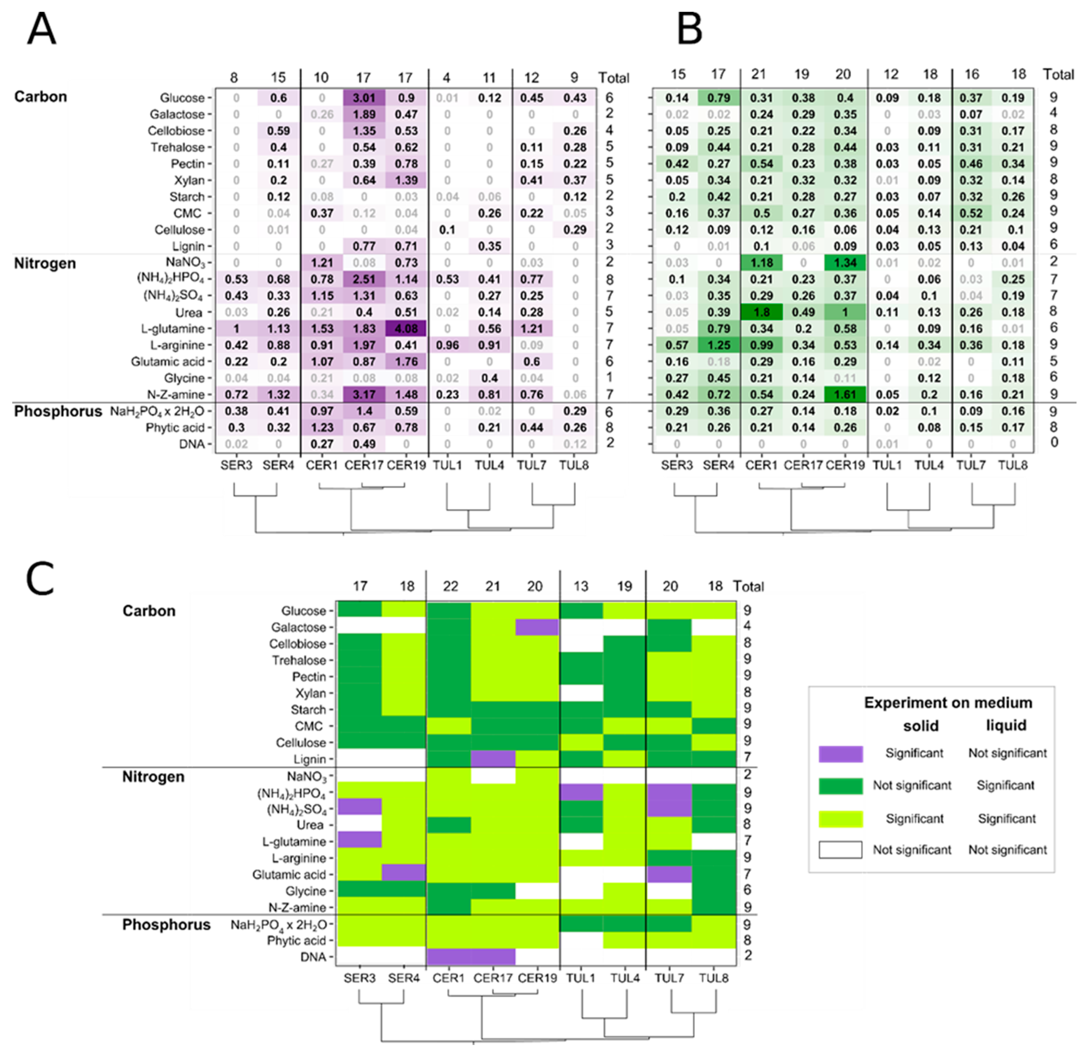
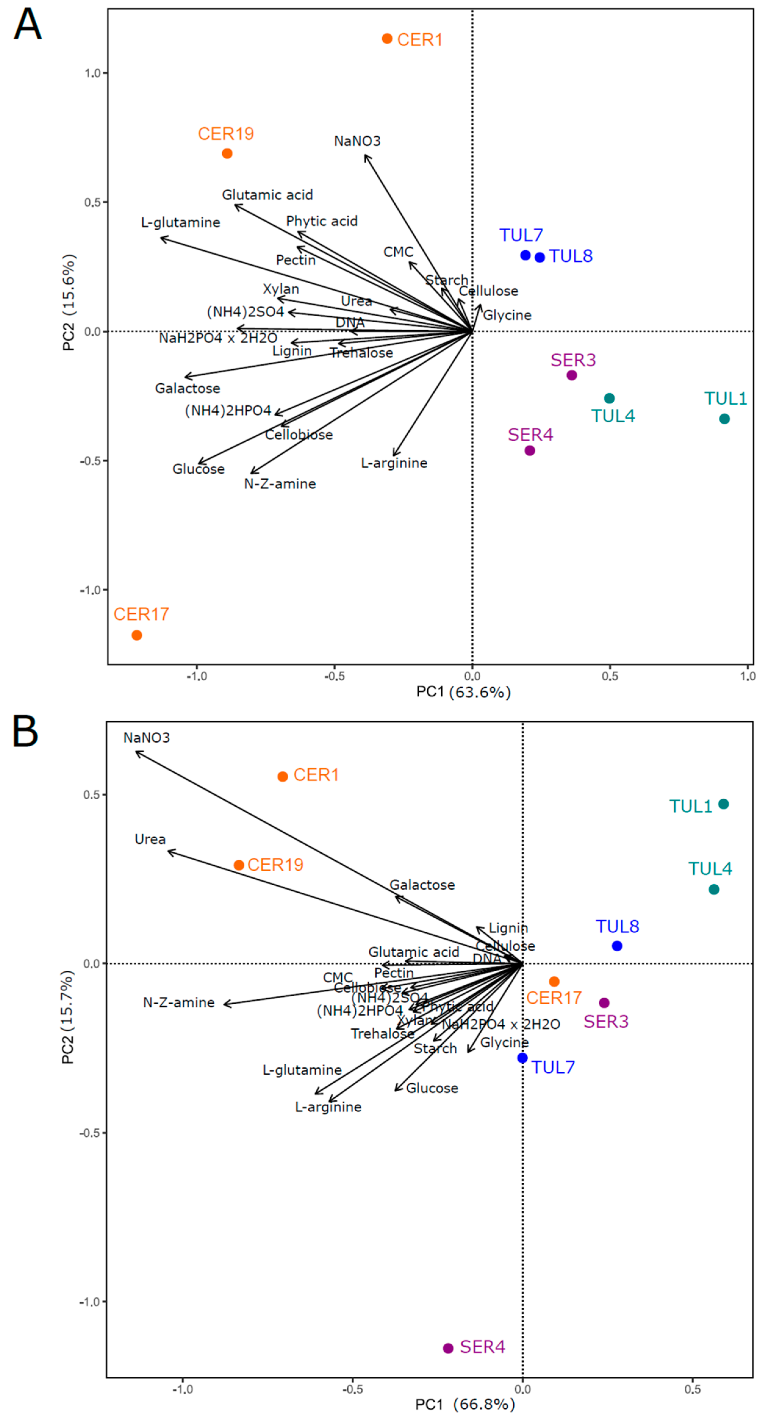
3.1. Fungal Biomass Production in Solid and Liquid Media

In general, fungal biomass production was higher in solid medium than in liquid medium for all isolates on most tested substrates (Figure 2A,B). Fungal group had a significant effect on fungal biomass production in both medium types, but this effect differed according to the interaction of the fungal group with a particular source type (C, N, P) (Table S2). While Ceratobasidiaceae produced a significantly higher fungal biomass than Tulasnellaceae clade A on all source types and both media types, the larger biomass of Ceratobasidiaceae than that of Serendipitaceae was achieved only in liquid medium (Table 2). Tulasnellaceae clade B produced a significantly higher fungal biomass than Tulasnellaceae clade A on all source types and both medium types, except for C source in liquid medium (Table 2). On solid medium, the variability in fungal biomass production between fungal groups and isolates was driven mainly by N substrates, and some C and P substrates such as glucose, galactose, inorganic phosphate and phytic acid (Figure 3A). In liquid medium, the major variability drivers were N substrates, particularly urea and sodium nitrate (Figure 3B).
This pattern, however, changed when we compared the isolates’ biomass grown on a particular substrate with that of the substrate-free control (Figure 2). While the fungal biomass produced on substrates in the liquid medium was mostly significantly higher than the biomass in the control, on the solid medium, the biomass frequently did not significantly differ between the substrate and the control. Moreover, some isolates showed no or limited biomass production on solid medium with particular nutrient sources.

3.2. Carbon Sources

Solid medium: Fungal isolates CER1, SER3, TUL1 and partially TUL4 (both Tulasnellaceae clade A) showed no or low capability to metabolise C substrates compared with the C-free control treatment (Figure 2A and Figure S1, Table S3). Isolates CER17 and CER19 effectively used all C substrates except for polysaccharides starch, CMC and cellulose. On the other hand, CER1 grew significantly only on CMC-rich substrate. While SER3 utilized none of the tested C substrates, SER4 produced significant biomass amounts on glucose-, cellobiose-, trehalose-, pectin-, xylan- and starch-rich medium. All Tulasnellaceae, except for TUL1, effectively metabolised glucose. Nonetheless, only TUL1 and TUL8 were able to degrade cellulose. The use of other C-sources by Tulasnellaceae was scarce, although TUL7 and TUL8 were able to produce significant biomass amounts on glucose, trehalose, pectin and xylan. Lignin use was demonstrated in the case of CER17, CER19 and SER4.
Liquid medium: The mean biomass increase of Ceratobasidiaceae was significantly higher compared to that of other fungal groups (Figure 2B and Figure S2, Table S2). Except for the non-significant biomass increase of CER17 on lignin-rich medium, all Ceratobasidiaceae effectively used all tested C sources (Table S3). Both Serendipitaceae OTUs showed difficulties in metabolising galactose and lignin. In general, Tulasnellaceae isolates were able to effectively utilize all tested C-substrates except for galactose, cellobiose and xylan.

3.3. Nitrogen Sources

In general, Ceratobasidiaceae demonstrated the highest biomass production detected in both medium types (Figure 2A,B). Some isolates, however, showed preference for either liquid or solid medium with a particular N-source. For example, CER1 produced the greatest fungal biomass when it metabolised urea (1.8 mg/day) in liquid medium, whereas its biomass increase on solid medium was not significantly different from that of the substrate-free control (Figures S3 and S4). Isolate CER19 gained 4.08 mg/day of biomass in L-glutamine-rich agar, which was the highest overall biomass production recorded, whereas in liquid medium, it reached only 0.58 mg/day (Figures S3 and S4). Tulasnellaceae clade A showed the lowest biomass increase of all fungal groups (Figure 2A,B, Table 2). A common trend of Serendipitaceae, Tulasnellaceae and CER17 in both media was the disability to digest nitrate (NaNO3).
Solid medium: The fungal biomass obtained on all tested N substrates differed significantly from that obtained in the substrate-free control, except for TUL8 with poor biomass production on all N substrates (Figure 2A and Figure S3, Table S3). However, there were apparent differences in substrate utilization among fungal groups and isolates. Ammonium phosphate ((NH4)2HPO4), L-glutamine, L-arginine and N-Z amine were the most suitable substrates for the growth of all fungal groups, with some exceptions, mainly from the Tulasnellaceae group. In contrast, glycine resulted in no or minimal biomass production except in TUL4. Isolate TUL8 did not effectively utilize any of the tested N substrates.
Liquid medium: All Ceratobasidiaceae effectively used all N sources except nitrate and glycine in case of CER17 and CER19, respectively (Figure 2B and Figure S4, Table S3). Isolate SER4 did not grow well on glutamic acid substrate and SER3 on ammonium sulphate (NH4)2SO4, urea and L-glutamine, respectively. Tulasnellaceae clade A and TUL7 produced no or small amounts of biomass on glutamic acid and (NH4)2SO4, respectively, and TUL1, together with TUL7, lacked the ability to metabolise amino acid glycine.

3.4. Phosphorus Sources

Two common trends were observed in both solid and liquid media: (i) inability to utilize DNA, except for CER1 and CER17 on solid medium; (ii) effective utilization of inorganic phosphate NaH2PO4 × 2H2O and organic phytic acid substrates by all isolates, except for Tulasnellaceae clade A and TUL7 from clade B on the NaH2PO4 × 2H2O substrate and TUL1 from clade A on phytic acid on solid medium (Figure 2A,B, Figures S5 and S6, Table S3).

3.5. Comparison of Radial Growth Rates on Solid Medium

When comparing the radial growth rates of the isolates on a particular source type (C, N, P) against substrate-free controls, we only observed significant growth of Ceratobasidiaceae and TUL8 isolates on N and P sources, respectively (Table S4). Specifically, most Ceratobasidiaceae isolates showed significant radial growth rates on NaNO3, (NH4)2HPO4, urea, L-glutamine, L-arginine; Serendipitaceae and Tulasnellaceae isolates showed occasional significant growth on a particular substrate, but without a consistent pattern (Table S4).

4. Discussion

Our data provided substantial evidence about the metabolic behaviour of nine isolates from the families Ceratobasidiaceae, Tulasnellaceae and Serendipitaceae cultivated simultaneously on solid and in liquid media. This is a unique feature because most studies have provided data from investigation of one orchid fungal family only [21,24,46] or several families but grown in liquid cultures only and a limited number of isolates (Nurfadilah et al. (2013) [22]—four OTUs in total). We also demonstrated a strong effect of Tulasnellaceae phylogenetic affinity (clade A vs. clade B) on nutrient acquisition, with isolates from clade A growing much slower, producing lower amount of biomass and utilizing a lower spectrum of nutrients than isolates from clade B. Isolates from clade A were studied for nutrient utilisation for the first time, which extends our knowledge on the fungal traits of the family Tulasnellaceae. In addition, our fungal isolates were obtained from six terrestrial orchid species from the northern hemisphere, thus our study broadens the current view of OMF nutrient utilization compared to available studies from Australia [22,23,24] and provides knowledge applicable to ex situ conservation of European orchids.

4.1. Carbon Utilization

After subtracting the biomass amount from that of the C-free control, almost all tested fungal OTUs showed significant biomass production in liquid medium, whereas the production on solid medium was only rarely significantly different from that of the control. Such a discrepancy could be explained (i) by the utilisation of a cellophane membrane as a C source by mycelium, and/or (ii) by the presence of additional C sources in the agar of the solid medium, such as cellulose, agarose and agaropectin. Indeed, most isolates were able to produce some biomass on solid medium and we have sometimes noticed visual decomposition of the cellophane membrane. One possibility to avoid the use of a cellophane membrane could be analysis of the ergosterol content [23,46,47], which is a fungus-specific membrane lipid. The effect of agar itself can be solved only by using liquid medium.
On solid medium, fungal biomass production on most mono-, di- and polysaccharides was significantly higher than that of the C-free control, mainly in Ceratobasidiaceae and Tulasnellaceae clade B and SER4 isolates. The isolate CER17 formed the greatest fungal biomass of all isolates on the monosaccharide’s glucose (3.01 mg/day) and galactose (1.89 mg/day), which is congruent with the findings of Nurfadilah et al. (2013) [22], demonstrating a high biomass production of a Ceratobasidium sp. (DQ028808.1; Figure 1C) on these carbon sources in liquid medium. In liquid medium, glucose, cellobiose, trehalose, pectin, xylan, starch, CMC and cellulose were the C sources utilised by the majority of the tested isolates. Generally, the polysaccharides pectin and CMC promoted the highest biomass production [48], while other studies [21,22,23] identified the polysaccharide xylan as a more efficient C-source.
The fungal groups largely varied in C-source utilization. Ceratobasidiaceae could metabolise the widest range of examined sugars, which might be related to their different nutritional modes, known as pathogenic, saprotrophic, ectomycorrhizal or endophytic fungi [7,29]. Veldre et al. (2013) [7] have suggested that autotrophic orchids form mycorrhizal associations with any available soil Ceratobasidiaceae, regardless of any pathogenicity they may have that might be restricted by the plant host, e.g., by the production of antifungal compounds [49]. Indeed, strains with confirmed pathogenicity are scattered along the Ceratobasidiaceae phylogenetic tree (Figure 1C) and in a recent study by Freestone et al. (2021) [50] one OTU appeared that contained both pathogenic and OMF forms. Tulasnellaceae clades differed significantly in their utilisation of tested C sources, with Tulasnellaceae clade B showing a significantly higher biomass production on the majority of the tested carbohydrates compared with clade A. Genomic and enzyme expression analyses of T. calospora from Tulasnellaceae clade B (96.3% identity between T. calospora voucher AY373298.1 and our TUL7) revealed numerous genes encoding the activation of carbohydrate-active enzymes (CAZymes), responsible for the degradation of lignocellulolytic compounds in plant cell walls [2,11]. However, the question of why Tulasnellaceae clade A does not use carbohydrates as efficiently as the sister group still needs to be explored. Serendipitaceae showed no or restricted biomass production on lignin and galactose, as shown in other studies [22,23,24]. Among micro-organisms, fungi are the most efficient lignin decomposers, but OMF from the family Ceratobasidiaceae have shown limited ability to utilize complex aromatic compounds, such as tannins, compared to ericoid mycorrhizal fungi ([21], and references therein); moreover, the extent of tannin degradation strongly varied among the isolates.

4.2. Nitrogen Utilization

The studied OMF isolates were able to utilize all tested N sources. Nitrogen is an essential element for fungal growth and biomass production [51]. It can be obtained either from mineral or organic compounds but is ultimately converted into inorganic NH4+ and amino acids (glutamate, glutamine, arginine), serving as storage N-containing compounds of the cells used in biosynthetic reactions [52]. Mycorrhizal fungi help their host plants with nitrogenous compound acquisition, and in the case of orchids, N represents the probable major nutrient transferred from OMF to the plants, as orchid tissue is highly N-enriched [53]. Cameron et al., (2006) [54] experimentally demonstrated that 78% of extra-radically supplied 15N-labelled glycine accumulated in the mycelial biomass of Ceratobasidium cornigerum compared to 20% in the roots and <3% in shoot tissues of the terrestrial orchid Goodyera repens.
Here, inorganic ammonium-containing substrates ((NH4)2HPO4, (NH4)2SO4) were effectively assimilated by the majority of the tested OTUs, confirming that directly accessible ammonium is a preferable N source for most filamentous fungi [52]. The uptake is mediated via specific ammonium membrane transporters, recognized in OMF, specifically in Serendipita vermifera and S. bescii [25,55]. Ammonium utilization by OMF isolates has been demonstrated in several in vitro studies [22,48]; however, its effective assimilation significantly varied among Serendipitaceae OTUs [24] or in T. calospora strains [25,56].
On the other hand, inorganic nitrate (NO3) and complex organic compounds are less frequently assimilated by fungal hyphae [57]. In our study, nitrate (NaNO3) stimulated biomass production only in two Ceratobasidiaceae isolates, CER1 and CER19, and the remaining OMF showed very poor or no growth. Similar findings have been obtained in other studies [22,56,58], where effective nitrate use was observed for Ceratobasidium spp. but not for other OMF genera. Vogt-Schilb et al. (2020) [59] demonstrated a higher abundance of Ceratobasidiaceae OTUs in meadows restored from arable fields than in natural grasslands, which aligns with the ruderal characteristics of the family and its ability to utilize nitrates from inorganic fertilisers. Recently, Figura et al. (2021) [60] observed no inhibitory effect of increasing nitrate concentrations on the germination success of Dactylorhiza majalis symbiotically cultivated with Ceratobasidium sp., but the authors found a strong inhibition when germinated with Serendipita sp. and Tulasnella spp. The inability of Serendipita indica and T. calospora to assimilate nitrate was confirmed when no functional nitrate transporters were identified [25,61]. The ability to utilize nitrates does not seem to be inherited by the whole Ceratobasidiaceae family, as isolate CER17 did not show this ability. This variation in nitrate utilization needs to be explored for a broader number of Ceratobasidiaceae isolates at both inter- and intra-specific levels.
Not all organic N-containing compounds tested in this study were good sources for utilization by OMF isolates. The simple amino acids L-arginine and L-glutamine, together with peptone N-Z amine, were effectively metabolised by most tested isolates, whereas glycine and glutamic acid were used poorly. Our findings agree with Stephen and Fung (1971) [62] and Nurfadilah et al., (2013) [22], who observed reasonable fungal biomass production on L-arginine and L-glutamine, but the greatest biomass yield was gained on glutamic acid-rich medium. Contrarily, Hadley and Ong (1978) [56] found glycine and urea to be suitable N sources for a T. calospora isolate, whereas L-glutamine and L-arginine were less suitable. Another isolate of T. calospora produced the highest biomass on L-glutamine, followed by glutamic acid [25]. Serendipitaceae isolates had the capacity to assimilate various N organic compounds such as amino acids, urea and L-arginine, but showed strong influence of isolate identity towards a particular organic N source [24,55]. In addition, we found substantial differences in N utilization by OMF when grown in liquid or on solid media. Most noticeably, urea and glycine were more efficiently utilized in liquid medium and with a wider range of tested fungal isolates. The differences between media types might stem from limited mobility of nutrients in solid media in comparison to liquid cultures [63].

4.3. Phosphorus Utilization

Most tested isolates effectively utilized inorganic orthophosphate (NaH2PO4 × 2 H2O) and phytic acid in both liquid and solid media, but, with some exceptions, did not utilise DNA (Figure 2A,B). Inorganic orthophosphate is a primary source of P and freely accessible to soil micro-organisms and plants, however, due to insufficient reserves in soil, it needs to be continually replenished by the mineralisation of organic P [64]. The uptake of orthophosphate from the soil environment by mycorrhizal fungi is mediated by specific membrane transporters, mainly located in the extra-radical mycelium [65]. They have been described in arbuscular and ectomycorrhizal fungi [66,67] and recently also in OMF, namely T. calospora and S. vermifera [2,68]. We observed effective NaH2PO4 × 2 H2O utilisation by the Ceratobasidiaceae and Serendipitaceae OTUs in both medium types compared to the P-free control, similar to other OMF studies [22,69]. On the contrary, Serendipitaceae isolates produced low biomass amounts on NaH2PO4 × 2 H2O in liquid cultures, which has been attributed to the limited P availability in Australian soils [24].
Phytic acid and DNA are organic P sources which are not directly available for soil micro-organisms or plants, and their utilization is conditioned by the microbial secretion of extracellular hydrolytic enzymes [70]. Numerous fungal phytases catalysing the hydrolysis of abundant phytic acids belong to the family of acid phosphatases, which are highly activated in saprotrophic fungi [71]. In the present study, we observed the highly effective use of phytic acid by all fungal isolates in both medium types, except for TUL1. Similar results have been achieved by Nurfadilah et al., (2013) [22]; however, the S. vermifera isolate produced the lowest biomass amount. By testing numerous Serendipita isolates, Oktalira (2020) [24] observed significant differences in mean fungal biomass among isolates within the same OTU.
Nucleic acids with phospholipids form a minor part of soil organic P; they need to be degraded by phosphodiesterases to become bioavailable. In saprotrophic fungi, the activity of these enzymes is secured by a lower number of genes, resulting in lower activities of phosphodiesterases compared to acid or alkaline phosphatases [71]. In our study, we revealed no or limited use of DNA by OMF in both medium types, apart from CER1 and CER17 on solid medium. The effective utilization of DNA by some Ceratobasidiaceae might be the result of their wide ecological amplitude, including pathogenic behaviour. Other studies have shown an ability of OTUs from all three orchid mycorrhizal families to use DNA, albeit with an effectiveness lower than for other P sources [22,24].

4.4. Disputable Use of Radial Growth Rate

The radial growth rate (or colony diameter) is a popular indirect measure of filamentous growth on solid media [72,73,74]. While the biomass production rate of the tested isolates on solid medium was significant in most cases, comparisons of radial growth rates on substrate and substrate-free controls yielded mostly non-significant results. The reason for such a discrepancy likely stems from the behaviour of filamentous fungi, exploring the habitat with rapidly growing, sparsely branched hyphae [75]. Therefore, we recommend avoiding radial growth rate as a measure for the assessment of substrate utilisation efficiency in filamentous fungi.

5. Conclusions

In the present study, we found significant inter- and intra-familiar variability and a strong effect of Tulasnellaceae phylogeny on C, N and P acquisition. The nutrient resource abilities of isolates strongly differed when grown in liquid or solid media, therefore we urge caution when generalizing knowledge from liquid cultures to ecology of OMF in situ. The main gaps in our current knowledge on OMF are in the ecology of enzyme production under various natural or semi-natural conditions, which could be explored in future by approaches used for arbuscular mycorrhizae [76,77].

Supplementary Materials

The following supporting information can be downloaded at: https://www.mdpi.com/article/10.3390/jof9010088/s1, Figures S1,S3,S5: Mean biomass (in mg/day) ±SD of the tested isolates in solid medium containing carbon, nitrogen and phosphorus sources, respectively; Figures S2,S4,S6: Mean biomass (in mg/day) ± SD of the tested isolates in liquid medium containing carbon, nitrogen and phosphorus sources, respectively; Table S1: Characteristics of studied compounds added into solid and liquid MMN media: carbon (C), nitrogen (N), and phosphorus (P) sources; Table S2: Generalized linear mixed models (GLMMs) testing the effects of the Fungal group, the Source type, and the interaction term by likelihood-ratio test (LRT) comparing with Chi2 distribution, Post-Hoc multiple comparison tests of the interaction term: testing the biomass differences among Fungal groups within a Source type; and testing the biomass differences among Source types within a Fungal group; Table S3: ANOVA with multiple comparisons of means: contrasts testing the biomass among all substrates against the substrate-free control in both solid and liquid medium types, and Dunnett’s Post-hoc tests: contrasts comparing the fungal biomass of particular C, N, P substrates with substrate-free control in both solid and liquid medium types; Table S4: ANOVA with multiple comparisons of means: contrasts testing the fungal growth rate among all substrates against the substrate-free control in both solid and liquid medium types, and Dunnett’s Post-hoc tests: contrasts comparing the fungal growth rate on particular C, N, P substrates with substrate-free control in both solid and liquid medium types.

Author Contributions

The research was designed by A.N. and J.J. The major part of the laboratory work was done by A.N., M.K., J.J., S.M., H.V.-S. and P.Š. performed the statistical analyses. C.C.P.d.P. and T.T. performed the phylogenetic analysis. A.N. and J.J. wrote the major part of the manuscript, with the contribution of all co-authors. All authors have read and agreed to the published version of the manuscript.

Funding

The work was supported by the Czech Science Foundation (project 18-11378S).

Institutional Review Board Statement

Not applicable.

Informed Consent Statement

Not applicable.

Data Availability Statement

Acknowledgments

We thank Milena Nováková and Joana Pimentel for help with laboratory work and Jan Říha for phylogenetic advice.

Conflicts of Interest

The authors declare that they have no conflict of interest.

References

  1. Smith, S.E.; Read, D.J. Mycorrhizal Symbiosis, 3rd ed.; Academic Press: London, UK, 2008. [Google Scholar]
  2. Kohler, A.; Kuo, A.; Nagy, L.G.; Morin, E.; Barry, K.W.; Buscot, F.; Canbäck, B.; Choi, C.; Cichocki, N.; Clum, A.; et al. Convergent losses of decay mechanisms and rapid turnover of symbiosis genes in mycorrhizal mutualists. Nat. Genet. 2015, 47, 410–415. [Google Scholar] [CrossRef] [PubMed]
  3. Rasmussen, H.N. Terrestrial Orchids. From Seed to Mycotrophic Plant, 1st ed.; Cambridge University Press: Cambridge, UK, 1995. [Google Scholar]
  4. Weiss, M.; Waller, F.; Zuccaro, A.; Selosse, M.A. Sebacinales—One thousand and one interactions with land plants. New Phytol. 2016, 211, 20–40. [Google Scholar] [CrossRef]
  5. Selosse, M.A.; Martos, F. Do chlorophyllous orchids heterotrophically use mycorrhizal fungal carbon? Trends Plant Sci. 2014, 19, 683–685. [Google Scholar] [CrossRef] [PubMed]
  6. Selosse, M.A.; Dubois, M.P.; Alvarez, N. Do Sebacinales commonly associate with plant roots as endophytes? Mycol. Res. 2009, 113, 1062–1069. [Google Scholar] [CrossRef] [PubMed] [Green Version]
  7. Veldre, V.; Abarenkov, K.; Bahram, M.; Martos, F.; Selosse, M.A.; Tamm, H.; Köljalg, U.; Tedersoo, L. Evolution of nutritional modes of Ceratobasidiaceae (Cantharellales, Basidiomycota) as revealed from publicly available ITS sequences. Fungal Ecol. 2013, 6, 256–268. [Google Scholar] [CrossRef]
  8. Bidartondo, M.I.; Bruns, T.D.; Weiß, M.; Sergio, S.; Read, D.J. Specialized cheating of the ectomycorrhizal symbiosis by an epiparasitic liverwort. Proc. R. Soc. Lond. B 2003, 270, 835–842. [Google Scholar] [CrossRef] [PubMed]
  9. Ceresini, P.C.; Costa-Souza, E.; Zala, M.; Furtado, E.L.; Souza, N.L. Evidence that the Ceratobasidium-like white-thread blight and black rot fungal pathogens from persimmon and tea crops in the Brazilian Atlantic Forest agroecosystem are two distinct phylospecies. Genet. Mol. Biol. 2012, 35, 480–497. [Google Scholar] [CrossRef] [Green Version]
  10. Mosquera-Espinosa, A.T.; Bayman, P.; Prado, G.A.; Gómez-Carabalí, A.; Otero, J.T. The double life of Ceratobasidium: Orchid mycorrhizal fungi and their potential for biocontrol of Rhizoctonia solani sheath blight of rice. Mycologia 2012, 105, 141–150. [Google Scholar] [CrossRef]
  11. Adamo, M.; Chialva, M.; Calevo, J.; De Rose, S.; Girlanda, M.; Perotto, S.; Balestrini, R. The dark site of orchid symbiosis: Can Tulasnella calospora decompose host tissues? Int. J. Mol. Sci. 2020, 21, 3139. [Google Scholar] [CrossRef]
  12. Suetsugu, K.; Matsubayashi, J.; Tayasu, I. Some mycoheterotrophic orchids depends on carbon from dead wood: Novel evidence from a radiocarbon approach. New Phytol. 2020, 227, 1519–1529. [Google Scholar] [CrossRef]
  13. van der Kinderen, G. A method for the study of field germinated seeds of terrestrial orchids. Lindleyana 1995, 10, 68–73. [Google Scholar]
  14. Boddy, L.; Frankland, J.C.; van West, P. Ecology of Saptrotrophic Basidiomycetes, 1st ed.; Elsevier Science: Amsterdam, The Netherlands, 2008. [Google Scholar]
  15. Baldrian, P. Enzymes of saprotrophic basidiomycetes. In Ecology of Satprotrophic Basidiomycetes, 1st ed.; Boody, L., Frankland, J.C., van West, P., Eds.; Elsevier Science: Amsterdam, The Netherlands, 2008; Chapter 2; pp. 19–42. [Google Scholar]
  16. Berg, B.; Laskowski, R. Litter Decomposition: A Guide to Carbon and Nutrient Turnover; Elsevier Science Publishing Co Inc.: San Diego, CA, USA, 2006. [Google Scholar]
  17. Kallenbach, C.; Frey, S.; Grandy, A. Direct evidence for microbial-derived soil organic matter formation and its ecophysiological controls. Nat. Commun. 2016, 7, 13630. [Google Scholar] [CrossRef] [Green Version]
  18. Stevenson, F.J. Organic forms of soil nitrogen. In Nitrogen in Agricultural Soils; Stevenson, F.J., Ed.; American Society of Agronomy: Madison, WI, USA, 1982; Volume 22, pp. 67–122. [Google Scholar]
  19. Richardson, A.E.; Simpson, R. Soil microorganisms mediating phosphorus availability update on microbial phosphorus. Plant Physiol. 2011, 156, 989–996. [Google Scholar] [CrossRef] [PubMed] [Green Version]
  20. Muindi, E.M. Understanding soil phosphorus. Int. J. Plant Soil Sci. 2019, 31, 1–18. [Google Scholar] [CrossRef] [Green Version]
  21. Midgley, D.J.; Jordan, L.A.; Saleeba, J.A.; McGee, P.A. Utilisation of carbon substrates by orchid and ericoid mycorrhizal fungi from Australian dry sclerophyll forests. Mycorrhiza 2006, 16, 175–182. [Google Scholar] [CrossRef] [PubMed]
  22. Nurfadilah, S.; Swarts, N.D.; Dixon, K.W.; Lambers, H.; Merritt, D.J. Variation in nutrient-acquisition patterns by mycorrhizal fungi of rare and common orchids explains diversification in a global biodiversity hotspot. Ann. Bot. 2013, 111, 1233–1241. [Google Scholar] [CrossRef] [PubMed] [Green Version]
  23. Mehra, S.; Morrison, P.D.; Coates, F.; Lawrie, A.C. Differences in carbon source utilisation by orchid mycorrhizal fungi from common and endangered species of Caladenia (Orchidaceae). Mycorrhiza 2017, 27, 95–108. [Google Scholar] [CrossRef]
  24. Oktalira, F.T. Diversity of Serendipitaceae Mycorrhizal Associations of Australian Terrestrial Orchids. Ph.D. Thesis, The Australian National University, Canberra, Australia, 2021. [Google Scholar]
  25. Fochi, V.; Chitarra, V.; Kohler, A.; Voyron, S.; Singan, V.R.; Lindquist, E.A.; Barry, K.W.; Girlanda, M.; Grigoriev, I.V.; Martin, F.; et al. Fungal and plant gene expression in the Tulasnella calosporaSerapias vomeracea symbiosis provides clues about nitrogen pathways in orchid mycorrhizas. New Phytol. 2017, 213, 365–379. [Google Scholar] [CrossRef] [Green Version]
  26. Freestone, M.; Linde, C.; Swarts, N.; Reiter, N. Ceratobasidium orchid mycorrhizal fungi reveal intraspecific variation and interaction with different nutrient media in symbiotic germination of Prasophyllum (Orchidaceae). Symbiosis 2022, 87, 255–268. [Google Scholar] [CrossRef]
  27. Cruz, D.; Suárez, J.P.; Kottke, I.; Piepenbring, M.; Oberwinkler, F. Defining species in Tulasnella by correlating morphology and nrDNA ITS-5.8S sequence data of basidiomata from a tropical Andean forest. Mycol. Prog. 2011, 10, 229–238. [Google Scholar] [CrossRef]
  28. Oberwinkler, F.; Riess, K.; Bauer, R.; Kirschner, R.; Garnica, S. Taxonomic re-evaluation of the Ceratobasidium-Rhizoctonia complex and Rhizoctonia butinii, a new species attacking spruce. Mycol. Prog. 2013, 12, 763–776. [Google Scholar] [CrossRef]
  29. González, D.; Rodriguez-Carres, M.; Boekhout, T.; Stalpers, J.; Kuramae, E.E.; Nakatani, A.K.; Vilgalys, R.; Cubeta, M.A. Phylogenetic relationships of Rhizoctonia fungi within the Cantharellales. Fungal Biol. 2016, 120, 603–619. [Google Scholar] [CrossRef] [Green Version]
  30. Gibbs, P.A.; Seviour, R.J.; Schmid, F. Growth of filamentous fungi in submerged culture: Problems and possible solutions. Crit. Rev. Biotechnol. 2000, 20, 17–48. [Google Scholar] [CrossRef] [PubMed]
  31. Pradeep, F.S.; Begam, M.S.; Palaniswamy, M.; Pradeep, B.V. Influence of culture media on growth and pigment production by Fusarium moniliforme KUMBF1201 isolated from Paddy Field Soil. World Appl. Sci. J. 2013, 22, 70–77. [Google Scholar]
  32. Viniegra-González, G.; Favela-Torres, E.; Aguilar, C.N.; Rómero-Gomez, S.J.; Díaz-Godínez, G.; Augur, C. Advantages of fungal enzyme production in solid state over liquid fermentation systems. Biochem. Eng. J. 2003, 13, 157–167. [Google Scholar] [CrossRef]
  33. Marx, D.H.; Bryan, W.C. Growth and ectomycorrhizal development of loblolly pine seedlings in fumigated soil infested with the fungal symbiont Pisolithus tinctorius. For. Sci. 1975, 21, 245–254. [Google Scholar] [CrossRef]
  34. White, T.J.; Bruns, T.; Lee, S.; Taylor, J.W. Amplification and direct sequencing of fungal genes for phylogenetics. In PCR Protocols: A Guide to Methods and Applications; Innis, M., Gelfand, D.H., Sninsky, J.J., White, T.J., Eds.; Academic Press Inc.: New York, NY, USA, 1990; pp. 315–322. [Google Scholar]
  35. Taylor, D.L.; McCormick, M.K. Internal transcribed spacer primers and sequences for improved characterization of basidiomycetous orchid mycorrhizas. New Phytol. 2008, 177, 1020–1033. [Google Scholar] [CrossRef]
  36. Těšitelová, T.; Jersáková, J.; Roy, M.; Kubátová, B.; Těšitel, J.; Urfus, T.; Trávníček, P.; Suda, J. Ploidy-specific symbiotic interactions: Divergence of mycorrhizal fungi between cytotypes of the Gymnadenia conopsea group (Orchidaceae). New Phytol. 2013, 199, 1022–1033. [Google Scholar] [CrossRef]
  37. Milne, I.; Wright, F.; Rowe, G.; Marshall, D.F.; Husmeier, D.; McGuire, G. TOPALi: Software for automatic identification of recombinant sequences within DNA multiple alignments. Bioinformatics 2004, 20, 1806–1807. [Google Scholar] [CrossRef] [PubMed] [Green Version]
  38. Katoh, K.; Misawa, K.; Kuma, K.; Miyata, T. MAFFT: A novel method for rapid multiple sequence alignment based on fast Fourier transform. Nucleic Acids Res. 2002, 30, 3059–3066. [Google Scholar] [CrossRef] [PubMed] [Green Version]
  39. Hall, T.A. BioEdit: A user-friendly biological sequence alignment editor and analysis program for Windows 95/98/NT. Nucleic Acids Symp. Ser. 1999, 41, 95–98. [Google Scholar]
  40. Tamura, K.; Stecher, G.; Kumar, S. MEGA11: Molecular Evolutionary Genetics Analysis version 11. Mol. Biol. Evol. 2021, 38, 3022–3027. [Google Scholar] [CrossRef] [PubMed]
  41. Felsenstein, J. Confidence limits on phylogenies: An approach using the bootstrap. Evolution 1985, 39, 783–791. [Google Scholar] [CrossRef]
  42. Schneider, C.A.; Rasband, W.S.; Eliceiri, K.W. NIH Image to ImageJ: 25 years of image analysis. Nat. Methods 2012, 9, 671–675. [Google Scholar] [CrossRef]
  43. R Core Team. R: A Language and Environment for Statistical Computing; R Foundation for Statistical Computing: Vienna, Austria, 2018; Available online: https://www.R-project.org/ (accessed on 1 February 2022).
  44. O’Hara, R.B.; Simpson, G.L.; Solymos, P.M.; Stevens, H.H.; Szoecs, E.; Wagne, H. Package “Vegan”: Community Ecology Package. R Package Version 2.5-1. 2018. Available online: https://CRAN.R-project.org/package=vegan (accessed on 1 February 2022).
  45. Grothendieck, G. LME4: Linear Mixed-Effects Models using ‘Eigen’ and S4. R Package Version 1.1-10. 2015. Available online: http://lme4.r-forge.r-project.org (accessed on 1 February 2022).
  46. Mehra, S. Nutritional and Genetic Diversity in Orchid Mycorrhizal Fungi from Caladenia Species. PhD Thesis, RMIT University, Victoria, Australia, December 2014. [Google Scholar]
  47. Gessner, M.O. Ergosterol as a measure of fungal biomass. In Methods to Study Litter Decomposition, 2nd ed.; Graça, M.A., Bärlocher, F., Gessner, M.O., Eds.; Springer: Dordrecht, The Netherlands, 2005; pp. 189–195. [Google Scholar]
  48. Wright, M.; Cross, R.; Cousens, R.D.; May, T.W.; McLean, C.B. The functional significance for the orchid Caladenia tentaculata of genetic and geographic variation in the mycorrhizal fungus Sebacina vermifera s. l. complex. Muelleria 2011, 29, 130–140. [Google Scholar] [CrossRef]
  49. Dearnaley, J.D.W.; Perotto, S.; Selosse, M.A. Structure and development of orchid mycorrhizas. In Molecular Mycorrhizal Symbiosis, 1st ed.; Martin, F., Ed.; John Wiley & Sons, Inc.: Hoboken, NJ, USA, 2017; pp. 63–86. [Google Scholar]
  50. Freestone, M.W.; Swarts, N.D.; Reiter, N.; Tomlinson, S.; Sussmilch, F.C.; Wright, M.M.; Holmes, G.D.; Phillips, R.D.; Linde, C.C. Continental-scale distribution and diversity of Ceratobasidium orchid mycorrhizal fungi in Australia. Ann. Bot. 2021, 128, 329–343. [Google Scholar] [CrossRef]
  51. Di Lonardo, D.P.; Van der Wal, A.; Harkes, P.; de Boer, W. Effect of nitrogen on fungal growth efficiency. Plant Biosyst. 2020, 154, 433–437. [Google Scholar] [CrossRef]
  52. Meti, R.S.; Ambarish, S.; Khajure, P.V. Enzymes of ammonia assimilation in fungi: An overview. Recent Res. Sci. Technol. 2011, 2, 28–38. [Google Scholar]
  53. Schweiger, J.M.I.; Bidartondo, M.I.; Gebauer, G. Stable isotope signatures of underground seedlings reveal the organic matter gained by adult orchids from mycorrhizal fungi. Fungal Ecol. 2017, 32, 870–881. [Google Scholar] [CrossRef]
  54. Cameron, D.D.; Leake, J.R.; Read, D.J. Mutualistic mycorrhiza in orchids: Evidence from plant-fungus carbon and nitrogen transfers in the green-leaved terrestrial orchid Goodyera repens. New Phytol. 2006, 171, 405–416. [Google Scholar] [CrossRef]
  55. Ray, P.; Abraham, P.E.; Guo, Y.; Giannone, R.J.; Engle, N.L.; Yang, Z.K.; Jacobson, D.; Hettich, R.L.; Tschaplinski, T.J.; Craven, K.D. Scavenging organic nitrogen and remodelling lipid metabolism are key survival strategies adopted by the endophytic fungi, Serendipita vermifera and Serendipita bescii to alleviate nitrogen and phosphorous starvation in vitro. Environ. Microbiol. Rep. 2019, 11, 548–557. [Google Scholar] [CrossRef] [PubMed] [Green Version]
  56. Hadley, G.; Ong, S.H. Nutritional requirements of orchid endophytes. New Phytol. 1978, 81, 561–569. [Google Scholar] [CrossRef]
  57. Tudzynski, B. Nitrogen regulation of fungal secondary metabolism in fungi. Front. Microbiol. 2014, 5, 656. [Google Scholar] [CrossRef] [Green Version]
  58. Dijk, E.; Eck, N. Effects of mycorrhizal fungi on in vitro nitrogen response of some Dutch indigenous orchid species. Canad. J. Bot. 1995, 73, 1203–1211. [Google Scholar] [CrossRef]
  59. Vogt-Schilb, H.; Těšitelová, T.; Kotilínek, M.; Sucháček, P.; Kohout, P.; Jersáková, J. Altered rhizoctonia assemblages in grasslands on ex-arable land support germination of mycorrhizal generalist, not specialist orchids. New Phytol. 2020, 227, 1200–1212. [Google Scholar] [CrossRef]
  60. Figura, T.; Tylová, E.; Jersáková, J.; Vohník, M.; Ponert, J. Fungal symbionts may modulate nitrate inhibitory effect on orchid seed germination. Mycorrhiza 2021, 31, 231–241. [Google Scholar] [CrossRef]
  61. Zuccaro, A.; Lahrmann, U.; Güldener, U.; Langen, G.; Pfiffi, S.; Biedenkopf, D.; Wong, P.; Samans, B.; Grimm, C.; Basiewicz, M.; et al. Endophytic life strategies decoded by genome and transcriptome analyses of the mutualistic root symbiont Piriformospora indica. PLoS Pathog. 2011, 7, e1002290. [Google Scholar] [CrossRef] [PubMed] [Green Version]
  62. Stephen, R.C.; Fung, K.K. Nitrogen requirements of the fungal endophytes of Arundina chinensis. Canad. J. Bot. 1971, 49, 407–410. [Google Scholar] [CrossRef]
  63. Hobbie, E.A.; Sánchez, F.S.; Rygiewicz, P.T. Carbon use, nitrogen use, and isotopic fractionation of ectomycorrhizal and saprotrophic fungi in natural abundance and 13C-labelled cultures. Mycol. Res. 2004, 108, 725–736. [Google Scholar] [CrossRef]
  64. Johri, A.K.; Oelmüller, R.; Dua, M.; Yadav, V.; Kumar, M.; Tuteja, N.; Varma, A.; Bonfante, P.; Persoon, B.L.; Stroud, R.M. Fungal association and utilization of phosphate by plants: Success, limitations, and future prospects. Front. Microbiol. 2015, 6, 984. [Google Scholar] [CrossRef] [Green Version]
  65. Jansa, J.; Finlay, R.; Wallander, H.; Smith, A.; Smith, S.E. Role of Mycorrhizal Symbioses in Phosphorus Cycling. In Phosphorus in Action: Soil Biology; Bünemann, E., Oberson, A., Frossard, E., Eds.; Springer: Berlin/Heidelberg, Germany, 2011; Volume 26. [Google Scholar]
  66. Harrison, M.J.; van Buuren, M.L. A phosphate transporter from the mycorrhizal fungus Glomus versiforme. Nature 1995, 378, 626–629. [Google Scholar] [CrossRef] [PubMed]
  67. Tatry, M.V.; Kassis, E.E.; Lambilliotte, R.; Corratgé, C.; Van Aarle, I.; Amenc, L.K.; Alary, R.; Zimmermann, S.; Sentenac, H.; Plassard, C. Two differentially regulated phosphate transporters from the symbiotic fungus Hebeloma cylindrosporum and phosphorus acquisition by ectomycorrhizal Pinus pinaster. Plant J. 2009, 57, 1092–1102. [Google Scholar] [CrossRef] [PubMed]
  68. Plassard, C.; Becquer, A.; Garcia, K. Phosphorus transport in mycorrhiza: How far are we? Trends Plant Sci. 2019, 24, 794–801. [Google Scholar] [CrossRef]
  69. Mujica, M.I.; Cisternas, M.; Claro, A.; Simunovic, M.; Pérez, F. Nutrients and fungal identity affect the outcome of symbiotic germination in Bipinnula fimbriata (Orchidaceae). Symbiosis 2020, 83, 91–101. [Google Scholar] [CrossRef]
  70. Jarosh, K.A.; Kandeler, E.; Frossard, E.; Bünemann, E.K. Is the enzymatic hydrolysis of soil organic phosphorus compounds limited by enzyme or substrate availability? Soil Biol. Biochem. 2019, 139, 107628. [Google Scholar] [CrossRef] [Green Version]
  71. Tedersoo, L.; Bahram, M. Mycorrhizal types differ in ecophysiology and alter plant nutrition and soil processes. Biol. Rev. 2019, 94, 1857–1880. [Google Scholar] [CrossRef]
  72. Linden, B.R. Comparison of radial growth rate of mycorrhizal fungi isolated from 43 species of northern orchids. Karstenia 1988, 28, 19–25. [Google Scholar] [CrossRef]
  73. Prosser, J.I. Kinetics of filamentous growth and branching. In The Growing Fungus; Gow, N.A.R., Gadd, G.M., Eds.; Chapman & Hall: London, UK, 1995; pp. 301–318. [Google Scholar]
  74. Zheng, W.; Lehmann, A.; Ryo, M.; Vályi, K.K.; Rilling, M.C. Growth rate trades off with enzymatic investment in soil filamentous fungi. Sci. Rep. 2020, 10, 11013. [Google Scholar] [CrossRef]
  75. Ivarsson, M.; Drake, H.; Bengtson, S.; Rasmussen, B. A cryptic alternative for the evolution of hyphae. Bioessays 2020, 42, 1900183. [Google Scholar] [CrossRef] [Green Version]
  76. Jansa, J.; Mozafar, A.; Frossard, E. Phosphorus acquisition strategies within arbuscular mycorrhizal fungal community of a single field site. Plant Soil 2005, 276, 163–176. [Google Scholar] [CrossRef] [Green Version]
  77. Bukovská, P.; Rozmoš, M.; Kotianová, M.; Gančarčíková, K.; Dudáš, M.; Hršelová, H.; Jansa, J. Arbuscular mycorrhiza mediates efficient recycling from soil to plants of nitrogen bound in chitin. Front. Microbiol. 2021, 12, 574060. [Google Scholar] [CrossRef] [PubMed]
Figure 1. Phylogenetic trees of fungal isolates from families Tulasnellaceae (two lineages, clades A and B (A), Serendipitaceae (B), and Ceratobasidiaceae (C). The black circles represent the studied fungal isolates. The open red circles in the Ceratobasidiaceae tree represent fungal strains with reported pathogenic abilities.
Jof 09 00088 g001aJof 09 00088 g001bJof 09 00088 g001c
Figure 2. Fungal biomass production (in mg/day; mean value) of nine fungal isolates grown on C, N and P substrates in solid (A) or liquid media (B). The biomass production on a particular substrate was subtracted from the substrate-free control (values significant at p < 0.05 are denoted in bold). The colour shades indicate the amount of biomass increase. (C) Comparison of significant biomass increases between solid and liquid media. The phylogenetic tree based on ITS sequences shows genetic relatedness of the isolates (see Figure 1). The values on the margins of the chart represent the total numbers of treatments (on the top) or isolates (on the right) in which a significant biomass increase was induced at least in one medium. See Table S1 for details of each substrate.
Jof 09 00088 g002
Figure 3. Principal components analysis of the fungal biomass of nine fungal isolates cultivated in solid (A) or in liquid (B) media, containing the 22 different carbon, nitrogen, and phosphorus sources (see Table S1 for details). The colour of the isolates indicates the fungal group: orange, purple, blue and green for Ceratobasidiaceae (CER1, 17, 19), Serendipitaceae (SER3, 4), Tulasnellaceae clade A (TUL1, 4) and Tulasnellaceae clade B (TUL7, 8), respectively.
Jof 09 00088 g003
Table 1. Description of fungal isolates. Fungal groups: SER—family Serendipitaceae, CER—family Ceratobasidiaceae, TUL-A and TUL-B—two clades within Tulasnellaceae, see Figure 1A–C. Sites of orchid host origin: CZ—Czech Republic (49.1292N; 13.6591E), F—France (43.9669N; 3.4028E). OTU—operational taxonomic unit.
OTU CodeFungal Group CodeOrchid HostSite Genbank
Accession
Number
Genetic Similarity with the Nearest Taxonomically Informative Blast
CER1CERNeotinea ustulataFMZ50299896.9% AF354063.1 Thanatephorus cucumeris
CER17CERAnacamptis morioFMZ50299989.0% AJ427402.1 Ceratobasisium anceps
CER19CEROphrys sphegodes
subsp. passionis
FMZ50300091.2% DQ097889.1 Ceratobasidium sp.
SER3SERNeottia ovataCZMZ50300197.1% EU625992.1 Sebacina vermifera
SER4SERNeottia ovataCZMZ50300295.8% NR144842.1 Serendipita herbamans
TUL1TUL-AAnacamptis pyramidalisFMZ50300397.9% AB369932.1 Epulorhiza sp.
TUL4TUL-AOrchis masculaFMZ50300497.9% MT489316.1 Tulasnella helicospora
TUL7TUL-BAnacamptis morioFMZ50300596.3% AY373298.1 Tulasnella calospora
TUL8TUL-BAnacamptis morioFMZ50300676.4% GU166413.1 Tulasnella irregularis
Table 2. Generalized linear mixed-effect models (GLMMs) comparing fungal biomass grown on three source types (carbon, nitrogen, phosphorus) in both solid and liquid media. Tukey post-hoc multiple comparison tests of the four fungal groups: Serendipitaceae (SER), Ceratobasidiaceae (CER), Tulasnellaceae (two clades TUL-A and TUL-B), within each source type; * p < 0.05, ** p < 0.01, *** p < 0.001.
Source Type
CarbonNitrogenPhosphorus
Media TypeMultiple Comparison Fungal Group vs. Source TypeEstimate (±SE)z-Valuep-ValueEstimate (±SE)z-Valuep-ValueEstimate
(±SE)
z-Valuep-Value
SolidCER vs. SER0.37 (0.17)2.100.150.26 (0.17)1.470.450.11 (0.18)0.620.92
CER vs. TUL−A0.78 (0.17)4.38<0.001 ***1.01 (0.17)5.64<0.001 ***0.55 (0.18)2.980.01 *
CER vs. TUL−B0.14 (0.17)0.820.840.44 (0.17)2.490.060−0.09 (0.18)−0.490.96
SER vs. TUL−A0.41 (0.19)2.090.150.74 (0.19)3.80<0.001 ***0.44 (0.20)2.150.14
SER vs. TUL−B−0.22 (0.19)−1.150.650.18 (0.19)0.930.79−0.21 (0.20)−1.020.74
TUL−A vs. TUL−B−0.63 (0.19)−3.240.006 **−0.56 (0.19)−2.870.02 *−0.65 (0.20)−3.170.008 **
LiquidCER vs. SER0.95 (0.28)3.380.003 **0.88 (0.28)3.090.01 *1.38 (0.31)4.36<0.001 ***
CER vs. TUL−A1.35 (0.28)4.80<0.001 ***1.36 (0.28)4.74<0.001 ***2.90 (0.32)8.95<0.001 ***
CER vs. TUL−B0.77 (0.28)2.740.030 *0.15 (0.28)0.540.951.13 (0.31)3.590.002 **
SER vs. TUL−A0.39 (0.30)1.270.580.48 (0.31)1.540.411.51 (0.35)4.30<0.001 ***
SER vs. TUL−B−0.18 (0.30)−0.590.93−0.72 (0.31)−2.310.09−0.25 (0.34)−0.730.88
TUL−A vs. TUL−B−0.57 (0.30)−1.870.24−1.20 (0.31)−3.83<0.001 ***−1.76 (0.34)−5.07<0.001 ***
Disclaimer/Publisher’s Note: The statements, opinions and data contained in all publications are solely those of the individual author(s) and contributor(s) and not of MDPI and/or the editor(s). MDPI and/or the editor(s) disclaim responsibility for any injury to people or property resulting from any ideas, methods, instructions or products referred to in the content.

