

Abstract
This paper presents experimental investigation of temperature scaling and threshold of instability in hot electron and bremsstrahlung radiation from the interaction of sub-nanosecond and kilo-joule class laser pulses with a tantalum foil target at Prague Asterix Laser System. The laser intensity was varied between  and
and  W · cm−2 at the target focus. The energy distribution functions of electrons were measured by an angular array of magnetic spectrometers indicating the electron temperature in the range between 30 keV and 70 keV. The bremsstrahlung spectrum was characterized using a scintillator-based calorimeter. In particular, we show the laser-energy scaling of the total flux of hot electrons in the forward and backward directions with respect to the laser vector, the conversion efficiency of the laser energy to the energy carried by hot electrons, and the temperature of hot electrons as well as the unfolded bremsstrahlung temperature using a Monte Carlo code consistent with signals of the scintillator detector. The scaling shows that the electron flux increases discontinuously with increasing laser intensity from
  W · cm−2 with consequent instability in the production of hot electrons and bremsstrahlung radiation.
Export citation and abstract BibTeX RIS
1. Introduction
Hot electrons generated by relativistic or sub-relativistic laser-solid interaction emit x-rays through bremsstrahlung mechanism [1–4]. Understanding the physics of the hot electron generation is important for many applications, such as sources of x-rays [5] and γ rays [6], ion acceleration [7] and fast ignition [8]. Precise knowledge of the energy distribution and temperature of electrons and x-rays generated is crucial for exploiting laser energy in various applications in plasma physics [9, 10] and high energy density physics [11].
The characteristics of hot electrons and bremsstrahlung spectra are scaled with laser energy, intensity, and target configuration [12, 13]. These scalings show that the mechanism of hot electrons production and bremsstrahlung emission depends on multiple factors, such as laser parameters and target geometries [14, 15]. Moreover, in many laser-plasma experiments, the analysis shows that two or even more acceleration mechanisms can contribute to the acceleration of electrons and ions [16, 17]. The particle acceleration critically depends on the properties of the target and laser pulse, namely its time course and contrast ratio, which cause the formation of pre-plasma and/or pre-target plasma produced by the leading edge of a laser pulse with duration above ten picoseconds. As a result, when considering the various properties of different target materials and different laser system, the occurrence of different acceleration mechanisms can change for different conditions of laser-target interaction. Moreover, the laser fields, polarization state, cluster field and external magnetic wiggler can also play an important role on particle acceleration during laser-cluster interaction [18]. In these studies, a scaling model [19] is always needed for characterizing the acceleration mechanism of such interactions.
The laser-plasma interaction at the moderate intensity is also affected by the stimulated Raman scattering (SRS) and two plasmon decay (TPD) [20–23]. The analysis of data obtained with the Prague Astrix Laser System (PALS) [24] suggests that SRS is driven in beam speckles with high local intensity and occurs in bursts, due to the presence of kinetic mechanisms saturating the SRS growth in the speckles [25]. At this point, the ponderomotive self-focusing of the most intense speckles with intensity above  W · cm−2 has been expected to occur at densities relevant for SRS and TPD processes. The threshold value of the ponderomotive self-focusing derived by Kruer [26] can be exceeded. Further self-focusing of the laser beam in the plasma can be produced by a relativistic mechanism [27]. The relativistic nonlinearity should appear immediately when the power of the incident laser exceeds the threshold required for self-focusing
  (
( ) in GW [28, 29], where ncr
is the critical density and the value of ne
should be determined experimentally. Femtosecond interferometry of the plasma produced by the PALS laser shows that
) in GW [28, 29], where ncr
is the critical density and the value of ne
should be determined experimentally. Femtosecond interferometry of the plasma produced by the PALS laser shows that  cm−3 can be produced in a layer of
  µm in thickness on the front surface of the irradiated target, 100 ps before the maximum laser power [15]. For the employed 1.315 µm radiation, the required
  500 GW is less than the 1.5 TW used in this experiment which implies that for higher intensities, the self focusing is possible. Simulations by Kovalev and Bychenko [30] have even shown that for the maximum intensity of laser radiation of I0(λ [µm])2 =
  W cm−2,
  (
( GW, which is lower than the above mentioned threshold value. In practice, however, plasma with a density ne
must be sufficiently thick to allow the creation of a self-focus channel [27]. Meeting this condition depends on a number of other parameters that are not sufficiently known. Under presented experimental condition the threshold intensity for both the ponderomotive and relativistic filamentation is reachable during the laser-plasma interaction [31–33]. In addition, the electron density structure can be also affected by the occurrence of intense spontaneous magnetic field [34]. It could occur in our experiments when the PALS laser system is able to deliver the power onto the target
GW, which is lower than the above mentioned threshold value. In practice, however, plasma with a density ne
must be sufficiently thick to allow the creation of a self-focus channel [27]. Meeting this condition depends on a number of other parameters that are not sufficiently known. Under presented experimental condition the threshold intensity for both the ponderomotive and relativistic filamentation is reachable during the laser-plasma interaction [31–33]. In addition, the electron density structure can be also affected by the occurrence of intense spontaneous magnetic field [34]. It could occur in our experiments when the PALS laser system is able to deliver the power onto the target  TW. The intensity threshold for classical thermal filamentation can also be reached, as stated by Kruer [26]. In addition, the self-focusing or defocusing of an intense laser beam in the plasma under combined effect of density ramp and higher-order axial electron temperature could also play an important role in the experiment [35].
The scaling law of hot electron and bremsstrahlung temperatures depends upon the dominant absorption mechanisms in different intensity ranges [9, 10, 36–38]. At moderate laser intensity below  W · cm−2, the scaling law [10, 37, 39, 40] for hot electron temperature is T
 (I
(I
 where I and λ are the intensity and wavelength of the laser respectively. However at laser intensity above
  W · cm−2 when the absorption mechanism is dominated by ponderomotive acceleration, the scaling [9, 13, 41] becomes T
 (I
(I
 . Apart from the temperature scaling, absorption mechanisms also influence the angular distributions of hot electrons and x-rays.
Bremsstrahlung radiation by fast electrons in non-relativistic laser-matter interaction is emitted as a result of the following processes: firstly, the conversion of laser energy into hot electron population through the processes of resonance absorption and parametric instabilities; secondly, generation of x-rays from hot electrons through the bremsstrahlung process in the target plasma [42]. An accurate measurements of hot electrons and x-rays are essential to draw meaningful conclusion and relevant physics from these studies [4].
The conventional diagnostics in laser-plasma experiment such as electron spectrometers have often been used to provide an indication on the distribution of fast electrons, however, this information is not sufficient because hot electrons lose energy while escaping the target plasma. Measuring bremsstrahlung radiation generated by such hot electrons [43, 44] inside the target provides the quantitative estimate that can be used to calculate the fast electron energy spectrum, conversion efficiency of laser energy into fast electrons and their angular distribution. In the past and current experiments at different laser facilities, electron spectrometers, filter stack or scintillator stack spectrometers were used simultaneously to measure the energy distribution and temperature scaling of hot electrons and x-rays [41, 44–46].
In the past studies of laser-solid interaction, experiments were carried out for the measurement of bremsstrahlung spectrum created by energetic electrons at the VULCAN facility at Rutherford Appleton Laboratory [47]. Characterization of multi-MeV bremsstrahlung x-ray sources created by a picosecond Nd:glass laser at SG-II upgrade facility was presented by Singh et al [44] where the laser absorption and effect of preformed plasma on the properties of bremsstrahlung x-ray emission was compared to the simulation results obtained from Monte Carlo codes [48–50]. Similar experiments were carried out using Titan and Alise laser to study the properties of fast electrons and bremsstrahlung produced by high-intensity laser-matter interaction [45, 51].
The optimal emission of x-rays in laser-solid interaction depends on achieving the highest possible conversion efficiency and electron temperature with the least amount of invested laser energy. The conversion efficiency of laser energy into electron energy and bremsstrahlung radiation relies on the laser energy, material and geometry of the target [52, 53]. Developing a clear analytical model of conversion efficiency is a bit complicated by the existence of competing energy loss mechanisms such as resonance absorption, wave-particle interaction, ionization and vacuum heating etc. These loss mechanisms are, moreover affected by different laser and plasma conditions. Recent analytical and simulation results suggest that target materials and thickness play a very important role in conversion efficiency and angular distribution of hot electrons and emitted x-rays [54].
In this experiment conducted at the PALS laser facility [24], we identify temperature scaling and threshold of instability in hot electrons and hard x-rays generation from laser-plasma interactions with solid targets (tantalum in this case) for laser intensities between  and
and  W · cm−2. However due to the nanosecond duration of the laser pulse, the peak of laser radiation interacts with the pre-target plasma, which is produced by the leading edge of a laser pulse which is sometimes inaccurately called a pre-pulse. Moreover, in non-relativistic regime the conversion efficiency of hot electron energy from laser energy are also discussed quantitatively. In order to increase the laser energy conversion into fast electrons, the thickness of tantalum target was varied for optimizing the emitted electron and photon energy distribution.
The paper is organized as follows. The experimental set-up and diagnostics are discussed in section 2. The detail description of the experimental finding is described in section 3. The simulation results using a Monte Carlo code are presented in section 4. The conclusion of this article is summarized in section 5.
2. Experimental set-up and diagnostics
2.1. Laser system and target configuration
The experiments were carried out using a single beam of iodine laser system at the PALS facility [24]. The layout of the experimental set-up is shown in figure 1. The laser delivered energy up to 1 kJ at the fundamental wavelength of 1.315 µm with pulse duration  ps at FWHM. In the target chamber, the laser is capable to deliver the energy up to 700 J per shot employing all five amplifiers. The repetition rate of the laser system is one shot per 30 min. The laser polarization was linear and horizontal. The laser beam was focused to a spot of diameter around
  m using a f/2 aspherical lens with focal length of 593 mm. The focused beam was incident on targets at normal incidence in the equatorial plane. The intensity of the laser pulse was varied in the range of
  –
– W · cm−2. The output beam of the laser system can be converted to the second or third harmonics using the non-linear crystals. The targets were chosen from different materials (low to high Z, where Z is the atomic number of the material); however, temperature scaling was performed using tantalum target. The thicknesses of the tantalum target was optimized by varying in the range between 5 µm and 100 µm.

Figure 1. Schematic of the experimental layout of the PALS target chamber.
Download figure:
Standard image High-resolution image2.2. Electron and hard x-ray diagnostics
Two main diagnostics were used in the experiment to measure the energy distribution of the fast electrons and the x-ray radiation emitted by the laser-solid interaction. The basic descriptions and design parameters of the diagnostics are discussed in the following subsections.
2.2.1. Multi-channel electron spectrometers.
The resonantly accelerated electrons escaping the target were measured with the multi-channel magnetic electron spectrometer on both sides of the targets [55, 56]. The compact model of the spectrometer measures electron energies in the range from 50 keV to 1.5 MeV using ferrite magnets having magnetic field of 95 mT. An extended model of spectrometer increases the measured energy range up to 21 MeV. Seven spectrometers were placed in the vacuum chamber around the target at a distance of  cm from the target at an angle of
  from the laser axis, respectively (see figure 1). In the spectrometer set-up, the electron beam collimator consists of polyoxymethylene plastic cylinder with outer diameter of 5 mm and 1 mm aperture size. The length of the plastic electron collimator is 15 mm which is designed to suppress the secondary radiation by absorbing the wide angle scattered electrons and photons inside the electron collimator [55]. A 1 mm aperture of each spectrometer subtended a solid angle of about
  sr at the target center.
The collimated electrons passing through the plastic collimator of the spectrometer are spectrally dispersed in a strong magnetic field and detected by an image plate (IP) of type BAS-SR manufactured by Fuji Film [57, 58]. The data were stored by scanning IP's using the Fujifilm BAS-1800II scanner. The IPs were absolutely calibrated as a detector [57, 59] to provide absolute electron flux for respective electron energies. Moreover, the multi-channel electron spectrometers provide a direct measurement of the angular distribution of hot electron temperature and maximum electron energy detected by the IP [55]. The innovative design of the plastic collimator and lead shielding were implemented in the geometry of the spectrometer to improve the signal-to-noise ratio of the measurements.
2.2.2. Scintillator-based calorimeter.
Hard x-rays generated by bremsstrahlung process were measured by the scintillator-based electromagnetic calorimeter [60, 61]. Unlike traditional filter stack spectrometers based on passive readouts of detector [4, 62], this diagnostic provides online data associated with each laser shot. As shown in figure 1, the hard x-rays were detected behind the target at angle 150∘ and 180∘ with respect to laser axis. Two stack of scintillators were placed outside the vacuum chamber at a distance of 84 cm from the target position. The x-rays emitted by the interaction passed through 1 cm thick glass viewport before entering between poles of permanent magnet of field strength of 0.4 T, which were placed at 10 cm distance from the glass plate not shown in the figure 1. The gap between two poles of the magnet was 3 cm. The charged particles were prevented from reaching the scintillators by being dispersed it in different direction in the magnetic field, thus they did not contribute to the measured x-ray signal. Downstream of the magnet and outside the view port, a lead shielding wall of thickness of 5 cm and an aperture of  cm2 was placed to collimate the x-ray beam on to the scintillator stacks. Each scintillator stack consisted of two types of scintillators (see figure 2). The front part of the stack included plastic (EJ-200) scintillators having low-density (1.03 g cm−3) organic crystals with a high light yield (
 photons MeV−1). These crystals are effective in resolving the low-energy components of the incident radiation. The second part of the stack consists of inorganic high-Z Bismuth Germanate (BGO) scintillators. High density (7.13 g cm−3) BGO scintillators are capable of effective interaction with the remaining energetic photons having light yield of 8000 photons MeV−1.

Figure 2. (a) The real image of the stack of scintillators. The first part of the stack is made of five plastic (EJ-200) scintillators and second part is consits of ten BGO scintillators (b) The raw signal is shown for each part of the scintillator stack. The arrow indicates the direction of x-rays incident on scintillator stack.
Download figure:
Standard image High-resolution imageEach crystal of the scintillator stack was wrapped in white polytetrafluoroethylene (PTFE) tape having total thickness around 0.7 mm. Shielding of crystal with PTFE improves the uniformity of the scintillation light and enhances the signal-to-noise ratio of the measurements. The array of the scintillators was placed in an adjustable 3D-printed plastic (polystyrene) cassette (see figure 2(a)). The cassete also separates crystals from each other to ensure the scintillation light is not transferred from one scintillator to another. The total length of the scintillator stack is 160 mm. Since the highest energy deposition is anticipated in the first BGO crystals, therefore first six BGO's crystals ( mm3) were decided to be half the thickness of the other crystals (
 mm3) to improve energy resolution at maximum intensity of the detected signal. One face of each crystal is uncovered to detect emitted photons by CMOS Manta-235G camera coupled with an objective having focal length of 8.5 mm. The set-up of both scintillators including two separate cameras were properly covered by a dense black fabric to prevent signal contamination by the external light.
The size, number and materials of the scintillating crystals were decided by the dedicated study of the Monte Carlo simulations performed in FLUKA [63–65]. The detector was initially designed to cover temperature range from 10 keV to 40 MeV [60] and calibrated with Cs-137 and Co-60 sources [61]. An example of the data collected from each scintillator is shown in the figure 2(b).
3. Experimental results
3.1. Characteristic of hot electrons and temperature scaling
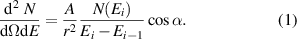
The energy distribution and temperature of hot electrons was measured by a multi-channel electron spectrometer installed at respective distances from front and back side of the tantalum target (see figure 1). The raw data of image plate signal along with electron energy spectrum is shown in figure 3. The energy distribution function (in units  ) is calculated using the analytical expression (1), where A is the area of the input aperture, r is the distance from the target, Ei
is the energy,
) is calculated using the analytical expression (1), where A is the area of the input aperture, r is the distance from the target, Ei
is the energy,  is the number of electrons detected in the given energy interval based on the imaging plate resolution and is given by the calibration of the imaging plate electron response. The importance of geometrical factor
  is to compensate the extra flux of electrons incoming at the angle to the detector.
The electron temperature  is determined by the slope of the energy distribution measured using multi-channel electron spectrometer which is shown by equation (2),
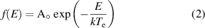
where,  =
= 
 and k is Boltzmann constant.
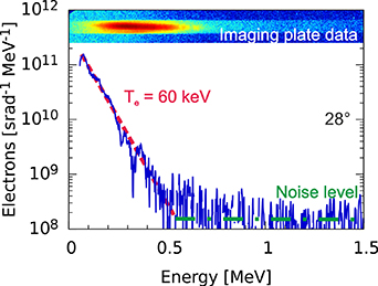
Figure 3. The energy distribution of escaped electrons from a single shot for Ta target of thickness of 10 µm. The measured temperature of electrons is 60 keV at angle 28∘ from the laser axis. The noise level is represented by dotted line in the electron spectrum. The measured signal on image plate detector is also shown on top of the figure.
Download figure:
Standard image High-resolution imageFor the  m thick target, the electron temperature estimated from the slope of the distribution is (60 ± 10) keV. Figure 4 shows the variation in electron temperature with different laser energy for the target thickness of
  m. The electron temperature estimated from the slope of the energy distribution increases for lower laser energy (
 J) however, temperature profile shows clearly transition to a saturation as shown by two different fitted lines (see figure 4). The electron temperature varies in the range from 20 keV to 60 keV and is almost constant for higher laser energy.

Figure 4. Hot electron temperature data measured for tantalum target of  m in thickness as a function of laser energy.
Download figure:
Standard image High-resolution imageThe profile of electron temperature at different laser intensities are compared with experimental and simulation results from sub-nanosecond and sub-picosecond laser systems (see figure 5). The theoretical expression for temperature scaling of hot electrons in high-contrast high-intensity laser-solid intercation [10] is shown by equation (3).
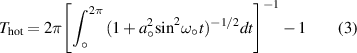
where, ![$a_{\circ} = [2I/(n_\mathrm{cr}m_\mathrm ec^3)]^{1/2}$](https://content.cld.iop.org/journals/0741-3335/64/10/105012/revision2/ppcfac8bf3ieqn44.gif) is the laser strength parameters and
is the laser strength parameters and  is the critical density of the plasma.
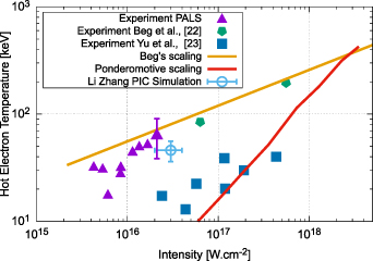
Figure 5. Intensity dependence of hot electron temperature from experiments of sub-nanosecond and sub-picosecond laser-matter interaction at different laser facilities together with the results presented in this paper (violet triangles). These measurements are compared with Beg scaling [39] (yellow line) and ponderomotive scaling [10] (red line).
Download figure:
Standard image High-resolution imageMoreover, the Beg scaling law [39] for the hot electron temperature associated with picosecond laser-solid interaction is represented by expression (4).

where I and λ are the intensity and wavelength of the laser respectively.
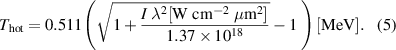
The analytical expression for ponderomotive scaling [66] relevant with relativistic laser intensities is shown by equation (5).
The first uncovering about the electron temperature is that T
 is considerably higher than the 'suprathermal' temperature, which should be less than 20 keV in our experiment [67, 68]. The calculated T
 from experimental data shows that the electron temperature scaling is mostly consistent with the Beg scaling observed for picosecond laser-matter interaction [39].
The experimental results by Kluge et al [19] also follows Beg scaling, however, past experimental results by Kluge et al [10] show that at lower laser intensities ( 1016 W cm−2), the electron temperature scaling varies in between Beg and ponderomotive scalings which are also in closer agreement with results shown in figure 5. It should be noted that the Beg scaling, corresponding to the resonance absorption mechanism, is observed for highest intensities which implies that the temporal contrast of the pulse might play important role at highest pulse energies. Moreover, ponderomotive scaling is usually valid for tightly focused laser beams and small plasma scale lengths which is contaray in our experiment having long plasma scale length (
 m). As noted in the manuscript, other experiments with high energy laser pulses (above 100 J) have also found electron temperatures higher than ponderomotive scaling. Higher electron temperatures can be attained by stochastic heating or direct laser-electron acceleration of electrons in a plasma with a large scale length.
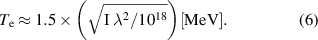
In addition, it is also well known that for under-dense plasma produced by target ablation from the laser prepulse, electron acceration occurs due to stochastic process and electron temperature is given by Pukhov scaling (see equation (6)) however, this scaling has some limitation once the plasma scale length is sufficiently large [69]. The electron temperature measured from laser interaction with a  m thick tantalum target is consistent with the expected energy lost by fast electrons while traversing
  m thick Ta target [70]. The electron spectrometer data thus shows that electrons with energies in the range of 0.5 MeV and 1 MeV were generated in the laser plasma interaction experiments for PALS parameters.
The total flux of hot electrons was measured by spectrometers in the front and back directions. Figures 6 and 7 represent the variation of electron flux with increasing laser energy for  m thick tantalum target in front and back directions respectively. The profiles of figures 6 and 7 show a threshold in laser energy and then step-like behavior of electron flux. The profiles of electron flux fitted with two linear lines with different slopes show a clear increase in electron flux, followed by a flat region and then increase again except the last two data points which can be attributed to statistical limitation in that particular two laser shots. In the front direction, the electron flux becomes significant for laser energy greater than 250 J. It is seen that electron flux increases with increasing laser energy. However, in the back direction of the target, the threshold in laser energy is around 350 J. The different energy threshold in front and backward direction could be attributed to the recirculation of electrons and reabsorption of x-ray photons inside the target plasma. The electron flux in the back direction has similar characteristic with increasing laser energy as in the front direction. It should be noted that similar threshold in laser energy is observed in the photon signal measured by the scintillator stack in the backward direction of the target, as will be presented later (see figure 12). The experimentally determined threshold energy in the range of
  J can be compared to the threshold power that should be exceeded to create relativistic self-focusing (RSF) in a plasma layer with
  cm−3 as discussed in the introduction section. In this case, P
 TW, as shown in figure 8 for laser energy ranging from 200 to 600 J. Figure 8 clearly shows that the relativistic self-focusing is out of the question for E
 J, but it can occur at higher laser energies. Even if RSF occurs, there will be no complete shrinkage of the focal spot to a diameter comparable to that of a laser wavelength [27] due to the probably insufficient thickness of the plasma layer with
  cm−3 at the target surface which is a part of the plasma expanding into vacuum. But there is limited shrinkage of the focal point, according to experimentally observed values of the maximum energy of protons reaching MeV values at maximum intensities on the target [71]. In this case, the focus diameter could shrink about ten times, to a value of
  m. It is obvious that the full development of the RSF will not occur, only its initial phase can occur, as indicated by figures 6–8.

Figure 6. The total electron flux in front direction of the tantalum target as a function of laser energy. A threshold in electron flux is observed around 250 J laser energy.
Download figure:
Standard image High-resolution image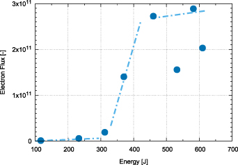
Figure 7. The total electron flux in back direction of the tantalum target as a function of laser energy. A similar threshold in electron flux is observed around 350 J laser energy as seen in front direction.
Download figure:
Standard image High-resolution image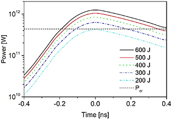
Figure 8. Time course of power for laser energy ranging from 200 to 600 J and critical power of relativistic self-focusing for laser wavelength of 1.315 µm and  cm−3.
cm−3.
Download figure:
Standard image High-resolution imageFor the interpretation of the presented results, it is important that the intensity dependence of temperature is far from the one that corresponds to the 'suprathermal' one [67, 68] but is very close to the Beg dependence, which is correlated with the extended Gibbon's model by Beg et al which was applied for 1 ps laser [39]. We also note that the maximum energy of ions accelerated under the presented experimental conditions [71] correlates well with the maximum energy of ions accelerated by electrons matching the Beg's scaling [39]. The interpretation of the results is difficult because different nonlinearities might play a role under the presented experimental conditions. It is very likely that the source of these difficulties is the fledgling RSF, however it has no chance of fully developing.
These experimental measurements indicate direct correlation between hot electron emission and bremsstrahlung emission. Furthermore, the maximum energy of the electrons was measured as a function of the laser energy for 10 µm thick Ta target. Figure 9 indicates that the profile of maximum electron energy is showing similar step-like behaviour as shown in figures 6 and 7.
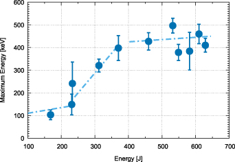
Figure 9. The maximum electron energy as a function of laser energy is observed for Ta targets with the use of the electron spectrometers.
Download figure:
Standard image High-resolution imageIn the case of 10 µm thick Ta target, integrating the measured electron spectra in angular direction to the entire 4π solid angle provides estimation of the total number of hot electrons per shot for laser energy about 600 J. The electron flux for energy greater than 50 keV is  , which corresponds to total charge of about
  C. The total energy carried with these electrons is limited to about 6 mJ, indicating a conversion efficiency from laser energy (
 J) to electrons of about
  . In addition, the measured profile of the conversion efficiency for different laser energy is shown in the figure 10. The result indicates that conversion efficiency is varying with increasing laser energy in the range between
  and
and  for laser energy from 100 J to 600 J, however, the characteristics of the profile resemble similar step-like behaviour as shown in the figures 6, 7 and 9.
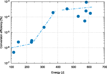
Figure 10. The conversion efficiency of laser energy to energy carried by fast electrons as a function of laser energy is shown for 10 µm thick Ta targets. The conversion efficiency is showing step like behaviour with laser energy.
Download figure:
Standard image High-resolution image3.2. Measurements and characterization of x-rays
The energy distribution of x-rays is measured using scintillator stack-based spectrometer. The location of the scintillation detector outside the interaction chamber affects the spectrum of the detected radiation because the lower-energy x-ray photons are absorbed by the chamber wall, in our case by the optical glass BK7 with a thickness of 10 mm. To define precisely the low energy spectrum, a 10 µm thick tantalum target was used in this case. Since the bremsstrahlung radiation emitted by the laser–plasma interaction is expected to follow Maxwellian distribution, a set of simulations of the scintillator stack interaction with photons of different temperatures were performed to create a response matrix. To estimate the temperature of the measured x-ray radiation, the difference between the experimental and simulated signals was minimized during a signal unfolding process. Figure 11 shows the profile of signal amplitude of each stack of 12 scintillators at angles (a) 150∘ and (b) 180∘ for the same laser shot delivering energy of 574 J on the target. A close agreement is observed between the experimental measurements and simulation results. This further confirms that the hot electrons with a mean energy in the range of 30–70 keV is the dominant component of accelerated electrons from laser-target interaction for the PALS parameters. Obviously, the maximum signal amplitude is almost 10 000 times higher than the minimum amplitude. Therefore, at lower laser intensities the lowest amplitudes are dominated by the noise.
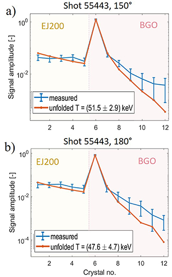
Figure 11. Experimental results of x-ray photons measured from two similar stack of scintillators mounted at (a) 150∘ and (b) 180∘ with respect to laser axis. The measured data are compared with FLUKA simulation using unfolded technique.
Download figure:
Standard image High-resolution imageThe photon temperature  which can represent the electron temperature was determined by the unfolding algorithm based on FLUKA simulations. The variation of photon temperature for laser intensities around
  W · cm−2 is shown in the figure 12. The temperature measurement is compared with Beg and Ponderomotive scaling. Although the lower sensitivity of the scintillation detector does not allow adequate measurements at lower intensities, it is clear from the figure 12 that the photon temperature profile match neither Beg scaling, nor ponderomotive scaling which is contrary to the electron temperature scaling as shown in the figure 5 that is in closer agreement with the Beg scaling. Such result is interesting since it indicates that hot electron scaling laws might not be applicable for the bremsstrahlung temperature prediction. The reason for the different temperature scaling of hot electrons and photons would be due to the different nature of bremsstrahlung and hot electron emission. The electron spectrometers located far from the target detect only hot electrons leaked from plasma through a potential barrier, while time integrating x-ray spectrometers detect radiation emitted by all electrons produced. Another important result is the observation of significant shot-to-shot variations in radiation temperature, which also occur in other target materials. This occurs indeed at higher laser energies, where the generation of fast electrons is unstable.
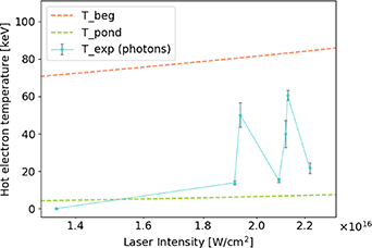
Figure 12. Hot electron temperature  determined by the unfolding algorithm based on FLUKA simulations at laser intensity of the order of
  W · cm−2. Upper and lower lines represent laser intensity dependence of temperature of hot electrons associated with Beg's and ponderomotive scaling, respectively.
Download figure:
Standard image High-resolution image4. Results of FLUKA simulations
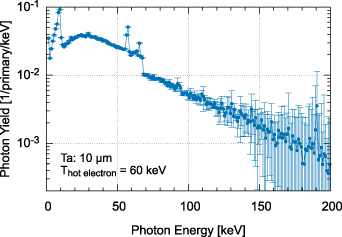
Figure 13 shows the expected x-ray energy spectrum from the experiment generated by Monte Carlo simulations. The simulations were performed with the FLUKA code as an input. The electron spectra used in simulation is limited by the fact that the hot electrons loose energy once they cross the potential barrier of the plasma. The electrons were injected along the direction of laser propagation in a form of a pencil-like beam. The energy of the x-ray photons are in the range of 10's of keV to 100 keV. The temperature of the photon distribution fitted with Maxwell distribution was  50 keV. The low energy photons with energy less than ~10 keV are significantly attenuated within the target. Based on the measured brightness of the scintillators, we assume that a target thickness in the range between 5 µm and 20 µm could be optimal for x-ray production in the energy range between 10 keV and 100 keV.
Figure 13. The photon spectra generated by Monte Carlo simulations for 10 µm thick tantalum target with electron distribution having 60 keV temperature. The profile indicates the simulated spectrum of photon energy is in the range between 10's keV and 100 keV. The low energy photons ( 10 keV) are mostly attenuated within the target.
Download figure:
Standard image High-resolution image
5. Conclusion
In summary, this paper presents the results from an experiment in which a nanosecond pulse ( ps) from an iodine laser was focused on tantalum thin foil targets at intensities in the range between
  and
and  W · cm−2. The hot electrons are characterized by measuring the energy spectrum for 10 µm Ta targets. The measured electron temperature using multi-channel electron spectrometer is observed in the range between 30 keV and 70 keV which implies that a mean energy of ~50 keV is the dominant component of accelerated electrons from laser-target interaction for the PALS and target parameters. The experimental investigation showed the temperature scaling and threshold in laser energy for hot electron emission. The electron energy spectrum and measured temperature were found to be consistent with Beg scaling law however, the unfolded temperature of the bremsstrahlung x-rays temperature is lower than the electron temperature, but fluctuates between values given by the Beg's and Ponderomotive scaling laws. This could be justified by ponderomotive and relativistic self-focusing, as well as classical thermal filamentation, because under our experimental conditions an appropriate intensity threshold is achievable [71]. In addition, the laser-plasma interaction at moderate intensity is also significantly affected by stimulated Raman scattering and dual plasmon decay. The presented results indicate that the simulated signal corresponding to the unfolded photon temperature using Monte Carlo code is consistent with measured signal of individual scintillator for each stack at two different angular direction. On contrary, temperature scaling of hot electrons escaping the plasma cannot be directly applied to the scaling of photons. Moreover, we observed that conversion efficiency from laser energy to hot electrons is showing step-like behaviour and it varies with increasing laser energy in the range between
  and
and  for laser energy in range between 100 J and 600 J, respectively. The experimental results associated with conversion efficiency and temperature scaling are important for the understanding of hot electron generation in long-pulse, high-intensity laser-solid interactions [72], such as those found in fast-ignition, high-brightness x-ray-generation experiments and laboratory astrophysics [41]. Furthermore, these measurements demonstrate that the x-ray radiography source [73, 74] can be produced with large scale kilo-joule nanosecond class laser systems. The production of high-fluence multi-keV sources of x-rays can be widely applicable for the radiography of dense plasma, material testing, nano-lithography, medicine and national security.
Data availability statement
The data generated and/or analysed during the current study are not publicly available for legal/ethical reasons but are available from the corresponding author on reasonable request. The data that support the findings of this study are available from the corresponding author upon reasonable request.
Acknowledgments
The research presented in this paper was supported by the Czech Republic- Ministry of Education, Youth and Sports—the projects: Prague Asterix Laser System (LM2018114) and creating and probing dense plasmas at the PALS facility (CZ ). The research leading to these results has received funding from the Czech Science Foundation (Grant No. 19-02545S) and from the Ministry of Education, Youth and Sports of the Czech Republic through the project 'Advanced Research Using High-Intensity Laser-Produced Photons and Particles' (CZ
 ). The support of CTU student support project 'Research on optical (nano) structures and laser plasma'
  is gratefully acknowledged. Moreover, this work has been carried out within the framework of the EUROfusion Consortium, funded by the European Union via the Euratom Research and Training Programme (Grant Agreement No. 101052200–EUROfusion). Views and opinions expressed are however those of the author(s) only and do not necessarily reflect those of the European Union or the European Commission. Neither the European Union nor the European Commission can be held responsible for them. The involved teams have operated within the framework of the Enabling Research Project: CfP-FSD-AWP21-ENR-01-CEA-02.