Article

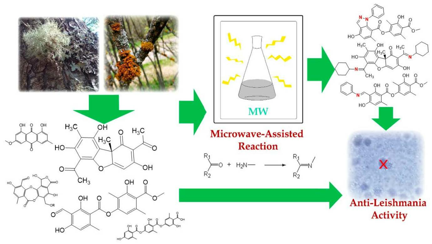
Microwave-Assisted Semisynthesis and Leishmanicidal Activity of Some Phenolic Constituents from Lichens

1
Instituto de Investigaciones Químicas IIQ, Universidad Mayor de San Andrés UMSA, Av. Villazón N° 1995, Box 303, La Paz 0201-0220, Bolivia
2
Instituto de Investigaciones Fármaco Bioquímicas, Área de Química Farmacéutica, Facultad de Ciencias Farmacéuticas y Bioquímicas, Universidad Mayor de San Andrés, Av. Saavedra N° 2224, Miraflores, Box 303, La Paz 0201-0220, Bolivia
3
Department of Food Technology, Engineering and Nutrition, Faculty of Engineering, Lund University, Box 117, SE-221 00 Lund, Sweden
4
Laboratory of Algal Biotechnology-Centre ALGATECH, Institute of Microbiology of the Czech Academy of Sciences, Opatovicky Mlyn, 37981 Trebon, Czech Republic
5
Faculty of Science, University of South Bohemia, Branišovská 1760, 37005 České Budějovice, Czech Republic
6
Departamento de Ciencias Químicas, Campus Viña del Mar, Universidad Andres Bello, Quillota 980, Viña del Mar 2520000, Chile
7
Departamento de Química, Facultad de Ciencias, Universidad de Chile, Box 653, Las Palmeras 3425, Ñuñoa, Santiago 8320000, Chile
*
Authors to whom correspondence should be addressed.
Separations 2023, 10(10), 524; https://doi.org/10.3390/separations10100524
Submission received: 16 August 2023 / Revised: 20 September 2023 / Accepted: 22 September 2023 / Published: 26 September 2023

Abstract

:
Leishmaniasis is considered one of the most untreated tropical diseases in the world. In this study, we investigated the in vitro leishmanicidal activity and cytotoxicity of various isolated lichen substances, including atranorin (1), usnic acid (2), gyrophoric acid (3), salazinic acid (4), galbinic acid (5), and parietin (6), and some semi-synthetic imine derivatives of usnic acid (7, 8, 9) and atranorin (10, 11, 12, 13). Imine condensation reactions with hydrazine and several amines were assisted by microwave heating, an efficient and eco-friendly energy source. The most interesting result was obtained for compound 2, which has high leishmanicidal activity but also high cytotoxicity. This cytotoxicity was mitigated in its derivative, 9, with better selectivity and high antileishmanic activity. This result may indicate that the usnic acid derivative (9) obtained using condensation with two cyclohexylamine groups is a promising lead compound for the discovery of new semisynthetic antiparasitic drugs.

Graphical Abstract

1. Introduction

Leishmaniasis is a group of diseases caused by protozoan parasites. More than 12 million people suffer from this disease, which is considered endemic in 88 countries around the world. Leishmaniasis is generally zoonotic in origin, including over 20 human pathogenic parasite species. Leishmaniasis is an intracellular infection, with different parasite species associated with various clinical forms of this disease. Many species cause skin ulcers and nodules leading to cutaneous leishmaniasis, which may be local or diffuse. Some species of these organisms can also affect the mucous membranes and cause lesions that disfigure the nose and cause mucocutaneous leishmaniasis. Other species damage internal organs and cause visceral leishmaniasis, which is fatal if not treated in time [1]. Leishmaniasis is considered by the WHO to be both a highly relevant endemic infectious disease and a public health threat. The pentavalent antimony preparations commonly used to treat this disease are limited. Therefore, new natural products could serve as alternatives to existing treatments [2] or could pave the way for the discovery of new leishmanicidal agents [3].
On the other hand, microwave-assisted organic synthesis facilitates more rapid reaction times and screening for chemical substances in order to discover potential new drugs with desirable properties [4]. Additionally, this technique is regarded as an eco-friendly source of energy in chemical synthesis, with better yields and higher purities when compared with traditional processes [5]. This approach has been employed to obtain new antileishmanial compounds such as natural-product-inspired functionalized spiro[indoline-3,2′-pyrrolidin]-2-one/spiro[indoline-3,3′-pyrrolizin]-2-one derivatives [6], γ-butyrolactones derivatives [7], methylene isoindolinone compounds [8], and 5-substituted benzylidene amino-2-butyl benzofuran-3-yl-4-methoxyphenyl methanones [9]. Keeping this in mind, we focused our interest on the condensation of amines with ketones to form imines, a reaction that has proven to be feasible under microwave conditions with promising results [10].
Taking this into account, our research has focused on natural products isolated from lichens. The classical definition states that lichens are organisms that arise from symbiosis between a fungus (mycobiont) and microscopic algae or cyanobacteria (photobiont). This symbiosis leads to a stable taxonomic unit with a long evolutionary history [11,12] that biosynthesizes some characteristic compounds called “lichen substances”, such as depsides, depsidones, dibenzofurans, anthraquinones, and pulvinic acid derivatives [13,14]. These compounds have a wide range of biological activities, such as antioxidant, anti-inflammatory, antiviral, analgesic, cytotoxic, antibiotic, antitumor, anti-HIV, antifungal, and antiproliferative properties [12,15]. This study aims to isolate lichen compounds, obtain new semisynthetic imine derivatives, and evaluate their leishmanicidal activity and cytotoxicity in vitro.

2. Materials and Methods

2.1. General

The reagents used for the derivatizations were aniline, 99.5%; phenylhydrazine, 97.0%; cyclohexylamine 95%; and naphthylamine, 99.0% (Sigma-Aldrich, St. Louis, MA, USA); absolute ethanol, 99.7% (Winkler, Lampa, Santiago, Chile); and chloroform, 99.99% (JT Baker, Puebla, Mexico). The deuterated solvents were purchased from Sigma-Aldrich (St. Louis, MA, USA). All NMR spectra were recorded using a spectrometer, the Bruker DRX 300 (Sao Paulo, Brazil, 300 MHz (1H) and 75 MHz (13C)), using TMS as the internal standard. MestReNova (Mestrelab Research, Santiago de Compostela, Coruña, Spain) was used to process the NMR spectra. Chemical shift values were referenced to the residual solvent signals for CDCl3 (δH/δC, 7.26/77.16) and DMSO (δH/δC, 2.50/39.51). Reactions requiring heating were performed using a microwave device (Monowave 450, Anton Paar, Buchs, Switzerland) with adjustable operating parameters. Finally, the melting point of the derivatives was determined with a fusiometer (Fisatom 430D, Sao Paulo, Brazil).

2.1.1. HPLC Parameters

A Thermo Scientific Dionex Ultimate 3000 UHPLC system equipped with a quaternary Series RS pump, and a PDA detector controlled by the Chromeleon 7.2 software (Thermo Fisher Scientific, Bremen, Germany), hyphenated, was used for analysis. A UHPLC C18 column (Acclaim, 150 mm × 4.6 mm ID, 5 µm, Thermo Fisher Scientific, Bremen, Germany) operated at 25 °C was employed. The PDA value was recorded from 200 to 800 nm, and the mobile phases were 0.1% formic aqueous solution (A) and acetonitrile (B). The gradient program (time (min), % B) was (0.00, 5); (5.00, 5); (10.00, 30); (15.00, 30); (20.00, 70); (25.00, 70); (35.00, 5). The flow rate was 1.00 mL min−1, and the injection volume was 10 μL. Samples (2 mg) were dissolved in methanol (10 mL) for the evaluation of purity.

2.1.2. MS Parameters

A Thermo Q Exactive focus orbitrap mass spectrometer with an electrospray ionization (ESI) source (Thermo, Bremen, Germany) was used for analysis. Sheath gas flow rate (N2), 75 units; aux. gas unit flow rate (N2), 20; capillary temperature, 400 °C; aux gas heater temperature, 500 °C; spray voltage, 2500 V; and S-lens RF level, 30. Full positive scan data were acquired at a resolving power of 70,000 FWHM (full width at half maximum) at m/z 200. For the compounds of interest, a scan range of m/z 100–1000 was chosen; the automatic gain control (AGC) was set at 3 × 10⁶ and the injection time was set to 200 ms. The scan rate was set at 2 scans/s−1. External calibration was performed using a calibration solution in positive and negative modes. For confirmation purposes, a targeted MS/MS analysis was performed using the mass inclusion list, with a 30 s time window, with an Orbitrap spectrometer operating both in positive and negative mode at 17,500 FWHM (m/z 200). The AGC target was set to 2 × 10⁵, with a maximum injection time of 20 ms. The precursor ions were filtered by a quadrupole, which operated at an isolation window of m/z 2. The fore vacuum, high vacuum, and ultrahigh vacuum were maintained at approximately 2 mbar, from 105 and below 1010 mbar. Collision energy (HCD cell) was operated at 30 kV. The mass tolerance window was set to 5 ppm for the two modes. Total analysis runtime was 1.0 min via direct injection.

2.2. Lichen Material

Lichen species were collected from different locations in Bolivia (May 2018) and Chile (September 2018). These places were chosen since there is a large population of these lichens. Sampling locations are listed in Table 1.

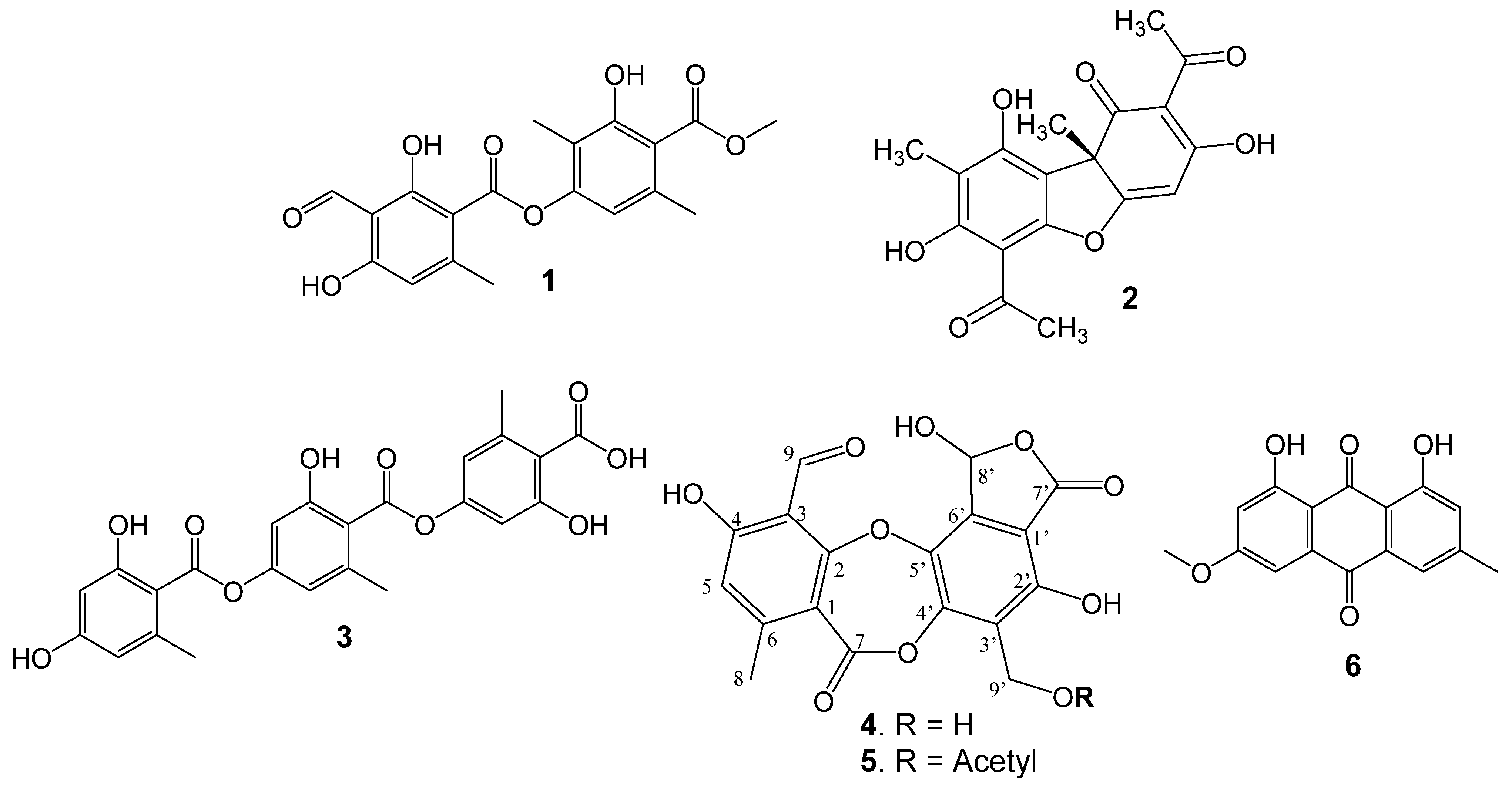
2.3. Isolation of Lichen Substances

Atranorin (1): This compound was isolated from S. ramulosum, as described in the literature [16]. In total, 100 g of dry lichen was extracted with EtOH (1.0 L) via maceration (48 h). The ethanolic extract (11.0 g) was eluted with a DCM:MeOH mixture (1:0; 9.5:0.5; 9:1; 8:2; 7:3; 6:4; 1:1; 0:1) to yield compound 1 (2.81 g): 1H-NMR (CDCl3, 300 MHz) δ (ppm); 2.71 (3H, s, CH3-9), 2.11(3H, s, CH3-8′), 2.56 (3H, s, CH3-9′), 4.00 (3H, s, CH3-O), 6.39 (1H, s, H-5), 10.37 (1H, s, H-8), 6.54 (1H, s, H-6′), 12.52 (1H, s, OH-2), 12.57 (1H, s, OH-4), 11.95 (1H, s, OH-3′). 13C-NMR (CDCl3, 75 MHz) δ (ppm); 102.2 (C-1), 169.1 (C-2), 108.5 (C-3), 167.5 (C-4), 112.8 (C-5), 152.4 (C-6), 169.7 (C-7), 193.0 (C-8), 25.6 (C-9), 152.4 (C-1′), 116.8 (C-2′), 162.9 (C-3′), 110.2 (C-4′), 139.9 (C-5′), 116.0 (C-6′), 172.2 (C-7′), 9.4 (C-8′), 24.0 (C-9′), 52.3 (CH3O-). This was confirmed via comparison with data from the literature [16,17].
Usnic acid (2): This natural product was isolated from X. lineola. Dry lichenic material (133.5 g) was extracted with EtOAc (0.9 L) for 48 h at room temperature; then, 80% of the solvent was removed under vacuum. The remaining 20% was left at room temperature to obtain a brown precipitate, which was filtered and washed with acetone followed by EtOH to yield compound 2 (3.01 g).
This natural product was also isolated from U. patagónica. Dry lichenic material (61.0 g) was extracted with acetone (1.0 L) for 72 h at room temperature; the extract was filtered; and the solvent was removed under vacuum. The obtained dry extract was then extracted with chloroform to collect yellow crystals corresponding to compound 2 (4.80 g): 1H-NMR (CDCl3, 300 MHz) δ (ppm); 1.78 (3H, s, CH3-15), 2.12 (3H, s, CH3-10), 2.68(3H, s, CH3-12), 2.69(3H, s, CH3-14), 6.00(1H, s, H-4), 11.04 (1H, s, OH-9),13.33 (1H, s, OH-7). 13C-NMR (CDCl3, 75 MHz) δ (ppm); 198.05 (C-1), 105.23 (C-2), 191.71 (C-3), 98.32 (C-4), 179.36 (C-4a), 155.20 (C-5a), 101.52 (C-6), 163.88 (C-7), 109.32 (C-8), 157.50 (C-9), 103.95 (C-9a), 59.07 (C-9b), 7.52 (C-10), 201.76 (C-11), 27.87 (C-12), 200.31 (C-13), 31.25 (C-14), 32.11 (C-15). This was confirmed via comparison with data from the literature [18].
Gyrophoric acid (3): This natural product was isolated from U. patagónica. Dry lichenic material (61.0 g) was extracted with acetone (1.0 L) for 72 h at room temperature; the extract was filtered; and the solvent was removed under vacuum. The dry extract obtained was then extracted with chloroform, and the remaining white amorphous solid was recrystallized in MeOH to yield compound 3 (3.50 g): 1H-NMR (DMSO-d6, 300 MHz) δ (ppm); 2.50 (3H, s, CH3-8), 2.51 (3H, s, CH3-8′), 2.52 (3H, s, CH3-8″), 6.36 (1H, d, J = 2.1 Hz, H-3), 6.38 (1H, d, J = 2.1 Hz, H-5), 6.72 (1H, d, J = 1.7 Hz, H-3′), 6.74 (1H, d, J = 1.7 Hz, H-5′), 6.76 (1H, d, J = 2.0 Hz, H-3″), 6.78 (1H, d, J = 2.0 Hz, H-5″). 13C-NMR (DMSO-d6, 75 MHz) δ (ppm); 109.3 (C-1), 160.9 (C-2), 101.4 (C-3), 162.0 (C-4), 110.7 (C-5), 141.2 (C-6), 168.0 (C-7), 21.2 (C-8), 118.9 (C-1′), 157.2 (C-2′), 118.0 (C-3′), 153.0 (C-4′), 115.3 (C-5′), 138.9 (C-6′), 166.6 (C-7′), 21.2 (C-8′), 118.2 (C-1″), 157.2 (C-2″), 108.0 (C-3″), 153.0 (C-4″), 115.1 (C-5″), 140.4 (C-6″), 171.4 (C-7″), 21.7 (C-8″). This was confirmed via comparison with data from the literature [17,19].
Salazinic acid (4): This natural product was isolated from P. paramoreliensis. Dry lichenic material (100.1 g) was extracted with EtOH (1.2 L) for 48 h at room temperature; the extract was filtered; and 85% of the solvent was removed under vacuum. The remaining 15% was left at room temperature to obtain a precipitate, which was filtered and washed with warm EtOH to yield compound 4 (2.60 g): 1H-NMR (DMSO-d6, 300 MHz) δ (ppm); 2.46 (3H, s, CH3-8), 4.66 (2H, s, CH2-9′), 10.46 (1H, s, H-9), 6.89 (1H, s, H-5), 6.81 (1H, s, H-8′), 8.31 (1H, s, OH-8′), 12.06 (1H, s, OH-2′). 13C-NMR (DMSO-d6, 75 MHz) δ (ppm); 112.43 (C-1), 164.03 (C-2), 111.14 (C-3), 164.47 (C-4), 117.89 (C-5), 152.74 (C-6), 160.76 (C-7), 21.88 (C-8), 193.20 (C-9), 110.12 (C-1′), 153.27 (C-2′), 123.92 (C-3′), 148.62 (C-4′), 138.57 (C-5′), 137.78 (C-6′), 166.35 (C-7′), 95.33 (C-8′), 53.16 (C-9′). This was confirmed via comparison with data from the literature [20,21].
Galbinic acid (5): This compound was isolated from U. cornuta [22]. From 50 g of dry lichen was macerated with methanol (1.2 L) for 24 h. The crude extract was submitted to flash chromatography and then CC using DCM/MeOH mixtures to yield compound 5 (400 mg): 1H-NMR (DMSO-d6, 300 MHz) δ (ppm); 2.47 (3H, s, CH3-8), 5.18 (2H, s, CH2-9′), 10.45(1H, s, H-9), 6.89 (1H, s, H-5), 6.82 (1H, s, H-8′), 8.34 (1H, s, OH-8′), 12.06 (1H, s, OH-2′), 1.99 (3H, s, CH3-O) [9]. 13C-NMR (DMSO-d6, 75 MHz) δ (ppm); 112.09 (C-1), 164.21 (C-2), 111.71 (C-3), 164.54 (C-4), 117.98 (C-5), 152.77 (C-6), 160.52 (C-7), 21.76 (C-8), 193.10 (C-9), 110.19 (C-1′), 156.73 (C-2′), 119.02 (C-3′), 149.34 (C-4′), 138.10 (C-5′), 136.55 (C-6′), 166.09 (C-7′), 95.68 (C-8′), 55.90 (C-9′), 170.04, 21.04 (Acetyl). This was confirmed via comparison with data from the literature [22].
Parietin (6): This natural product was isolated from T. chrysophthalmus. Dry lichenic material (39.9 g) was extracted with acetone (0.7 L) for 30 h at room temperature; then, 80% of the solvent was removed under vacuum. The remaining 20% was cooled to 0 °C in an ice bath, yielding a precipitate that was filtered and washed with warm EtOAc to obtain compound 6 (1.05 g): 1H-NMR (DMSO-d6, 300 MHz) δ (ppm); 2.44 (3H, s, CH3-15), 3.93 (3H, s, CH3-16), 6.69 (1H, d, J = 2.48, H-10), 7.07 (1H, s, H-2), 7.34 (1H, d, J = 2.52, H-8) 7.61 (1H, s, H-5), 12.11 (1H, s, OH-1), 12.30 (1H, s, H-10). 13C-NMR (DMSO-d6, 75 MHz) δ (ppm); 162.48 (C-1), 124.49 (C-2), 148.45, (C-3),121.27, (C-4), 133.19 (C-5), 182.01 (C-6), 135.23 (C-7), 108.23 (C-8), 166.54 (C-9), 106.73 (C-10), 165.18 (C-11), 110.22 (C-12), 190.78, (C-13), 133.65 (C-14), 22.15 (C-15), 56.08 (C-16). This was confirmed via comparison with data from the literature [23,24].

2.4. Semi-Synthesis of Derivatives

(S)-5-hydroxy-1,4,5b,7-tetramethyl-3,9-diphenyl-5b,9-dihydrofuro [2,3-e:4,5-f’]bis(indazole)-6(3H)-one (7): Compound 2 (502 mg, 1.46 mmol) was dissolved in an Erlenmeyer flask with EtOH (96 °GL, 20 mL) and then phenylhydrazine (0.5 mL, 5.08 mmol) was added. The mixture was irradiated with microwaves (170 W) for 15 min at intervals of 20 s; during the rest periods, the Erlenmeyer flask was cooled at 0 °C in an ice bath. After the completion of the reaction, the mixture was left in the ice bath at 0 °C for 1 h to precipitate the crude product. The solid was filtered and washed with EtOH, yielding the following: 511 mg (1.05 mmol, 71.9%), mp 285 °C, 1H-NMR (CDCl3, 300 MHz) δ (ppm); 1.82 (3H, s, CH3-15), 2.21 (3H, s, CH3-10), 2.61 (3H, s, CH3-12), 2.46 (3H, s, CH3-14), 6.18 (1H, s, H-4), 11.61 (1H, s, OH-9), 7.59–6.94 (10H, H- [a, b, c, d, a’, b’, c’ d’]). 13C-NMR (CDCl3, 75 MHz) δ (ppm); 196.43 (C-1), 108.29 (C-2), 148.59 (C-3), 88.15 (C-4), 173.75 (C-4a), 143.98 (C-5a), 100.15 (C-6), 1652.38 (C-7), 108.29 (C-8), 158.07 (C-9), 103.65 (C-9a), 60.52 (C-9b), 8.20 (C-10), 151.40 (C-11), 13.34 (C-12), 145.65 (C-13), 15.16 (C-14), 30.58(C-15), 138.09 (C-a), 123.96 (C-b), 129.43 (C-c), 128.58 (C-d), 153.23 (C- a’), 113.02 (C-b’), 129.65 (C-b’), 120.94 (C-d’). ESI-MS: 489.19 ([M + H]+, m/z); (calcd. for C30H25N4O3+, 489.19).
(S,E)-6-acetyl-3,7,9-trihydroxy-8,9b-dimethyl-2-(1-(phenylimino)ethyl)dibenzo[b,d] furan-1(9bH)-one (8): A mixture of compound 2 (877 mg, 2.55 mmol), aniline (0.5 mL, 5.48 mmol), and EtOH (10 mL) was irradiated with microwaves (170 W) at 30 s intervals for 12 min in an Erlenmeyer flask, and the mixture was cooled at 0 °C in an ice bath between each heating interval. At the end of the reaction time, the mixture was cooled in an ice bath at 0 °C for 60 min. After this time, a yellow amorphous solid formed, which was recovered via filtration and washed with EtOH:H2O 1:1, yielding: 863 mg of product 8 (2.06 mmol, 80.8%), mp 226 °C, 1H-NMR (CDCl3, 300 MHz) δ (ppm); 1.77 (3H, s, CH3-15), 2.12 (3H, s, CH3-10), 2.59 (3H, s, CH3-12), 2.71 (3H, s, CH3-14), 5.89 (1H, s, H-4), 11.88 (1H, s, OH-9), 13.39 (1H, s,OH-7), 15.08 (1H, s,OH-3), 7.73–7.21 (5H, H-[a, b, c, d]). 13C-NMR (CDCl3, 75 MHz) δ (ppm); 198.84 (C-1), 102.65 (C-2), 191.03 (C-3), 102.26 (C-4), 174.78 (C-4a), 155.80 (C-5a), 101.39 (C-6), 163.56 (C-7), 108.18 (C-8), 158.22 (C-9), 104.96 (C-9a), 57.55 (C-9b), 7.52 (C-10), 173.98 (C-11), 20.65 (C-12), 200.70 (C-13), 31.35 (C-14), 31.92 (C-15), 136.06 (C-a), 125.75 (C-b), 129.70 (C-c), 128.28 (C-d). ESI-MS: 420.13 ([M + H]+, m/z); (calcd. for C24H22NO6+, 420.14).
(S)-2-((E)-1-(cyclohexylimino)ethyl)-6-(1-(cyclohexylimino)ethyl)-3,7,9-trihydroxy-8,9b-dimethyldibenzo[b,d]furan-1(9bH)-one (9): A mixture of compound 2 (802 mg, 2.33 mmol), cyclohexylamine (0.6 mL, 5.23 mmol), and EtOH (25 mL) was irradiated with microwaves (170 W) in an Erlenmeyer flask for 15 min at intervals of 20 s, and the mixture was cooled to 0 °C in an ice bath between each heating interval. At the end of the reaction time, the mixture was cooled for 2 h at 0 °C in an ice bath. After this time, a yellow amorphous solid formed, which was recovered via filtration and washed with EtOH, yielding: 1042 mg of product 9 (2.06 mmol, 88.4%), mp 235 °C, 1H-NMR (CDCl3, 300 MHz) δ (ppm); 1.69 (3H, s, CH3-15), 2.13 (3H, s, CH3-10), 2.57 (3H, s, CH3-12), 2.64 (3H, s, CH3-14), 5.72 (1H, s, H-4), 11.50 (1H, s, OH-9), 13.57 (1H, s, OH-7),3.75, 3.72 (2H, CH=N), 1.95-1.25 (20H, H- [a, b, c, a’, b’, c’]). 13C-NMR (CDCl3, 75 MHz) δ (ppm); 198.81 (C-1), 102.02 (C-2), 190.83 (C-3), 101.32 (C-4), 168.66 (C-4a), 154.69 (C-5a), 98.97 (C-6), 163.71 (C-7), 108.91 (C-8), 155.45 (C-9), 103.50 (C-9a), 54.30 (C-9b), 8.12 (C-10), 171.50 (C-11), 17.59 (C-12), 167.06 (C-13), 18.33 (C-14), 32.72 (C-15), 54.30 (C-a), 33.29 (C-b), 25.35 (C-c), 24.47 (C-d), 52.41 (C-a’), 32.72 (C-b’), 25.08 (C-b’), 24.03 (C-d’). ESI-MS m/z: 507.28 ([M + H]+, m/z); (calcd. for C30H39N2O5+, 507.29).
3-hydroxy-4-(methoxycarbonyl)-2,5-dimethylphenyl 4-hydroxy-6-methyl-1-phenyl-1H-indazole-7-carboxylate (10): Compound 1 (1080 mg, 2.88 mmol) was dissolved in an Erlenmeyer flask with EtOH (96 °GL, 20 mL) and then phenylhydrazine (0.5 mL, 5.1 mmol) was added. The mixture was irradiated with microwaves (170 W) for 3 min at 12 s intervals. During the rest periods, the Erlenmeyer flask was cooled to 0 °C in an ice bath. The reaction was monitored using TLC. When the reaction was complete, the mixture was cooled at 4 °C for 30 min to precipitate the crude product. The solid was filtered, yielding: 1101 mg of compound 10 (2.5 mmol, 86.2%), mp 187 °C, 1H-NMR (CDCl3, 300 MHz) δ (ppm); 2.69 (3H, s, CH3-9), 2.12 (3H, s, CH3-8′), 2.56 (3H, s, CH3-9′), 4.00 (3H, s, CH3-O), 6.50 (1H, s, H-5), 8.38 (1H, s, H-8), 6.55 (1H, s, H-6′), 12.11 (1H, s, OH-4), 11.97 (1H, s, OH-3′), 7.55–6.97 (6H, m, 1″-4″ aromatic). 13C-NMR (CDCl3, 75 MHz) δ (ppm); 102.79 (C-1), 163.72 (C-2), 105.39 (C-3), 162.88 (C-4), 112.77 (C-5), 144.35 (C-6), 170.23 (C-7), 136.83 (C-8), 25.03 (C-9), 152.35 (C-1′), 116.91 (C-2′), 162.84 (C-3′), 110.05 (C-4′), 139.77 (C-5′), 116.25 (C-6′), 172.29 (C-7′), 9.39 (C-8′), 24.06 (C-9′), 52.32 (CH3O-), 143.41 (C-1″), 112.54 (C-2″), 129.57 (C-3″), 120.84 (C-4″). ESI-MS m/z: 447.15 ([M + H]+, m/z); (calcd. for C25H23N2O6+, 447.16).
3-hydroxy-4-(methoxycarbonyl)-2,5-dimethylphenyl (E)-2,4-dihydroxy-6-methyl-3-((phenylimino)methyl)benzoate (11): A mixture of compound 1 (325 mg, 0.87 mmol), aniline (0.15 mL, 1.58 mmol), and EtOH (10 mL) was irradiated with microwaves (170 W) in an Erlenmeyer flask for 3 min at 15 s intervals. Between each heating interval, the mixture was cooled to 0 °C in an ice bath. The reaction was monitored using TLC. Once complete, the reaction mixture was cooled for 60 min at 0 °C in an ice bath for 60 min. After this time, a yellow amorphous solid formed, which was recovered via filtration, corresponding to 345 mg of product 11: (0.77 mmol, 88.5%), mp 167 °C, 1H-NMR (CDCl3, 300 MHz) δ (ppm); 2.67 (3H, s, CH3-9), 2.13 (3H, s, CH3-8′), 2.57 (3H, s, CH3-9′), 4.00 (3H, s, CH3-O), 6.41 (1H, s, H-5), 9.17 (1H, s, H-8), 6.55 (1H, s, H-6′), 15.65 (1H, s, OH-2), 12.67 (1H, s, OH-4), 11.97 (1H, s, OH-3′), 7.49-7.28 (6H, m, a-d aromatic). 13C-NMR (CDCl3, 75 MHz) δ (ppm); 100.22 (C-1), 168.04 (C-2), 105.99 (C-3), 170.24 (C-4), 115.27 (C-5), 148.38 (C-6), 171.94 (C-7), 156.54 (C-8), 25.41 (C-9), 152.35 (C-1′), 116.93 (C-2′), 162.87 (C-3′), 110.04 (C-4′), 139.74 (C-5′), 116.27 (C-6′), 172.28 (C-7′), 9.37 (C-8′), 24.05 (C-9′), 52.30 (CH3O-), 144.79 (C-a), 120.64 (C-b), 129.65 (C-c), 127.24 (C-d). ESI-MS m/z: 450.15 ([M + H]+, m/z); (calcd. for C25H24NO7+, 450.16).
3-hydroxy-4-(methoxycarbonyl)-2,5-dimethylphenyl (E)-2,4-dihydroxy-6-methyl-3-((naphthalen-2-ylimino)methyl)benzoate (12): A mixture of compound 1 (505 mg, 1.35 mmol), 1-naphthylamine 300 mg, 2.10 mmol), and EtOH (15 mL) was irradiated with microwaves (170 W) at 15 s intervals for 5 min in an Erlenmeyer flask. Between each heating interval, the mixture was cooled to 0 °C in an ice bath. The reaction was monitored using TLC. Once complete, the reaction mixture was cooled for 60 min at 0 °C in an ice bath. After this time, a yellow amorphous solid formed, which was recovered via filtration, yielding: 592 mg of product 13 (1.19 mmol, 88.1%), mp 177 °C, 1H-NMR (CDCl3, 300 MHz) δ (ppm); 2.71 (3H, s, CH3-9), 2.13 (3H, s, CH3-8′), 2.56 (3H, s, CH3-9′), 3.99 (3H, s, CH3-O), 6.50 (1H, s, H-5), 9.27 (1H, s, H-8), 6.56 (1H, s, H-6′), 15.80 (1H, s, OH-2), 12.57 (1H, s, OH-4), 11.97 (1H, s, OH-3′), 7.92-7.22 (10H, m, a-j aromatic) 13C-NMR (CDCl3, 75 MHz) δ (ppm); 101.07 (C-1), 170.18 (C-2), 106.58 (C-3), 167.53 (C-4), 114.61 (C-5), 148.49 (C-6), 170.30 (C-7), 158.23 (C-8), 25.42 (C-9), 152.32 (C-1′), 116.90 (C-2′), 162.85 (C-3′), 110.07 (C-4′), 139.76 (C-5′), 116.25 (C-6′), 172.27 (C-7′), 9.37 (C-8′), 24.02 (C-9′), 52.29 (CH3O), 143.49 (C-a), 114.48 (C-b), 126.75 (C-c), 127.28 (C-d), 134.04 (C-e), 128.10 (C-f), 125.92 (C-g), 126.80 (C-h), 127.78 (C-i), 122.69 (C-j). ESI-MS m/z: 500.16 ([M + H]+, m/z); (calcd. for C29H26NO7+, 500.17).
Ethyl (E)-2,4-dihydroxy-6-methyl-3-((phenylimino)methyl)benzoate (13): Compound 1 (500 mg, 1.34 mmol) was dissolved in a mixture of ethanol:chloroform (2:1) (15 mL), and aniline (0.2 mL, 2.19 mmol) was added; then, the reaction was sonicated for 30 min at room temperature in an ultrasonic bath. The mixture was then left at room temperature overnight, which enabled the crystallization of 13 into yellow needles, yielding: 329 mg (1.10 mmol, 82.1%). mp 117 °C, 1H-NMR (CDCl3, 300 MHz) δ (ppm); 1.45 (3H, t, J = 7.08, CH3-11), 2.54 (3H, s, CH3-9), 4.42 (2H, q, J = 7.04, CH2-10), 6.32 (1H, s, H-5), 9.15 (1H, s, H-8), 13.03 (1H, s, OH-2), 15.36 (1H, s, OH-4), 7.28-7.47 (5H, m, 1′-4′aromatic). 13C-NMR (CDCl3, 75 MHz) δ (ppm); 101.96 (C-1), 169.63 (C-2), 106.03(C-3), 166.71 (C-4), 113.87 (C-5), 146.00 (C-6), 172.7 (C-7), 157.18 (C-8), 25.13 (C-9), 61.44 (C-10), 14.25 (C-11), 148.28 (C-1′), 120.82 (C-2′), 129.52 (C-3′), 126.91 (C-4′). ESI-MS m/z: 300.10 ([M + H]+, m/z); (calcd. for C17H18NO4+, 300.12).
All synthesized compounds were identified via qualitative analysis using a combination of both TLC and HPLC, and we observed the purity of the compounds. In relation to the compounds containing an imine group (8, 9, and 1113), one E isomer was detected. After TLC and HPLC analysis, only one compound was observed and confirmed with NMR analysis. Rotation is common in imine groups, with the E isomer being favored owing to steric effects. It was not possible for it to show up clearly on X-rays since not all compounds were crystalline material. Compounds 79 were eluted at retention times (Rt) of 26.9, 27.0, and 26.5 min on HPLC, respectively, while compounds 1013 were eluted at Rts of 27.0, 26.7, 27.3, and 28.8 min. TLC analysis (SiO2) was conducted using dichloromethane, and a spot was observed under UV light (254–366 nm). The analysis was determined via the retention factor (Rf). Compounds 713 showed Rf values of 0.45, 0.57, 0.42, 0.18, 0.33, 0.12, and 0.10 on TLC, respectively.

2.5. Evaluation against Leishmania Parasites

Promastigotes of Leishmania-Leishmania: L. amazonensis, clone 1, NHOM-BR-76-LTB-012 (Lma), Leishmania-Viannia: L. braziliensis, M2904 C192 RJA (M2904). Strains were cultured in Schneider’s insect medium (pH 6.2) supplemented with 10% fetal bovine serum (FBS) and incubated in 96-microwell plates at 26 °C. The biological assay was performed as described elsewhere [25]. Briefly, promastigotes in the logarithmic growth phase at a concentration of 3 × 10⁶ parasites/mL were treated with samples dissolved in DMSO (1%) at different concentrations (3.1–100 μg/mL). Miltefosine (3.1–100 μg/mL, Profounda Company, Orlando, FL, USA) and total alkaloid extracts from Galipea longiflora (CAT, 3.1–100 μg/mL) [26] were used as control drugs. The total alkaloid extract, referred to as CAT y/o AEE, was used as a positive natural extract control for Leishmania and other protozoa parasites as quoted from different authors [27]. The main component of the G. longiflora of the active fraction was 2-phenyl-quinoline, but the bulk total alkaloids are cited as an active natural reference since the values of other synthetic drugs are active at very low concentrations when compared with raw extracts. The leishmanicidal model used was based on promastigotes, and activity values were quoted against cytotoxicity against RAW cells, indicating whether these substances present some selective activity. The microwell plates were incubated at 26 °C for 72 h, the optical density of each well was determined, and the IC50 values were calculated. The assays were performed in triplicate.

2.6. Cytotoxicity

The RAW 264.7 cell line (Murine Macrophages, Carlsbad, CA, USA) was used and grown in DMEM-HG medium (Dulbeccos Modified Eagle’s Medium-High Glucose, Grand Island, NY, USA), supplemented with 1% streptomycin–penicillin, sodium bicarbonate (2.2 g/L), and 10% fetal bovine serum in sterile 25 cm2 culture boxes incubated under standard conditions (5% CO2 in air, 37 °C, and 100% relative humidity). The culture medium was changed every 72 h. The biological assay was performed with cells at a concentration of 5 × 10⁴ cells/mL incubated for 24 h and then treated with samples dissolved in DMSO (1%) at different concentrations. The microwell plates were incubated for 72 h; then, resazurine (2 mM, 10 μL/well) prepared in PBS (2.86 g/L Na2HPO4, 0.31g/L NaH2PO4, pH = 7.4) was added and incubated for another 3 h. The fluorescence of each well was measured (excitation 540 nm and emission 590 nm), and IC50 values were calculated. Assays were performed in triplicate.

2.7. 96-Well Plate Preparation

After 72 h of incubation and cell confluence of approximately 95%, the cells were mechanically detached (scraped), the cell suspension was counted in a Neubauer chamber, and the population was adjusted to 5 × 10⁴ cells/mL and distributed into 96-well plates (100 µL). They were incubated for 24 h. Then, the concentrations of the compounds (100 µL) (200-100-50-25-12.5-6.2 ug/mL) were added to the indicated wells. DMEM-HG medium blank (100 µL) plus study drugs (100 µL) were the cell growth controls. Plates were incubated at 37 °C for 72 h.

2.8. IC50 by Fluorometric Method (Rezasurin)

After 72 h of incubation at 37 °C, resazurin (2 mM) was added to each well (10 µL) and incubated for another 3 h under the same conditions. Plates were read in a SYNERGY spectrofluorometer at 540 nm excitation and 590 nm emission (Santa Clara, CA, USA). The Gen5 program (Bio tek) and linear trend calculations using Microsoft Excel (Version 2110) were used to determine the mean inhibitory concentration (IC50).

2.9. Selective Index

Selective index values were calculated as the ratio between cell LD50 values and parasite IC50 values (SI = LD50/IC50).

3. Results and Discussion

3.1. Lichen Compounds

Depside atranorin (1) was isolated from Stereocaulon ramulosum. This known dibenzofuran usnic acid (2) was isolated from various species, that is, Xanthoparmelia lineola and Usnea patagonica; tridepside gyrophoric acid (3) was isolated from U. patagonica; depsidones salazinic acid (4) and galbinic acid (5) were isolated from Parmotrema paramoreliensis and Usnea cornuta, respectively; and finally, the anthraquinone called parietin (6) was isolated from Teloschistes chrysophthalmus. All these structures (Figure 1) were elucidated via NMR spectroscopy (Supplementary Material).

3.2. Microwave Semisynthesis

Microwaves (MWs) have been used in lichen chemistry, especially for the extraction of lichen substances, with analytical proposals [28,29,30]. On the other hand, MW-assisted reactions have been widely and successfully used for the derivatization of natural products in pharmaceutical research, with the advantage of being an environmentally friendly tool that offers improved reaction times and yields [31]. Moreover, MW heating improves the results and experimental procedures for the formation of imine and Schiff bases [10], which are interesting functional groups for lichen compounds given their potential biological activities [32].
Two lichen substances were selected as starting materials given their fast derivatization from the lichens under study. They are common compounds in lichens: usnic acid (2) was used to obtain compounds 7, 8, and 9, and atranorin (1) was used to obtain compounds 10, 11, 12, and 13 (Figure 2A). The condensation of usnic acid (2) with various nitrogen-containing compounds has been described previously under conventional heating with aromatic and aliphatic amines and phenylhydrazines [11,33,34,35,36,37] and atranorin (1), which were condensed using hydrazides under conventional heating [38]. The nitrogen-based reagents that were used in this study were pheynylhydrazine, aniline, 2-naphtilamine, and cyclohexylamine (Figure 2B). Preliminary experiments with aliphatic amines have shown complex reaction mixtures, possibly related to degradation; therefore, we focused our study on cyclic and aromatic compounds. An additional advantage was that the adducts with cyclic amines were much easier to isolate and purify via crystallization.
The condensation reaction between 2 and phenylhydrazine provided compound 7 with a good yield (71.9%). This product contains two heterocyclic pyrazoline moieties, which have not been reported for conventional heating. This last product had a pyrazoline ring and an imine intermediate that could not cyclize even in the presence of a large excess of phenylhydrazine [11]. Although the reaction products were different, the reaction times could be compared, showing that MW heating is much faster than conventional heating. Under MWs, the reaction was completed in only 15 min, but under conventional heating, it took 2 h without completing product 7 [11]. Compound 8 was obtained via the condensation of 2 with aniline. The formation of imine adducts has been studied and compared under MW conditions and conventional heating. The authors reported a reaction between 2 and several aromatic amines. The reported reaction conditions were an irradiation power of 60 W for 20 min, a usnic acid (2):aromatic amine ratio of 1:1, and ethanol as solvent. On the other hand, our conditions were an irradiation power of 170 W for 6 min (intervals of 30 s), a usnic acid (2):aromatic amine ratio of 1:1.8, and ethanol as solvent. In some cases where aniline is used as an aromatic amine, the reported yield was 64.8%, and our procedure provided a yield of 80.8%. As expected, more irradiation power and a higher usnic acid (2):aniline ratio, as employed in our procedure, can improve yields [39]. Finally, compound 9 was the condensation product of 2 with cyclohexylamine. This product has not been reported previously, but reports of coupling with aliphatic amines under conventional heating have been published [33,34,35,36,37,39,40]. Under conventional heating, the yields are nearly 80%, but under MW irradiation, the yield is nearly 90%. However, the most important difference is the reaction time, which was significantly improved from an average of 1.5 h under conventional heating to 12 min under MWs.
The condensation of 1 with phenylhydrazine yielded compound 10 (86.2%). The reaction provided similar results to 7, forming a pyrazoline ring, but with regioselectivity in this case. To explain this regioselectivity, a reaction mechanism is proposed in Figure 3 in which an aromatic nucleophilic substitution of the hydroxyl group from the phenol by the nitrogen of the coupled hydrazine is facilitated by a 1,4 addition to a vicinal α,β-unsaturated carbonyl group. It has been reported that the addition of hydrazines to 1 under conventional heating provides intermediate imines [38]. Therefore, the formation of the pyrazoline ring in product 10 may indicate that only MW irradiation can provide the necessary energy to complete the reaction. Compound 11 was obtained via the condensation of 1 with aniline. Unlike the other products, there is no information about this reaction. The result shows that MW heating provides a high yield (88.5%) in a short reaction time (3 min). The same result was observed when the reaction was repeated with 2-naphtilamine to obtain compound 12, for which the yield and reaction time were also higher (87.9%, 5 min).
Finally, compound 13 was an artifact produced by the chemical reaction of compound 11. To obtain a homogeneous mixture in ethanol:chloroform, compound 1 and aniline were sonicated for 30 min. After this time, yellow needle crystals, identified as 13, were observed. Even considering that the reaction was not planned, the yield was high (82.1%). The formation of 13 can be explained as follows: ultrasound methods have been shown to be a fast and efficient method of producing Schiff bases [41]. The additional transesterification of orsellinic acid with an ethoxy group in 3-formyl orsellinic acid can be facilitated via ultrasound in the same way as the transesterification of fatty acids under ultrasound for the production of biodiesel [42].

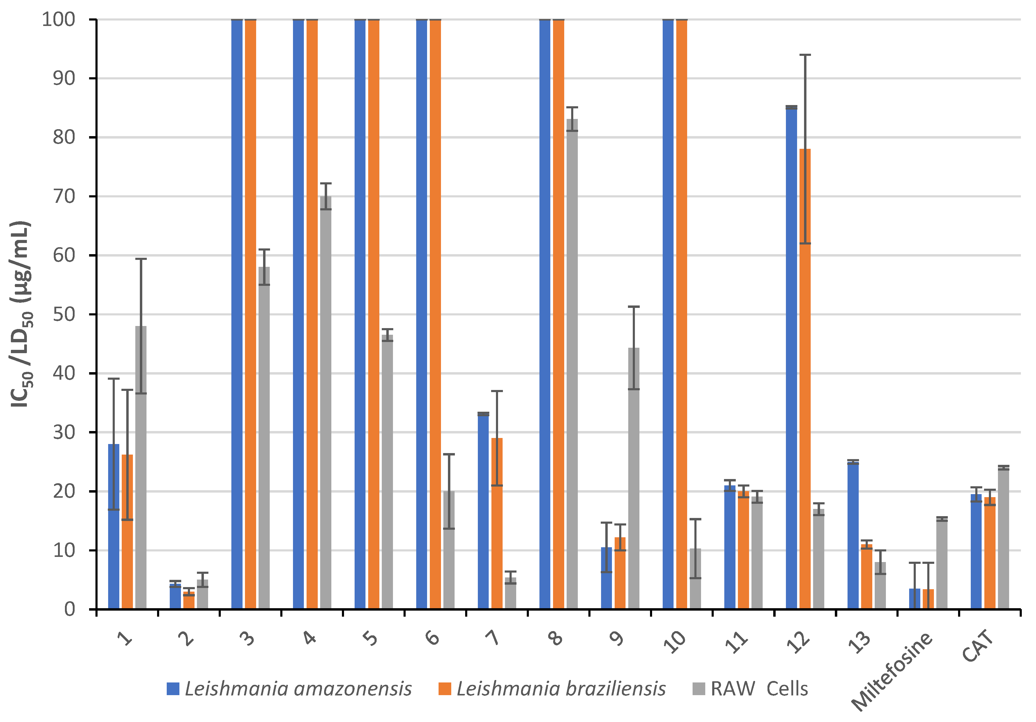
3.3. Leishmania Activity and Cytotoxicity

Previous studies on lichen compounds with antileishmanial activity have been poorly explored. Similar studies have shown that depside isodivaricatic acid, depsidonas 1′-chloropannarine, and pannarine have good activity against Leishmania amazonensis, L. brasiliensis, and L. infantum [43]. Usnic acid, one of the most representative and accessible lichen compounds, is a candidate for the treatment of infections caused by L. major, L. infantum, L. tropica [44,45], L. donovani [44], L. brasiliensis, and L. infantum chagasi [2], but its use as a treatment is not recommended because it has hepatoxic effects [46].
Gyroporic acid (3), salazinic acid (4), galbinic acid (5), parietin (6), and derivatives 8 and 10 showed no activity against Leishmania parasites, suggesting that they are more cytotoxic than the antiparasitic ones (Table 2). Atranorin (1) showed moderate activity, whereas usnic acid (2) showed strong activity against both Leishmania strains. The SI for 1 was 1.8 for both strains. The SI for 2 was 1.2 for L. amazonensis and 1.7 for L. braziliensis. These results may indicate that these active natural products could be promising lead compounds for the synthesis of leishmanicidal compounds. The semisynthetic derivatives 10, 11, 12, and 13 obtained from 1 showed a significant increase in cytotoxicity or loss of leishmanicidal activity (Figure 4). Interestingly, the semisynthetic derivative 9 from 2 provided a better SI value (about four) than its precursor, suggesting that 2 could be an interesting drug leader providing selective and highly active leishmanicidal compounds. In Table 2, only compounds 1 and 9 could be compared with Miltefosine and CAT since all others are mainly cytotoxic and cannot be classified as leishmanicidal.
The reason why these reported compounds exert activity remains unknown. There are many natural products that show activity, but no studies for understanding the mechanisms of action have been performed. Some antifungal agents (such as Amphotericine-B) show anti-leishmanial activity because fungi and leishmania parasites share some cell wall characteristics [47], but other compounds, such as Glucantime® [48], have a very different mechanism of action. Therefore, detailed studies on the mechanisms of action of these lichen compounds should be carried out.

4. Conclusions

Our results show that novel derivatives were semi-synthesized via the condensation of lichen compounds with amines. The reactions were carried out under MW conditions following green chemistry procedures. They proved to be favorable for the formation of the products in high yields, especially in short reaction times. Although the results for leishmanicidal activity were not encouraging given the cytotoxicity of the compounds, we believe that usnic acid is still a promising lead compound for the development of new antiparasitic drugs if selectivity is improved. Additionally, semisynthetic derivative 9 provided a better SI than 2, suggesting that aliphatic imines can have better selectivity and activity. We suggest carrying out further research on this issue.

Supplementary Materials

The following supporting information can be downloaded at: https://www.mdpi.com/article/10.3390/separations10100524/s1, Figure S1: 13C-NMR (DMSO-d6, 75 MHz) spectrum of galbinic acid (5); Figure S2: 1H-NMR (CDCl3, 300 MHz) spectrum of compound (7); Figure S3: 13C-NMR (CDCl3, 75 MHz) spectrum of compound (7); Figure S4: 1H-NMR (CDCl3, 300 MHz) spectrum of compound (8); Figure S5: 13C-NMR (CDCl3, 75 MHz) spectrum of compound (8); Figure S6: 1H-NMR (CDCl3, 300 MHz) spectrum of compound (9); Figure S7: 13C-NMR (CDCl3, 75 MHz) spectrum of compound (9); Figure S8: 1H-NMR (CDCl3, 300 MHz) spectrum of compound (10); Figure S9: 13C-NMR (CDCl3, 75 MHz) spectrum of compound (10); Figure S10: 1H-NMR (CDCl3, 300 MHz) spectrum of compound (11); Figure S11: 13C-NMR (CDCl3, 75 MHz) spectrum of compound (11); Figure S12: 1H-NMR (CDCl3, 300 MHz) spectrum of compound (12); Figure S13: 13C-NMR (CDCl3, 75 MHz) spectrum of compound (12); Figure S14: 1H-NMR (CDCl3, 300 MHz) spectrum of compound (13); Figure S15: 13C-NMR (CDCl3, 75 MHz) spectrum of compound (13).

Author Contributions

Conceptualization, G.C., A.G. and C.A.; Lichen collection, G.C., J.L.V., B.S. and C.A.; Methodology, G.C., R.V., P.C.-P., J.A.B. and J.L.V.; in vitro leishmania assays, A.G. and E.S.; writing—original draft preparation, G.C. and R.V.; writing—review and editing, J.C., D.B.-P., A.G. and C.A. All authors have read and agreed to the published version of the manuscript.

Funding

This research was funded by Agencia Nacional de Investigación y Desarrollo de CHILE (Fondecyt Regular under Grant N° 1230414).

Institutional Review Board Statement

Not applicable.

Informed Consent Statement

Not applicable.

Data Availability Statement

Data of the compounds are available from the authors.

Acknowledgments

A cordial thanks to Yonny Flores and Maribel Lozano from NMR Laboratory—IIQ-UMSA for their collaboration in recording the NMR spectra and to UMSA-ASDI: Biomolecules–Antiparasitic project for covering biological evaluations.

Conflicts of Interest

The authors declare no conflict of interest.

References

  1. WHO. Control of the Leishmaniases: Report of a Meeting of the WHO Expert Commitee on the Control of Leishmaniases; Report No. 9789241209496; World Health Organization: Geneva, Switzerland, 2010.
  2. Da Luz, J.; de Oliveira, E.; Martins, M.; da Silva, N.; Alves, L.; dos Santos, F.; da Silva, L.; Silva, E.; de Medeiros, P. Ultrastructural Analysis of Leishmania infantum chagasi Promastigotes Forms Treated In Vitro with Usnic Acid. Sci. World J. 2015, 2015, 617401–617408. [Google Scholar] [CrossRef]
  3. Cheuka, P.; Mayoka, G.; Mutai, P.; Chibale, K. The Role of Natural Products in Drug Discovery and Development against Neglected Tropical Diseases. Molecules 2017, 22, 58. [Google Scholar] [CrossRef]
  4. Kappe, C.; Dallinger, D. The impact of microwave synthesis on drug discovery. Nat. Rev. Drug Discov. 2006, 5, 51–63. [Google Scholar] [CrossRef]
  5. Leadbeater, N. Microwave Heating as a Tool for Sustainable Chemistry; CRC Press: Boca Raton, FL, USA, 2010; ISBN 9780429075339. [Google Scholar]
  6. Sahu, N.; Sharma, R.; Suhas, K.; Joshi, J.; Prakash, K.; Sharma, R.; Pratap, R.; Hu, X.; Kaur, S.; Jain, M.; et al. Natural-Product-Inspired Microwave-Assisted Synthesis of Novel Spirooxindoles as Antileishmanial Agents: Synthesis, Stereochemical Assignment, Bioevaluation, SAR, and Molecular Docking Studies. Molecules 2023, 28, 4817–4836. [Google Scholar] [CrossRef]
  7. Pinatto-Botelho, M.; Crotti, A.; Souza, J.; Magalhães, L.; Donate, P. Microwave-assisted synthesis and antileishmanial activity of 3-methoxycarbonyl-γ-butyrolactone derivatives. J. Braz. Chem. Soc. 2014, 25, 1331–1337. [Google Scholar] [CrossRef]
  8. Jellali, H.; Amri, N.; Mukhrish, Y.; Al Nasr, I.; Koko, W.; Khan, T.; Deniau, E.; Sauthier, M.; Ghalla, H.; Hamdi, N. Copper-Catalyzed Asymmetric Hydroboration Reaction of Novel Methylene Isoindolinone Compounds through Microwave Irradiation and Their Antileishmanial and Antitoxoplasma Activities. ACS Omega 2023, 8, 23067–23077. [Google Scholar] [CrossRef]
  9. Patil, S.; Bollikonda, S.; Patil, R.; Sangshetti, J.; Bobade, A.; Asrondkar, A.; Reddy, P.; Shinde, D. Microwave-assisted synthesis of novel 5-substituted benzylidene amino-2-butyl benzofuran-3-yl-4-methoxyphenyl methanones as antileishmanial and antioxidant agents. Bioorg. Med. Chem. 2018, 28, 482–487. [Google Scholar] [CrossRef]
  10. Chakraborty, M.; Baweja, S.; Bhagat, S.; Chundawat, T. Microwave Assisted Synthesis of Schiff Bases: A Green Approach. Int. J. Chem. React. Eng. 2012, 10, 1–4. [Google Scholar] [CrossRef]
  11. Luzina, O.; Polovinka, M.; Salakhutdinov, N.; Tolstikov, G. Chemical modification of usnic acid: III.* Reaction of (+)-usnic acid with substituted phenylhydrazines. Russ. J. Org. Chem. 2009, 45, 1783–1789. [Google Scholar] [CrossRef]
  12. Calcott, M.; Ackerley, D.; Knight, A.; Keyzers, R.; Owen, J. Secondary metabolism in the lichen symbiosis. Chem. Soc. Rev. 2018, 47, 1730–1760. [Google Scholar] [CrossRef]
  13. Jha, B.; Shrestha, M.; Pandey, D.; Bhattarai, T.; Bhattarai, H.; Paudel, B. Investigation of antioxidant, antimicrobial and toxicity activities of lichens from high altitude regions of Nepal. BMC Complement. Altern. Med. 2017, 17, 282. [Google Scholar] [CrossRef] [PubMed]
  14. Salgado, F.; Albornoz, L.; Cortéz, C.; Stashenko, E.; Urrea-Vallejo, K.; Nagles, E.; Galicia-Virviescas, C.; Cornejo, A.; Ardiles, A.; Simirgiotis, M.; et al. Secondary Metabolite Profiling of Species of the Genus Usnea by UHPLC-ESI-OT-MS-MS. Molecules 2018, 23, 54. [Google Scholar] [CrossRef] [PubMed]
  15. Bhattacharyya, S.; Deep, P.; Singh, S.; Nayak, B. Lichen Secondary Metabolites and Its Biological Activity. Am. J. PharmTech Res. 2016, 6, 28–44. [Google Scholar]
  16. Vila, J.; Mollinedo, P.; Flores, Y.; Sterner, O. 1,3,7-Trimethylguanine from the lichen Stereocaulon ramulosum. Rev. Bol. Quim. 2008, 25, 1–3. [Google Scholar]
  17. Edwards, H.G.M.; Newton, E.M.; Wynn-Williams, D.D. Molecular structural studies of lichen substances II: Atranorin, gyrophoric acid, fumarprotocetraric acid, rhizocarpic acid, calycin, pulvinic dilactone and usnic acid. J. Mol. Struct. 2003, 651–653, 27–37. [Google Scholar] [CrossRef]
  18. Rashid, M.; Majid, M.; Quader, M. Complete NMR assignments of (+)-usnic acid. Fitoterapia 1999, 70, 113–115. [Google Scholar] [CrossRef]
  19. Narui, T.; Sawada, K.; Takatsuki, S.; Okuyama, T.; Culberson, C.F.; Culberson, W.L.; Shibata, S. NMR assignments of depsides and tridepsides of the lichen family umbilicariaceae. Phytochemistry 1998, 48, 815–822. [Google Scholar] [CrossRef]
  20. Eifler-Lima, V.L.; Sperry, A.; Sinbandhit, S.; Boustie, J.; Tomasi, S.; Schenkel, E. NMR spectral data of salazinic acid isolated from some species of Parmotrema. Magn. Reson. Chem. 2000, 38, 472–474. [Google Scholar] [CrossRef]
  21. Vila, J.; Mollinedo, P.; Sterner, O. Spectroscopic studies of lichen depsides and depsidones. Rev. Bol. Quim. 2011, 28, 28–34. [Google Scholar]
  22. Castañeta, G.; Sepulveda, B.; Vargas, R.; Garcia-Beltran, O.; Simirgiotis, M.; Areche, C. A sustainable application for the extraction of lichen metabolites from Usnea cornuta: Nontargeted metabolomics and antioxidant activity. Nat. Prod. Res. 2022, 37, 2076–2082. [Google Scholar] [CrossRef]
  23. Edwards, H.G.M.; Newton, E.M.; Wynn-Williams, D.D.; Coombes, S.R. Molecular spectroscopic studies of lichen substances 1: Parietin and emodin. J. Mol. Struct. 2003, 648, 49–59. [Google Scholar] [CrossRef]
  24. Canaviri, P.; Bergquist, K.-E.; Vila, J. Estudio fitoquímico del liquen Teloschistes flavicans. Rev. Bol. Quim. 2006, 23, 9–12. [Google Scholar]
  25. Arévalo-Lopéz, D.; Nina, N.; Ticona, J.C.; Limachi, I.; Salamanca, E.; Udaeta, E.; Paredes, C.; Espinoza, B.; Serato, A.; Garnica, D.; et al. Leishmanicidal and cytotoxic activity from plants used in Tacana traditional medicine (Bolivia). J. Ethnopharmacol. 2018, 216, 120–133. [Google Scholar] [CrossRef] [PubMed]
  26. Campos-Buzzi, F.; Fracasso, M.; Clasen, B.K.; Ticona, J.C.; Gimenez, A.; Cechinel-Filho, V. Evaluation of antinociceptive effects of Galipea longiflora alkaloid extract and major alkaloid 2-phenylquinoline. Methods Find. Exp. Clin. Pharmacol. 2010, 32, 707–711. [Google Scholar] [CrossRef]
  27. Calla-Magarinos, J.; Gimenez, A.; Troye-Blomberg, M.; Fernandez, C. An Alkaloid Extract of Evanta, Traditionally Used as Anti-leishmania Agent in Bolivia, Inhibits Cellular Proliferation and Interferon-γ Production in Polyclonally Activated Cells. Scan. J. Immunol. A 2009, 69, 251–258. [Google Scholar] [CrossRef]
  28. Bonny, S.; Hitti, E.; Boustie, J.; Bernard, A.; Tomasi, S. Optimization of a microwave-assisted extraction of secondary metabolites from crustose lichens with quantitative spectrophotodensitometry analysis. J. Chromatogr. A 2009, 1216, 7651–7656. [Google Scholar] [CrossRef]
  29. Calla-Quispe, E.; Robles, J.; Areche, C.; Sepulveda, B. Are Ionic Liquids Better Extracting Agents Than Toxic Volatile Organic Solvents? A Combination of Ionic Liquids, Microwave and LC/MS/MS, Applied to the Lichen Stereocaulon glareosum. Front. Chem. 2020, 8, 450. [Google Scholar] [CrossRef]
  30. Komaty, S.; Sauvager, A.; Bazureau, J.; Tomasi, S.; Paquin, L. Efficiency and selectivity of ionic liquids in microwave-assisted extraction of major lichen phenolic compounds: A scalable process with recycling of ionic liquids. Phytochem. Anal. 2021, 32, 592–600. [Google Scholar] [CrossRef]
  31. Majhi, S.; Das, D. Chemical derivatization of natural products: Semisynthesis and pharmacological aspects—A decade update. Tetrahedron 2021, 78, 131801–131823. [Google Scholar] [CrossRef]
  32. Kajal, A.; Bala, S.; Kamboj, S.; Sharma, N.; Saini, V. Schiff Bases: A Versatile Pharmacophore. J. Catal. 2013, 2013, 893512. [Google Scholar] [CrossRef]
  33. Kutney, J.; Sanchez, I. Studies in the usnic acid series. I. The condensation of (+)-usnic acid with aliphatic and aromatic amines. Can. J. Chem. 1976, 54, 2795–2803. [Google Scholar] [CrossRef]
  34. Bazin, M.; Lamer, A.; Delcros, J.; Rouaud, I.; Uriac, P.; Boustie, J.; Corbel, J.; Tomasi, S. Synthesis and cytotoxic activities of usnic acid derivatives. Biorg. Med. Chem. 2008, 16, 6860–6866. [Google Scholar] [CrossRef] [PubMed]
  35. Luzina, O.; Sokolov, D.; Pokrovskii, M.; Pokrovskii, A.; Bekker, O.; Danilenko, V.; Salakhutdinov, N. Synthesis and Biological Activity of Usnic Acid Enamine Derivatives. Chem. Nat. Compd. 2015, 51, 646–651. [Google Scholar] [CrossRef]
  36. Victor, K.; Boris, L.; Athina, G.; Anthi, P.; Marija, S.; Marina, K.; Oliver, R.; Marina, S. Design, synthesis and antimicrobial activity of usnic acid derivatives. MedChemComm 2018, 9, 870–882. [Google Scholar] [CrossRef]
  37. Samuelsen, L.; Hansen, P.; Vang, O. Derivatives of usnic acid cause cytostatic effect in Caco-2 cells. Nat. Prod. Res. 2021, 35, 4953–4959. [Google Scholar] [CrossRef]
  38. Duong, T.-H.; Paramita Devi, A.; Tran, N.-M.-A.; Phan, H.-V.-T.; Huynh, N.-V.; Sichaem, J.; Tran, H.-D.; Alam, M.; Nguyen, T.-P.; Nguyen, H.-H.; et al. Synthesis, α-glucosidase inhibition, and molecular docking studies of novel N-substituted hydrazide derivatives of atranorin as antidiabetic agents. Bioorg. Med. Chem. Lett. 2020, 30, 127359. [Google Scholar] [CrossRef]
  39. Nguyen, G.; Luu, K.; Nguyen, P. Preparation of some derivatives of (+)-usnic acid with aromatic amines under microwave irradiation condition. STDJ 2015, 18, 113–120. [Google Scholar] [CrossRef]
  40. Luzina, O.; Polovinka, M.; Salakhutdinov, N.; Tolstikov, G. Chemical modification of usnic acid—2. Reactions of (+)-usnic acid with amino acids. Russ. Chem. Bull. 2007, 56, 1249–1251. [Google Scholar] [CrossRef]
  41. Kargar, H.; Behjatmanesh-Ardakani, R.; Fallah-Mehrjardi, M.; Torabi, V.; Munawar, K.; Ashfaq, M.; Tahir, M. Ultrasound-based synthesis, SC-XRD, NMR, DFT, HSA of new Schiff bases derived from 2-aminopyridine: Experimental and theoretical studies. J. Mol. Struct. 2021, 1233, 130105. [Google Scholar] [CrossRef]
  42. Armenta, R.; Vinatoru, M.; Burja, A.; Kralovec, J.; Barrow, C. Transesterification of Fish Oil to Produce Fatty Acid Ethyl Esters Using Ultrasonic Energy. JAOCS 2007, 84, 1045–1052. [Google Scholar] [CrossRef]
  43. Schmeda-Hirschmann, G.; Tapia, A.; Lima, B.; Pertino, M.; Sortino, M.; Zacchino, S.; Rojas de Arias, A.; Feresin, G. A new antifungal and antiprotozoal depside from the andean lichen Protousnea poeppigii. Phytother. Res. 2008, 22, 349–355. [Google Scholar] [CrossRef] [PubMed]
  44. Fournet, A.; Ferreira, M.; de Arias, A.; de Ortiz, S.; Inchausti, A.; Yalaff, G.; Quilhot, W.; Fernandez, E.; Hidalgo, M. Activity of Compounds Isolated from Chilean Lichens Against Experimental Cutaneous Leishmaniasis. Comp. Biochem. Physiol. C Pharmacol. Toxicol. 1997, 116, 51–54. [Google Scholar] [CrossRef] [PubMed]
  45. Derici, M.; Cansaran-Duman, D.; Taylan-Özkan, A. Usnic acid causes apoptotic-like death in Leishmania major, L. infantum and L. tropica. 3 Biotech 2018, 8, 384. [Google Scholar] [CrossRef] [PubMed]
  46. Kwong, S.; Wang, C. Review: Usnic acid-induced hepatotoxicity and cell death. Environ. Toxicol. Pharmacol. 2020, 80, 103493. [Google Scholar] [CrossRef]
  47. Balasegaram, M.; Ritmeijer, K.; Lima, M.A.; Burza, S.; Ortiz, G.; Milani, B.; Gaspani, S.; Potet, J.; Chappuis, F. Liposomal amphotericin B as a treatment for human leishmaniasis. Expert Opin. Emerg. Drugs 2012, 17, 493–510. [Google Scholar] [CrossRef]
  48. Iranpour, S.; Hosseinzadeh, A.; Alipour, A. Efficacy of miltefosine compared with glucantime for the treatment of cutaneous leishmaniasis: A systematic review and meta-analysis. Epidemiol. Health 2019, 41, e2019011. [Google Scholar] [CrossRef]
Figure 1. Chemical structure of the isolated lichen compounds.
Separations 10 00524 g001
Figure 2. (A) Chemical structure of semisynthetic compounds. (B) Nitrogen-based reagents used for the derivatization.
Separations 10 00524 g002
Figure 3. Proposed reaction mechanism for the formation of a pyrazoline ring for compounds 7 and 10.
Separations 10 00524 g003
Figure 4. In vitro leishmanicidal activity (IC50) and cytotoxicity in RAW line cell (LD50) against Leishmania amazonensis and Leishmania braziliensis.
Separations 10 00524 g004
Table 1. Lichen species from Bolivia and Chile.
LichenProvidence (Site)Jurisdiction
Usnea patagonica *Sud Yungas (Pongo)La Paz
Stereocaulon ramulosum *Sud Yungas (Unduavi)La Paz
Teloschistes chrysophthalmus *Murillo (Cota-Cota)La Paz
Xanthoparmelia lineola *Omasuyos (Apuvillque)La Paz
Usnea Cornuta ⁑ChillanVIII region
Parmotrema paramoreliensis ⁑LongaviVII region
* The lichens were identified at the Herbario Nacional de Bolivia at Universidad Mayor de San Andrés (UMSA), La Paz, Bolivia. ⁑ The lichens were identified at the Biology Department of the Universidad Metropolitána de Ciéncias de la Educación (UMCE), Santiago, Chile.
Table 2. In vitro leishmanicidal activity and cytotoxicity in RAW line cell.
CompoundLeishmania amazonensis
IC50 (µg/mL)
SILeishmania braziliensis
IC50 (µg/mL)
SIRAW
Cells
LD50 (µg/mL)
128.0 ± 11.11.726.2 ± 111.848.0 ± 11.4
24.3 ± 0.51.23.0 ± 0.61.75.0 ± 1.2
3>100->100-58.0 ± 3.0
4>100->100-70.0 ± 2.2
5>100->100-46.5 ± 1.0
6>100->100-20.0 ± 6.3
733.1 ± 6.50.229.0 ± 8.00.25.4 ± 1.0
8>100->100-83.1 ± 2.0
910.5 ± 0.34.212.2 ± 2.23.644.3 ± 7.0
10>100->100-10.3 ± 5.0
1121.0 ± 2.00.920.0 ± 1.01.019.1 ± 1.0
1285.1 ± 130.278.0 ± 160.217.0 ± 1.0
1325 ± 2.00.311.0 ± 0.20.78.0 ± 2.0
Miltefosine3.5 ± 0.64.43.4 ± 1.04.515.3 ± 0.3
CAT19.5 ± 1.41.219.0 ± 2.41.324.0 ± 0.3
SI: Selective index.
Disclaimer/Publisher’s Note: The statements, opinions and data contained in all publications are solely those of the individual author(s) and contributor(s) and not of MDPI and/or the editor(s). MDPI and/or the editor(s) disclaim responsibility for any injury to people or property resulting from any ideas, methods, instructions or products referred to in the content.

