Communication

Synthesis and In Vitro Evaluation of Novel Dopamine Receptor D2 3,4-dihydroquinolin-2(1H)-one Derivatives Related to Aripiprazole

1
National Institute of Mental Health, Topolova 748, 250 67 Klecany, Czech Republic
2
Department of Chemistry, University of Hradec Kralove, Rokitanskeho 62, 500 03 Hradec Kralove, Czech Republic
3
CZ-OPENSCREEN: National Infrastructure for Chemical Biology, Department of Informatics and Chemistry, Faculty of Chemical Technology, University of Chemistry and Technology Prague, Technicka 5, 166 28 Prague, Czech Republic
4
Institute of Physiology, Czech Academy of Sciences, Videnska 1083, 142 20 Prague, Czech Republic
5
Biomedical Research Centre, University Hospital Hradec Kralove, Sokolska 581, 500 05 Hradec Kralove, Czech Republic
*
Authors to whom correspondence should be addressed.
Biomolecules 2021, 11(9), 1262; https://doi.org/10.3390/biom11091262
Submission received: 15 July 2021 / Revised: 19 August 2021 / Accepted: 20 August 2021 / Published: 24 August 2021

Abstract

:
In this pilot study, a series of new 3,4-dihydroquinolin-2(1H)-one derivatives as potential dopamine receptor D2 (D2R) modulators were synthesized and evaluated in vitro. The preliminary structure–activity relationship disclosed that compound 5e exhibited the highest D2R affinity among the newly synthesized compounds. In addition, 5e showed a very low cytotoxic profile and a high probability to cross the blood–brain barrier, which is important considering the observed affinity. However, molecular modelling simulation revealed completely different binding mode of 5e compared to USC-D301, which might be the culprit of the reduced affinity of 5e toward D2R in comparison with USC-D301.

1. Introduction

Dopamine primarily mediates its effect through activation of dopamine receptors (DRs) [1]. There are a total of five DR subtypes that are members of the G-protein-coupled receptors (GPCRs) [2], and are further divided into two classes according to the structure. The D1-like family includes D1Rs and D5Rs, whereas D2–4Rs belong to the D2-like family [1,2,3]. The main difference between both families is that D1-like family activate adenylate cyclase (AC), which leads to production of cyclic adenosine monophosphate (cAMP), whereas D2-like stimulates AC activity.
Dopamine D2 type receptors (D2Rs) are integral membrane receptors coupled to G proteins with three extracellular loops, seven transmembrane domains, and three intracellular loops [4,5]. D2Rs are present in two isoforms, short D2S and long D2L, which differ by the insertion of 29 amino acids in the third intracellular loop on D2LRs [6]. The “short” version is exclusively expressed presynaptically as an autoreceptor, whereas, the “long” one is mainly found at the postsynaptic cells [7]. Furthermore, both isoforms can inhibit intracellular cyclic adenosine monophosphate via Gi [4]. The highest levels of D2Rs in the human brain are expressed within striatum, the olfactory tubercle, and the nucleus accumbens, but it can be also found in the ventral tegmental area, substantia nigra, septum, amygdala, cortical areas, and hippocampus. D2Rs are involved in working memory, reward motivation functions, and regulation of locomotion [2,8]. D2Rs are the main target in the treatment of schizophrenia [9]. Indeed, antagonists or partial agonists of D2Rs are the main representatives for schizophrenia. Besides, they can be applied in the therapy of depression and anxiety [9,10,11,12,13].
Schizophrenia is a serious mental disorder that affects up to 1% of the population worldwide [9]. It is believed, that environmental factors (i.e., viral infection, early childhood trauma, and obstetrical complications) increase the expression of certain genes to enhance risk factors for schizophrenia out-break [14,15]. The symptoms of schizophrenia include three groups, namely (i) positive (hallucinations, disorganized speech and delusions), (ii) negative (diminished expressiveness and reduced motivation), and (iii) cognitive (impaired memory, decreased speed of mental processing and executive functions) [14]. The pathophysiology of schizophrenia is poorly understood, however, it is assumed, that dysfunction of the dopamine mesolimbic pathway is responsible for positive symptoms and mesocortical pathway for negative symptoms [9,16]. Cognitive symptoms have been suggested to result from decreased levels of dopamine in cortical areas [17]. The current approaches for the management of schizophrenia involve three generations of antipsychotics. The first antipsychotics are mainly D2R antagonists, the second exhibit multi-target antagonism with significant antagonism at serotonin 5-HT2A type receptor (5-HT2AR), and the third possess a multi-target profile with partial agonism at D2R [9]. All licensed neuroleptics exhibited D2R affinity at therapeutic doses crucial for their mechanism of action [18,19,20,21]. In addition to D2Rs affinity, antipsychotic drugs show activity toward other dopamine (D1, D3, and D4) and serotonin (especially 5-HT1A, 5-HT2A, and 5-HT2C) receptors. However, selective D1, D3, and D4 receptor antagonists are not so effective in providing antipsychotic response, and D1Rs agonists exhibited limited clinical efficacy [22,23,24,25,26,27]. The evidence of 5-HT1ARs agonism/partial agonism is inconclusive and the role of the 5-HT2ARs antagonism is neither necessary nor a major contributor to antipsychotic effect [28,29,30]. Furthermore, 5-HT2CRs antagonism has been associated mainly with side-effects including weight gain, diabetes, and sexual disturbances [30,31]. On the other hand, pimavanserin (an inverse agonist at 5-HT2ARs) has recently been approved for the treatment of psychosis associated with Parkinson’s disease [32]. According to various studies, there is no benefit even when choosing different combinations of antipsychotics in 13–50% of all patients [33]. In addition, the current medication reduces mainly positive symptoms and causes severe side-effects such as diabetes, sexual dysfunction, weight gain, confusion, blurred vision, sedation, or dizziness, which also typically occurs during the treatment of depressive or anxiety disorders [34,35,36,37,38]. Thus, there is an enormous need to develop new effective and safe neuroleptic drugs since CNS disorders are also considered among the most expensive medical conditions (the total cost of disorders of the brain was estimated 798 billion EUR in Europe in 2010) [39].
In this pilot study, we designed and synthesized a novel series of compounds based on the approved drug aripiprazole characterized by the effect via D2R [40], in combination with structural properties of USC-D301, highly selective D2R ligand [41]. Considering this effect, we have performed in vitro evaluation of novel 3,4-dihydroquinolin-2(1H)-one derivatives (Figure 1) for their affinity to D2Rs, predicted their CNS availability, and established their cytotoxicity profile in order to estimate the clinical relevance of our observations. The D2Rs affinity is also corroborated by the in silico simulation.

2. Materials and Methods

2.1. Chemistry

The chemicals were purchased from Sigma-Aldrich Co., LLC (Prague, Czech Republic) and were used without additional purification. Analytical thin-layer chromatography was carried out using plates coated with silica gel 60 with the fluorescent indicator F254 (Merck, Prague, Czech Republic). The thin-layer chromatography (TLC) plates were visualized by exposure to ultraviolet light (254 nm) or by the detection reagent ninhydrin. Column chromatography was performed using silica gel 100 at atmospheric pressure (70–230-mesh ASTM, Fluka, Prague, Czech Republic). The NMR spectra were all recorded on a Varian S500 spectrometer (500 MHz for 1H and 126 MHz for 13C). Chemical shifts are reported in δ ppm referenced to residual solvent signals (for 1H NMR and 13C NMR: chloroform-d (CDCl3; 7.26 (D) or 77.16 (C) ppm). A CEM Explorer SP 12 S was used for the MW-assisted reaction. The final compounds were analyzed by LC-MS with a Dionex Ultimate 3000 RS UHPLC system coupled with a Q Exactive Plus Orbitrap mass spectrometer (Thermo Fisher Scientific, Bremen, Germany) to obtain high-resolution mass spectra. Gradient LC analysis with UV detection (254 nm) confirmed >95% purity.

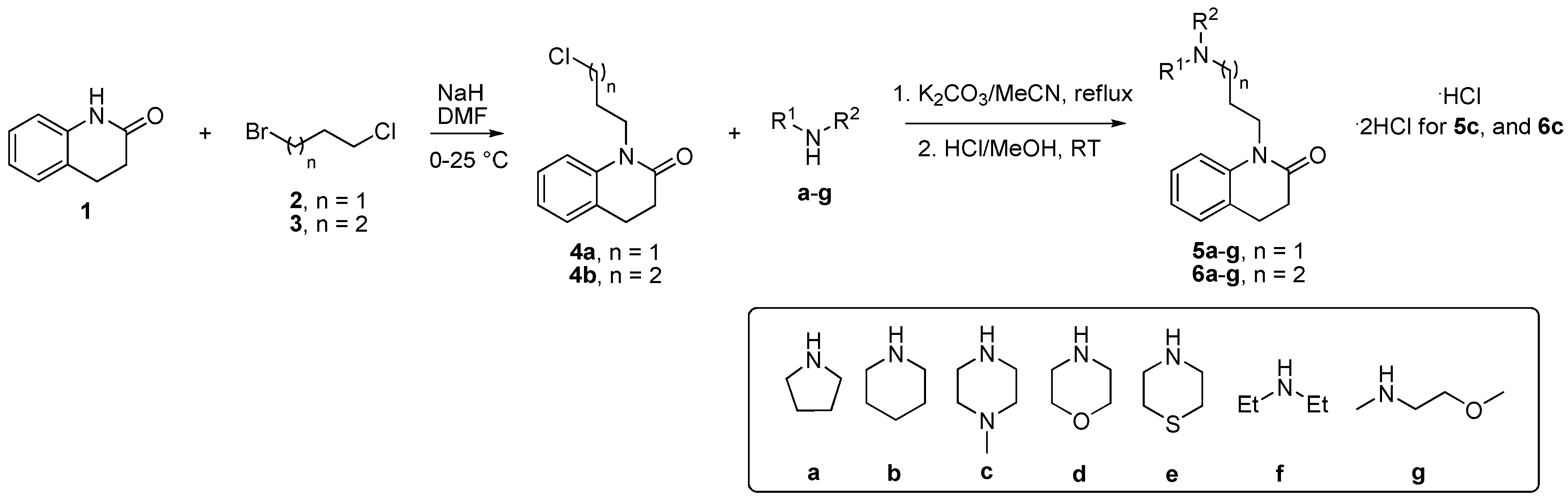
2.1.1. Preparation of 1-(3-chloropropyl)-3,4-dihydroquinolin-2(1H)-one (4a)

To a stirred solution of 3,4-dihydroquinolin-2-(1H)-one (1) (2.7 mmoL) and 60% NaH (272 mg) in DMF (20 mL), 1-bromo-3-chloropropane (2) (3.0 mmoL) was added in drop-by-drop manner under ice-cooled condition. After the addition of 2, the reaction mixture was stirred for 4 h at room temperature (r.t.) [42]. After the completion of the reaction (monitored by TLC), the mixture was diluted with toluene (30 mL) and concentrated under reduced pressure. This operation was done three times. To the resulting mixture, DCM (200 mL) and distilled water (100 mL) were added, and it was vigorously stirred at r.t. for 30 min. The organic phase was then separated, dried over Na2SO4, filtered and concentrated under reduced pressure. The crude residue was purified by silica gel chromatography (DCM:EtOAc = 4:1 v/v).
The product was isolated as yellowish oil in 89% yield (540 mg); 1H NMR of 4a agrees with the literature-reported spectra [43]. 1H NMR (500 MHz, CDCl3) δ 7.28–7.24 (m, 1H), 7.17 (dq, J = 7.4, 1.2 Hz, 1H), 7.07 (dd, J = 8.2, 1.1 Hz, 1H), 7.01 (td, J = 7.4, 1.1 Hz, 1H), 4.12–4.07 (m, 2H), 3.62 (t, J = 6.3 Hz, 2H), 2.89 (dd, J = 8.7, 6.1 Hz, 2H), 2.69–2.60 (m, 2H), 2.19–2.10 (m, 2H).

2.1.2. Preparation of 1-(4-chlorobutyl-3,4-dihydroquinolin-2(1H)-one (4b)

To a stirred solution of 3,4-dihydroquinolin-2-(1H)-one (1) (6.1 mmoL) and 60% NaH (624 mg) in DMF (18 mL), 1-bromo-4-chlorobutane (3) (12 mmoL) was added in a drop-by-drop manner under ice-cooled condition. After the addition of 3, the reaction mixture was stirred at r.t. overnight [42,44,45]. After the completion of the reaction (monitored by TLC), the mixture was diluted with toluene (30 mL) and concentrated under reduced pressure. This operation was done three times. To the resulting mixture, EtOAc (300 mL) and distilled water (100 mL) were added, and it was vigorously stirred at r.t. for 30 min. The organic phase was then separated, dried over Na2SO4, filtered and concentrated under reduced pressure. The crude residue was purified by silica gel chromatography (DCM:EtOAc = 98:2 v/v).
The product was isolated as yellowish oil in 70% yield (1.0 g); 1H NMR (500 MHz, CDCl3) δ 7.27–7.23 (m, 1H), 7.20–7.14 (m, 1H), 7.01 (ddd, J = 8.5, 5.8, 1.2 Hz, 2H), 3.98 (t, J = 7.0 Hz, 2H), 3.58 (t, J = 6.1 Hz, 2H), 2.89 (dd, J = 8.7, 6.1 Hz, 2H), 2.67–2.58 (m, 2H), 1.82 (tdd, J = 9.0, 7.4, 4.9 Hz, 4H). 13C NMR (126 MHz, CDCl3) δ 170.4, 139.4, 128.2, 127.6, 126.7, 122.9, 114.8, 44.7, 41.2, 32.0, 29.9, 25.7, 24.7.

2.1.3. General Procedure for the Preparation of Final Compounds 5a-g and 6a-g

To a stirred solution of appropriate analogue 4a,b (0.5 mmoL) and amine a-g (1.5 mmoL) in MeCN (5 mL), K2CO3 (1.5 mmoL) was added and the reaction mixture was stirred for overnight at reflux [46]. After the completion of the reaction (monitored by TLC), the mixture was diluted with CHCl3 (30 mL), the solid was filtered off and the residue was concentrated under reduced pressure. The crude product was purified by silica gel chromatography (DCM:MeOH = 95:5 v/v). Final compounds (5a-g, 6a-g) were prepared as hydrochlorides by mixing with small portion of hydrochloric acid (37% aq.) in MeOH at r.t.
1-(3-(Pyrrolidin-1-yl)propyl)-3,4-dihydroquinolin-2(1H)-one (5a), Colorless oil. Yield: 56% (72 mg); 1H NMR (500 MHz, CDCl3) δ 7.21 (td, J = 7.8, 1.6 Hz, 1H), 7.13 (dd, J = 7.5, 1.5 Hz, 1H), 7.05 (d, J = 8.1 Hz, 1H), 6.97 (t, J = 7.3 Hz, 1H), 4.01–3.95 (m, 2H), 2.86 (dd, J = 8.7, 6.1 Hz, 2H), 2.65–2.53 (m, 8H), 1.90 (p, J = 7.5 Hz, 2H), 1.79 (h, J = 3.1 Hz, 4H). 13C NMR (126 MHz, CDCl3) δ 170.3, 139.6, 128.1, 127.6, 126.6, 122.8, 115.0, 54.2, 53.7, 40.6, 32.0, 26.6, 25.6, 23.5. LC-MS: calc m/z = 259.180232 for C16H23N2O+; found [M+H]+ = 259.1802; 99% purity.
1-(3-(Piperidin-1-yl)propyl)-3,4-dihydroquinolin-2(1H)-one (5b), Colorless oil. Yield: 64% (87 mg); 1H NMR (500 MHz, CDCl3) δ 7.24–7.19 (m, 1H), 7.14 (d, J = 7.3 Hz, 1H), 7.08 (d, J = 8.2 Hz, 1H), 6.98 (t, J = 7.4 Hz, 1H), 3.96 (t, J = 7.5 Hz, 2H), 2.87 (dd, J = 8.7, 6.1 Hz, 2H), 2.62 (dd, J = 8.7, 6.1 Hz, 2H), 2.45–2.39 (m, 6H), 1.87 (p, J = 7.5 Hz, 2H), 1.61 (p, J = 5.7 Hz, 4H), 1.43 (p, J = 5.9 Hz, 2H). 13C NMR (126 MHz, CDCl3) δ 170.3, 139.7, 128.0, 127.6, 126.6, 122.8, 115.1, 56.4, 54.6, 40.6, 32.0, 25.8, 25.7, 24.6, 24.3. LC-MS: calc m/z = 273.1961 for C17H25N2O+; found [M+H]+ = 273.1945; 98% purity.
1-(3-(4-Methylpiperazin-1-yl)propyl)-3,4-dihydroquinolin-2(1H)-one (5c), Yellowish oil. Yield: 51% (72 mg); 1H NMR (500 MHz, CDCl3) δ 7.21 (td, J = 7.8, 1.6 Hz, 1H), 7.14 (dd, J = 7.4, 1.5 Hz, 1H), 7.06 (dd, J = 8.2, 1.0 Hz, 1H), 6.98 (td, J = 7.4, 1.1 Hz, 1H), 4.04–3.89 (m, 2H), 2.87 (dd, J = 8.7, 6.1 Hz, 2H), 2.66–2.57 (m, 2H), 2.50 (s, 8H), 2.42 (t, J = 7.2 Hz, 2H), 2.30 (s, 3H), 1.82 (p, J = 7.2 Hz, 2H). 13C NMR (126 MHz, CDCl3) δ 170.3, 139.7, 128.1, 127.5, 126.6, 122.8, 115.0, 55.6, 55.1, 53.0, 45.9, 40.5, 32.0, 25.7, 24.8. LC-MS: calc m/z = 288.2070 for C17H26N3O+; found [M+H]+ = 288.2065; 98% purity.
1-(3-Morpholinopropyl)-3,4-dihydroquinolin-2(1H)-one (5d), Yellowish oil. Yield: 45% (62 mg); 1H NMR (500 MHz, CDCl3) δ 7.21 (td, J = 7.8, 1.6 Hz, 1H), 7.14 (dd, J = 7.4, 1.5 Hz, 1H), 7.06 (d, J = 7.9 Hz, 1H), 6.98 (td, J = 7.4, 1.1 Hz, 1H), 4.01–3.95 (m, 2H), 3.70 (t, J = 4.7 Hz, 4H), 2.87 (dd, J = 8.7, 6.1 Hz, 2H), 2.67–2.59 (m, 2H), 2.48–2.36 (m, 6H), 1.83 (p, J = 7.3 Hz, 2H). 13C NMR (126 MHz, CDCl3) δ 170.3, 139.7, 128.1, 127.5, 126.6, 122.8, 114.9, 67.0, 56.2, 53.8, 40.5, 32.0, 25.7, 24.4. LC-MS: calc m/z = 275.1754 for C16H23N2O2+; found [M+H]+ = 275.1749; 95% purity.
1-(3-Thiomorpholinopropyl)-3,4-dihydroquinolin-2(1H)-one (5e), Colorless oil. Yield: 47% (68 mg); 1H NMR (500 MHz, CDCl3) δ 7.23 (td, J = 7.9, 1.6 Hz, 1H), 7.15 (dd, J = 7.5, 1.4 Hz, 1H), 7.05 (d, J = 8.1 Hz, 1H), 6.99 (t, J = 7.4 Hz, 1H), 3.99–3.93 (m, 2H), 2.88 (dd, J = 8.7, 6.1 Hz, 2H), 2.76–2.66 (m, 8H), 2.66–2.59 (m, 2H), 2.45 (t, J = 7.2 Hz, 2H), 1.83 (p, J = 7.2 Hz, 2H). 13C NMR (126 MHz, CDCl3) δ 170.3, 139.7, 128.1, 127.5, 126.6, 122.9, 114.9, 56.4, 55.1, 40.5, 32.0, 27.9, 25.7, 24.3. LC-MS: calc m/z = 291.1526 for C16H23N2OS+; found [M+H]+ = 291.1520; 95% purity.
1-(3-(Diethylamino)propyl)-3,4-dihydroquinolin-2(1H)-one (5f), Colorless oil. Yield: 45% (59 mg); 1H NMR (500 MHz, CDCl3) δ 7.23 (td, J = 7.8, 1.6 Hz, 1H), 7.17–7.12 (m, 1H), 7.06 (d, J = 8.1 Hz, 1H), 6.99 (t, J = 7.4 Hz, 1H), 4.00–3.93 (m, 2H), 2.87 (dd, J = 8.7, 6.1 Hz, 2H), 2.66–2.54 (m, 8H), 1.89–1.80 (m, 2H), 1.09–1.02 (m, 6H). 13C NMR (126 MHz, CDCl3) δ 170.4, 139.6, 128.1, 127.6, 126.6, 122.9, 115.0, 50.3, 46.9, 40.7, 32.0, 25.7, 24.6, 11.4. LC-MS: calc m/z = 261.1961 for C16H25N2O+; found [M+H]+ = 261.1958; 98% purity.
1-(3-((2-Methoxyethyl)(methyl)amino)propyl)-3,4-dihydroquinolin-2(1H)-one (5g), Yellowish oil. Yield: 52% (72 mg); 1H NMR (500 MHz, CDCl3) δ 7.25–7.20 (m, 1H), 7.17–7.12 (m, 1H), 7.06 (d, J = 8.1 Hz, 1H), 6.98 (tt, J = 7.4, 1.0 Hz, 1H), 3.99–3.93 (m, 2H), 3.49 (td, J = 5.7, 1.2 Hz, 2H), 3.34 (d, J = 0.7 Hz, 3H), 2.87 (dd, J = 8.7, 6.1 Hz, 2H), 2.65–2.57 (m, 4H), 2.55–2.48 (m, 2H), 2.30 (d, J = 1.4 Hz, 3H), 1.88–1.80 (m, 2H). 13C NMR (126 MHz, CDCl3) δ 170.3, 139.7, 128.1, 127.6, 126.6, 122.8, 115.0, 70.5, 59.0, 56.7, 55.5, 42.7, 40.6, 32.0, 25.7, 24.9. LC-MS: Calc m/z = 277.1911 for C16H25N2O2+; found [M+H]+ = 277.1910; 98% purity.
1-(4-(Pyrrolidin-1-yl)butyl)-3,4-dihydroquinolin-2(1H)-one (6a), Colorless oil. Yield: 59% (71 mg); 1H NMR (500 MHz, CDCl3) δ 7.25–7.18 (m, 1H), 7.14 (d, J = 7.3 Hz, 1H), 7.02–6.95 (m, 2H), 3.93 (d, J = 6.7 Hz, 2H), 2.90–2.83 (m, 2H), 2.74 (dt, J = 7.2, 3.6 Hz, 4H), 2.69–2.64 (m, 2H), 2.64–2.58 (m, 2H), 1.87 (h, J = 2.7 Hz, 4H), 1.69 (dq, J = 5.9, 3.4 Hz, 4H). 13C NMR (126 MHz, CDCl3) δ 170.4, 139.4, 128.1, 127.6, 126.6, 122.9, 114.9, 55.7, 54.0, 41.5, 32.0, 25.6, 25.1, 25.0, 23.5. LC-MS: Calc m/z = 273.1961 for C17H25N2O+; found [M+H]+ = 273.1957; 99% purity.
1-(4-(Piperidin-1-yl)butyl)-3,4-dihydroquinolin-2(1H)-one (6b), Colorless oil. Yield: 61% (87 mg); 1H NMR (500 MHz, CDCl3) δ 7.29–7.24 (m, 1H), 7.18 (d, J = 7.3 Hz, 1H), 7.08 (d, J = 8.1 Hz, 1H), 7.02 (t, J = 7.4 Hz, 1H), 4.00–3.94 (m, 2H), 2.90 (dd, J = 8.7, 6.2 Hz, 2H), 2.65 (dd, J = 8.8, 6.1 Hz, 2H), 2.44 (dd, J = 17.9, 10.2 Hz, 6H), 1.67 (qt, J = 12.0, 4.9 Hz, 8H), 1.48 (p, J = 5.9 Hz, 2H). 13C NMR (126 MHz, CDCl3) δ 170.3, 139.6, 128.1, 127.5, 126.6, 122.8, 115.0, 58.6, 54.5, 41.9, 32.0, 25.7, 25.1, 24.3, 23.8. LC-MS: Calc m/z = 287.2118 for C18H27N2O+; found [M+H]+ = 287.2118; 97% purity.
1-(4-(4-Methylpiperazin-1-yl)butyl)-3,4-dihydroquinolin-2(1H)-one (6c), Yellowish oil. Yield: 69% (104 mg); 1H NMR (500 MHz, CDCl3) δ 7.22 (td, J = 7.8, 1.6 Hz, 1H), 7.13 (dd, J = 7.4, 1.5 Hz, 1H), 7.05–7.00 (m, 1H), 6.97 (td, J = 7.4, 1.1 Hz, 1H), 3.92 (dd, J = 8.7, 6.5 Hz, 2H), 2.86 (dd, J = 8.7, 6.1 Hz, 2H), 2.64–2.57 (m, 2H), 2.53–2.46 (m, 8H), 2.42–2.35 (m, 2H), 2.29 (s, 3H), 1.65 (ddd, J = 15.1, 9.0, 7.0 Hz, 2H), 1.60–1.51 (m, 2H). 13C NMR (126 MHz, CDCl3) δ 170.2, 139.6, 128.1, 127.5, 126.6, 122.8, 115.0, 57.8, 55.0, 52.9, 45.9, 41.9, 32.0, 25.6, 25.0, 24.0. LC-MS: calc m/z = 302.2227 for C18H28N3O+; found [M+H]+ = 302.2228; 98% purity.
1-(4-Morpholinobutyl)-3,4-dihydroquinolin-2(1H)-one (6d), Yellowish oil. Yield: 64% (92 mg); 1H NMR (500 MHz, CDCl3) δ 7.23 (td, J = 7.8, 1.6 Hz, 1H), 7.15 (dd, J = 7.5, 1.5 Hz, 1H), 7.03 (d, J = 8.2 Hz, 1H), 6.99 (t, J = 7.4 Hz, 1H), 3.98–3.91 (m, 2H), 3.73 (t, J = 4.8 Hz, 4H), 2.87 (dd, J = 8.7, 6.1 Hz, 2H), 2.62 (dd, J = 8.7, 6.1 Hz, 2H), 2.49–2.44 (m, 4H), 2.41 (t, J = 7.4 Hz, 2H), 1.73–1.63 (m, 2H), 1.63–1.55 (m, 2H). 13C NMR (126 MHz, CDCl3) δ 170.3, 139.6, 128.1, 127.5, 126.7, 122.9, 115.0, 66.8, 58.3, 53.6, 41.8, 32.0, 25.7, 24.9, 23.5. LC-MS: calc m/z = 288.1838 for C17H24N2O2; found [M+H]+ = 289.1911; 96% purity.
1-(4-Thiomorpholinobutyl)-3,4-dihydroquinolin-2(1H)-one (6e), Colorless oil. Yield: 58% (88 mg); 1H NMR (500 MHz, CDCl3) δ 7.23 (td, J = 7.9, 1.6 Hz, 1H), 7.15 (dd, J = 7.5, 1.5 Hz, 1H), 7.03 (d, J = 8.2 Hz, 1H), 6.99 (t, J = 7.4 Hz, 1H), 3.97–3.90 (m, 2H), 2.87 (dd, J = 8.7, 6.1 Hz, 2H), 2.76–2.65 (m, 8H), 2.65–2.59 (m, 2H), 2.41 (t, J = 7.3 Hz, 2H), 1.65 (ddd, J = 15.1, 8.8, 6.8 Hz, 2H), 1.56 (ddd, J = 14.8, 8.3, 6.0 Hz, 2H). 13C NMR (126 MHz, CDCl3) δ 170.3, 139.6, 128.1, 127.5, 126.7, 122.8, 115.0, 58.6, 55.0, 41.9, 32.0, 28.0, 25.7, 25.0, 23.6. LC-MS: Calc m/z = 305.1682 for C17H25N2OS+; found [M+H]+ = 305.1683; 95% purity.
1-(4-(Diethylamino)butyl)-3,4-dihydroquinolin-2(1H)-one (6f), Yellowish oil. Yield: 49% (67 mg); 1H NMR (500 MHz, CDCl3) δ 7.23 (td, J = 7.8, 1.6 Hz, 1H), 7.15 (dd, J = 7.4, 1.5 Hz, 1H), 7.03 (d, J = 8.2 Hz, 1H), 6.99 (td, J = 7.4, 1.1 Hz, 1H), 3.97–3.91 (m, 2H), 2.87 (dd, J = 8.7, 6.1 Hz, 2H), 2.67–2.55 (m, 6H), 2.55–2.50 (m, 2H), 1.66 (ddd, J = 14.9, 8.9, 6.9 Hz, 2H), 1.61–1.54 (m, 2H), 1.06 (t, J = 7.2 Hz, 6H). 13C NMR (126 MHz, CDCl3) δ 170.3, 139.6, 128.1, 127.6, 126.7, 122.8, 115.0, 52.3, 46.8, 41.9, 32.0, 25.7, 25.2, 24.0, 11.3. LC-MS: Calc m/z = 275.2118 for C17H27N2O+; found [M+H]+ = 275.2119; 96% purity.
1-(4-((2-Methoxyethyl)(methyl)amino)butyl)-3,4-dihydroquinolin-2(1H)-one (6g), Yellowish oil. Yield: 59% (86 mg); 1H NMR (500 MHz, CDCl3) δ 7.23 (td, J = 7.8, 1.6 Hz, 1H), 7.14 (dd, J = 7.4, 1.5 Hz, 1H), 7.03–6.94 (m, 2H), 3.97–3.90 (m, 2H), 3.48 (td, J = 5.7, 1.0 Hz, 2H), 3.33 (s, 3H), 2.87 (dd, J = 8.7, 6.1 Hz, 2H), 2.65–2.55 (m, 4H), 2.48–2.41 (m, 2H), 2.28 (d, J = 1.1 Hz, 3H), 1.73–1.61 (m, 2H), 1.56 (ddd, J = 15.3, 8.5, 5.9 Hz, 2H). 13C NMR (126 MHz, CDCl3) δ 170.3, 139.6, 128.1, 127.5, 126.7, 122.8, 115.0, 70.6, 59.0, 57.7, 56.8, 42.6, 42.0, 32.0, 25.7, 25.1, 24.4. LC-MS: Calc m/z = 291.2067 for C17H27N2O2+; found [M+H]+ = 291.2068; 99% purity.

2.2. Molecular Studies

2.2.1. Docking Simulation

Molecular docking of ligands was performed using the Molecular Operating Environment (MOE) software package [47]. The receptor used was pdb structure 6CM4, which contains a structure of the atypical antipsychotic drug and D2R antagonist risperidone bound to the D2R [48]. An induced fit docking protocol was used, with the Triangle Matcher used for placement (10.000 placements), London dG scoring function used for initial scoring (100 conformations retained), refinement with Amber10:EHT force field, and rescoring with GBVI/WSA dG (100 conformations retained and ranked by docking score). Before docking, compounds were prepared by protonation at physiological pH and energy minimization with Amber10:EHT force field.

2.2.2. Thermodynamic Integration and Free Energy Calculations

Final docked conformations of risperidone, aripiprazole, USC-D301, and 5e were generated using the preceding method. For thermodynamic integration standard settings were used (Figure S60, Supplementary Material). The used force field was Amber10:EHT. Preparation of the molecular system was done using MOE, the thermodynamic free energy calculation was performed using the PMEMD package of the AMBER molecular dynamics toolkit [47].

2.3. BBB Score Prediction

Blood–brain barrier (BBB) score of newly developed compounds was calculated using an algorithm defined by Gupta et al. [49]. A MarvinSketch software (ChemAxon Ltd., v. 20.15.0; https://www.chemaxon.com) was used to predict some of the physicochemical descriptors like number of aromatic ring, number heavy atoms, MWHBN (a descriptor comprising molecular weight, hydrogen bond donor, and hydrogen bond acceptors), topological polar surface area, and pKA.

2.4. Biology Evaluation

2.4.1. D2 Receptor Binding Affinity–Transfection and Membrane Preparation

Chinese hamster ovary cells (CHO cells) were used for the binding experiments. About 24 h before transfection, CHO cells were plated on a 10 cm Petri dish at a density of 1.5 × 10⁶ cells and cultivated in 10 mL DMEM/Ham’s F12 supplemented with 10% heat-inactivated fetal bovine serum (FBS).
For transfection, the desired amount of linear polyethyleneimine (PEI) 25_K (Polysciences, Eppelheim, Germany) and DNA (dopamine receptor (D2) wild type, cDNA resource centre, Bloomsberg, PA, USA) were diluted separately in phosphate buffer saline (PBS). After 20 min, DNA was added to PEI solution, mixed and left for an additional 30 min. Final concentration of reactants was 6 µg plasmid DNA and 18 µg PEI per ml. PEI/DNA complex was added (1 mL/dish) and the plates were incubated for another 24 h. Then, the medium was replaced with fresh DMEM/Ham’s F12 with 10% FBS and incubated for an additional 24 h. Cells were maintained at 37 °C in a 5% CO2-humidified atmosphere.
About 48 h after transfection, cells were washed with PBS, mechanically detached from the dish with a plastic scraper in the ice-cold PBS, and centrifuged for 3 min at 250× g. Pellet was suspended in the ice-cold homogenization medium (100 mM NaCl, 20 mM Na-HEPES, 10 mM EDTA, pH 7.4) and homogenized on ice by two 30-s strokes using Polytron homogenizer (Ultra-Turrax; Janke & Kunkel GmbH & Co. KG, IKA-Labortechnik, Staufen, Germany) with a 30-s pause between strokes. Cell homogenates were centrifuged for 5 min at 1000× g. The supernatant was collected and centrifuged for 30 min at 30,000× g. Pellets were suspended in incubation medium (100 mM NaCl, 20 mM Na-HEPES, 10 mM MgCl2, pH 7.4), left for 30 min at 4 °C and centrifuged again for 30 min at 30,000× g. The membrane pellets were kept at −80 °C until use.

2.4.2. D2R Binding Affinity–Radioligand Experiment

All radioligand binding experiments were optimized and carried out as described by El-Fakahany and Jakubik [50]. Dissociation constant KD of [3H]-spiperone to D2Rs was determined in saturation binding experiment. Saturation experiments were performed in 800 μL volume containing: 400 μL of the membrane suspension and 400 μL of the radioligand in six increasing concentrations (ranging from 31 to 1000 pM).
Affinity of the tested compounds was determined in competition experiments with 180 pM [3H]-spiperone, that corresponds to triple KD value ([3H]-spiperone, Kd = 60.1 ± 2.79 pM (n = 6)). The examined compounds were diluted in incubation buffer and tested in six concentrations (ranging from 0.1 nM–1 mM). The reactions were performed in 400 µL volume containing: 100 µL of the radioligand, 100 µL of tested substances dilution, and 200 µL of the membranes.
Nonspecific binding was determined in the presence of 10 μM unlabeled sulpiride. Membrane suspensions from both saturation and binding experiments (approximately 10 μg of membrane proteins per sample) were incubated in 96-well plates for 1 h at 25 °C in the incubation medium (100 mM NaCl, 20 mM Na-HEPES,10 mM MgCl2, pH = 7.4) in a shaking incubator (25 °C; PST-60HL, Biosan, Riga, Latvia).
The binding reactions were terminated by filtration of the membranes through APFC filter plate (Millipore, Prague, Czech Republic) pre-soaked with 0.5% PEI and washed with ice-cold distilled water using a Brandel cell harvester (Brandel, Gaithersburg, MD, USA). Then, filters with labelled membranes were dried. After 24 h, scintillation cocktail (Rotiszint eco plus, Carl Roth) was added to each sample and radioactivity was quantified by liquid scintillation spectrometry using Wallac Microbeta scintillation counter (Wallac, Turku, Finland). Competition binding experiments were performed per triplicate and all experiments were performed three times. Protein concentration was determined by the Lowry method in the Peterson modification [51].

2.4.3. D2 Receptor Binding Affinity–Data Analysis

[3H]NMS Saturation Binding

The equilibrium dissociation constant (KD) and maximum binding capacity (BMAX) were determined in the saturation experiments. Non-specific binding in the presence of 10 µM sulpiride was subtracted to determine specific binding. Free concentration of [3H]-spiperone was calculated by subtraction of values of specific binding from the final concentration of [3H]-spiperone calculated from measurements of added radioactivity. Equation (1) was fitted to the data.
y = B M A X     x K D + x
where y is the specific binding at free concentration x. KD values are expressed as µM and BMAX values as pmol of binding sites per mg of membrane protein.

Competition Binding

The binding of tested agonists was determined in competition experiments with 180 pM [3H]-Spiperone fitting of Equation (2)
y = 100 100     x x   +   IC 50
where y is the specific radioligand biding at concentration x of competitor expressed as a percent of binding in the absence of a competitor, IC50 is the concentration causing 50% inhibition of radioligand binding. Inhibition constants KI for analyzed agonists were calculated as:
K I = IC 50 1 + [ D ] K D
where IC50 is the concentration causing 50% inhibition of [3H]-spiperone binding calculated according to Equation (2) from competition binding data, [D] is the concentration of [3H]NMS used, and KD is its equilibrium dissociation constant calculated according to Equation (1) from saturation binding data.

2.4.4. MTT Assay

Standard MTT (3-[4,5-dimethylthiazol-2-yl]-2,5-diphenyltetrazolium bromide) assay (Sigma Aldrich, Prague, Czech Republic) was used according to the manufacturer´s protocol on the CHO-K1 (Chinese hamster ovary, ECACC, Salisbury, UK) in order to compare the effect of different compounds within the series. The cells were cultured according to ECACC recommended conditions and seeded in a density of 8000 per well as described previously [52]. Briefly, tested compounds were dissolved in DMSO (Sigma Aldrich, Prague, Czech Republic) and subsequently in the growth medium (F–12) so that the final concentration of DMSO did not exceed 1% (v/v). Cells were exposed to a tested compound for 24 h. Then the medium was replaced by a medium containing 10 μM of MTT and cells were allowed to produce formazan for another approximately 3 h under surveillance. Thereafter, medium with MTT was sucked out and crystals of formazan were dissolved in DMSO (100 µL). Cell viability was assessed spectrophotometrically by the amount of formazan produced. Absorbance was measured at 570 nm with 650 nm reference wavelength on Synergy HT (BioTek, Winooski, VT, USA). IC50 was then calculated from the control-subtracted triplicates using non-linear regression (four parameters) of GraphPad Prism 9 software. Final IC50 and SEM values were obtained as a mean of three independent measurements.

2.4.5. PAMPA Assay

PAMPA is a high-throughput screening tool applicable for prediction of the passive transport of potential drugs across the BBB [53]. In this study, it has been used as a non-cell-based in vitro assay carried out in a coated 96-well membrane filter. The filter membrane of the donor plate was coated with PBL (Polar Brain Lipid, Avanti, AL, USA) in dodecane (4 µL of 20 mg/mL PBL in dodecane) and the acceptor well was filled with 300 µL of PBS pH 7.4 buffer (VA tested compounds were dissolved first in the DMSO/phosphate-buffered saline mixture (maximum 0.5% v/v of DMSO) and subsequently diluted with phosphate-buffered saline (pH 7.4) to final concentrations of 40–100 μM in the donor wells). Concentration of DMSO did not exceed 0.5% (v/v) in the donor solution. About 300 µL of the donor solution was added to the donor wells (VD) and the donor filter plate was carefully put on the acceptor plate so that coated membrane was “in touch” with both donor solution and acceptor buffer. Test compound diffused from the donor well through the lipid membrane (Area = 0.28 cm2) to the acceptor well. The concentration of the drug in both donor and the acceptor wells were assessed after 3, 4, 5, and 6 h of incubation in quadruplicate using the UV plate reader Synergy HT (Biotek, Winooski, VT, USA) at the maximum absorption wavelength of each compound. Besides that, solution of theoretical compound concentration, simulating the equilibrium state established if the membrane were ideally permeable was prepared and assessed as well. Concentration of the compounds in the donor and acceptor well and equilibrium concentration were calculated from the standard curve and expressed as the permeability (Pe) according to the equation:
P e = C × l n ( 1 [ d r u g ]   a c c e p t o r [ d r u g ]   e q u i l i b r i u m )   w h e r e   C = ( V D × V A ) ( V D × V A )   A r e a × T i m e

3. Results

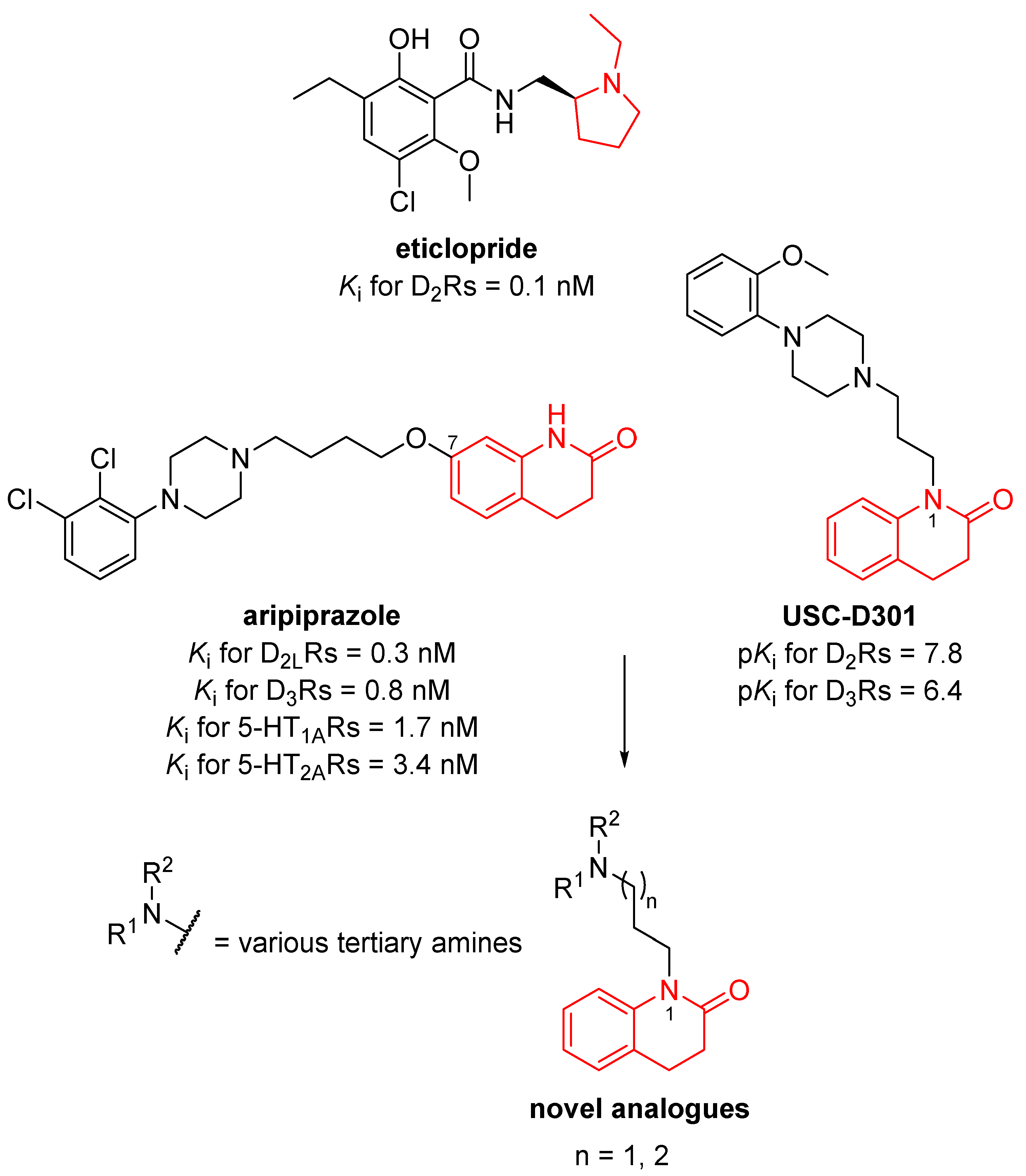
3.1. Design of Novel Compounds

Aripiprazole (Figure 1), a partial D2Rs agonist, belongs to the third generation of antipsychotic drugs and has been approved by the Food and Drug Administration (FDA) agency for the use as an adjunctive medication in the treatment of depressive and bipolar disorders [9,11,54,55,56]. Aripiprazole has a unique, biased, mode of action comprising partial agonism for Gαi/o and a robust antagonism for Gβγ signaling and an antagonism or a partial agonism for β-arrestin-2 signaling [57,58]. Furthermore, if extracellular concentration of dopamine levels are high (e.g., in mesolimbic areas), aripiprazole competes with dopamine and acts as a partial antagonist. On the other hand, in the presence of low dopamine concentration (e.g., dopamine areas that are involved in working memory), aripiprazole can activate other receptors. Thus, aripiprazole can be classified as a “dopamine stabilizer” [9,59,60]. In addition, aripiprazole is a D3 and 5-HT1A receptors partial agonist and 5-HT2ARs antagonist [54,61]. In its structure, aripiprazole combines 3,4-dihydro-7-hydroxyquinolin-2(1H)-one fragment attached at position 7- to 2,3-dichlorophenyl piperazine and thus is a member of large group of antipsychotics, so called 1,4-disubstituted arylpiperazines. The biological activity of this subgroup of compounds is encoded by an aromatic warhead, which controls intrinsic activity, and an amine moiety, which is responsible for the formation of a hydrogen bond to the crucial residue Asp3.32 in the transmembrane helix 3 of D2R [62]. A linker controls subtype selectivity; 3-methylene linker was found suitable for D2R selectivity [63,64]. Aromatic/heteroaromatic appendage on the opposite site of the ligand orchestrates receptor affinity [62]. In the past decade, many compounds have been generated containing 2,3-dichlorophenylpiperazine fragment with unique pharmacological profile exhibiting high D2Rs affinity. From the extensive SAR, it was deduced that the central linker has only moderate impact on affinity but huge effect on functional activity at D2Rs [65,66,67,68]. Besides various substitutions made to the central linker, it has been shown that lipophilic appendages strongly influence functional and subtype selectivity [68,69,70]. 3,4-Dihydroquinolin-2(1H)-one scaffold-containing ligands have shown to possess an affinity to D2Rs as well. In this case, the nature of the central linker showed a moderate impact on D2Rs affinity [71]. Modifications within the amine moiety influenced D2Rs affinity and functional selectivity [72]. Besides, substitutions in aromatic warheads also strongly affected D2Rs affinity and functional and subtype selectivity [71,72]. Recently, some 3,4-dihydroquinolin-2-(1H)-one scaffold-containing compounds exhibited high D4Rs selectivity over other D2-like family receptors [73]. These findings show that small structural modifications within one region of the molecule based on aripiprazole can tune significantly the properties of the ligand.
In the study of Lopéz et al., researchers tethered arylpiperazine-like core (different phenylpiperazine moieties) with 3,4-dihydroquinolin-2(1H)-one moiety (red color, Figure 1) at position 1- [41]. USC-D301 (Figure 1) is an example of small molecule exhibiting strong D2R antagonism and high selectivity over D3Rs [41]. On the other hand, eticlopride (Figure 1) is a substituted benzamide analog without 1,4-disubstituted arylpiperazine fragment exhibiting very high affinity for D2Rs [74]. Thus, we wanted to explore the effect of novel prepared analogs containing various tertiary amines with 3,4-dihydroquinolin-2(1H)-one fragment connected by aliphatic linker at position 1- of the quinolinone core towards the D2Rs as the main targets. We are aware of the fact that other dopamine receptors (especially D3R and D4R) are of importance for the complexity of the antipsychotic action, as observed also in case of aripiprazole [54], however, this was not the aim of this study. Molecular imaging studies have revealed that striatal D2Rs antagonism is essential in vivo for therapeutic doses of all neuroleptics [19,75,76,77,78,79,80], and D2Rs affinity of antipsychotic drugs is the crucial for their antipsychotic efficacy. The aliphatic linkers (1,3-propane-diyl and 1,4-butane-diyl) for new ligands were chosen based on previous studies [41,44]. These new derivatives were evaluated for their D2R antagonistic properties with the emphasis on the structure–activity relationships regarding the type of amine and length of the linker.

3.2. The Synthesis of Novel Compounds

The syntheses of novel analogues are depicted in Scheme 1. Nucleophilic addition of 1-bromo-3-chloropropane (2) or 1-bromo-4-chlorobutane (3) to the starting compound 3,4-dihydro-2(1H)-quinolinone (1, Scheme 1) in the presence of sodium hydride produced intermediates 4a,b [42]. These derivatives were obtained in excellent yields (>70%). The final compounds 5a-g and 6a-g were prepared again by nucleophilic addition of the appropriate amine (a-g) to 4a,b in the presence of K2CO3 [46]. All the reactions exhibited good yields (>45%). The final products were analyzed by 1H and 13C NMR, HRMS and LC-MS analysis revealing the purity > 95%.

3.3. Binding Affinities of Novel Compounds at D2Rs and Their Cytotoxicities

The results of the affinity of 5a-g and 6a-g for D2R are summarized in Table 1. In general, 1,3-propane-diyl derivatives 5a-g exhibited slightly better D2R antagonism than their 1,4-butane-diyl counterparts 6a-g. The aliphatic analogues 5f,g and 6g possessed slightly lower D2R antagonism than the molecules with cyclic amines 5a-e and 6a-c,e. Morpholine-containing compound 6d showed the lowest D2R antagonism from all cyclic amine derivatives (5a-e and 6a-e). On the other hand, thiomorpholine analogue 5e had the strongest antagonistic behavior at D2Rs from all prepared final compounds. Interestingly, the final derivatives 5a-g, 6a-g exhibited very low cytotoxicity in MTT (3-[4,5-dimethylthiazol-2-yl]-2,5-diphenyltetrazolium bromide) assay using Chinese hamster ovary cell lines (Table 1) so that the relatively lower affinity toward D2R is compensated by low toxicity allowing higher dosing.

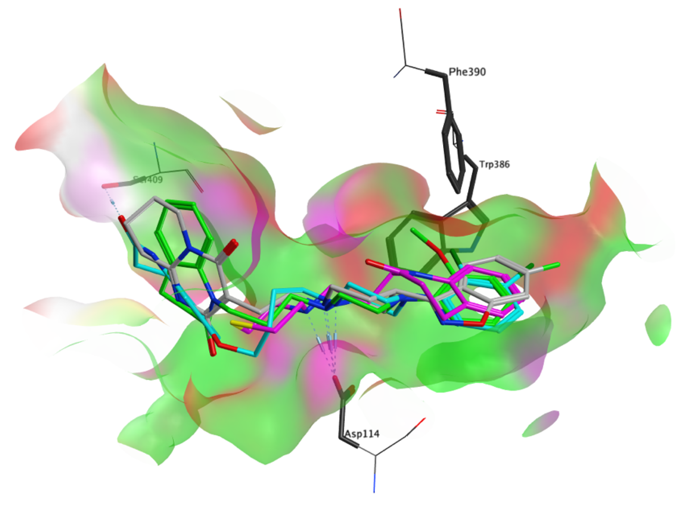
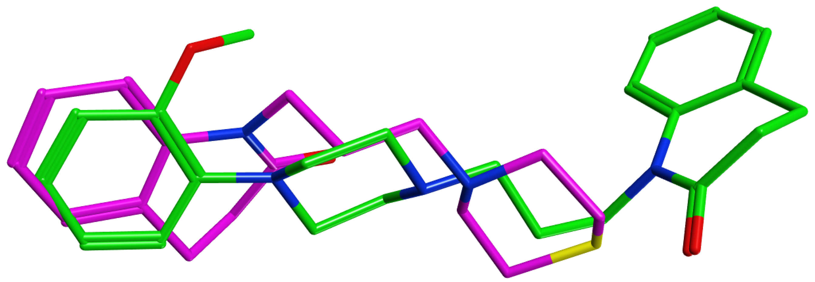
3.4. Molecular Modelling Studies

A molecular modelling study was undertaken to help explain differences in binding strength. Quantitative results are summarized in Table 2, the docking score (S) and the calculated relative binding energy (ddG) from the thermodynamic integration free energy calculation follow the same tendency as the measured affinities. Docking of USC-D301 and 5e revealed strongly differing binding poses, despite their high molecular similarity (Figure 2). The positively charged nitrogen of all docked ligands was recognized by Asp114 via a strong ionic ammonium-carboxylate interaction. A pi–pi interaction with Phe390 was also apparent, with the isoxazole, 2,3-dichlorophenyl and 2-methoxyphenyl fragment of risperidone, aripiprazole, and USC-D301, respectively. Surprisingly, the lowest scoring docking pose of 5e did not overlap with USC-D301 (Figure 3). Owing to its smaller size and thiomorpholine cap, there is no aromatic group at that position that can undergo the stacking interaction with Phe390 or other nearby residues, likely leading to a strong penalty to the binding energy. Instead, the molecule adopts a flipped conformation that places the benzolactam fragment in this pocket, where the fused benzene ring is not positioned well to undergo stacking with Phe390, Trp386, and others.

3.5. Central Nervous System Availability Prediction and Study for Novel Compounds

Before the synthesis, we have calculated the so-called BBB score to predict the compound’s CNS availability. Indeed, all the compounds displayed high values above 5.0 (5.2–5.4) which is indicative of their high potential to cross BBB. The prediction was then confirmed by the data from parallel artificial membrane permeation (PAMPA) assay pointing out their potential to cross the BBB by passive diffusion (5a-g and 6a-g Pe (× 10−6 cm s−1) = 7.0–24) (Table 3). The validation of PAMPA has been performed using standard compounds whose availability or unavailability was experimentally predicted in vitro and confirmed in vivo [53,82].

4. Conclusions

In summary, a series of 3,4-dihydroquinolin-2(1H)-one analogues, inspired by aripiprazol was designed and synthesized. The substitutions of the amine group revealed a negligible impact on D2R affinity. Although the binding affinities at D2Rs of new analogues are much weaker compared to aripiprazole, they are very close to the binding affinity, for instance, of memantine acting as N-methyl-d-aspartate receptor antagonist, a well-established drug for the treatment of Alzheimer’s disease [83,84]. Out of these ligands, 5e possessed the highest D2R affinity, very low cytotoxicity profile, and the highest probability to cross the BBB. Molecular modeling simulation revealed completely different binding mode of 5e compared to USC-D301, which might be the culprit of the reduced affinity of 5e toward D2R. The subject of further investigation of these compounds will be to assess their affinity toward other members of the D2-like receptors family and other GPCRs, especially 5-HT1A and 5-HT2ARs. Since aripiprazole has few of the typical adverse effects of other antipsychotics, such as extrapyramidal symptoms, hyperprolactinemia, weight gain, metabolic disorders, and sedation given to its unique biased activity and/or partial agonistic/antagonistic actions on D2Rs supplemented by the action on other dopamine receptor subtypes (mainly D3R) and 5-HT receptor subtypes (mainly 5-HT1A, 5-HT2A) [54], the ongoing study must determine the functional affinity of newly developed compounds at these receptors to evaluate the real antipsychotic effect and clinical potential [85].

Supplementary Materials

The following are available online at https://www.mdpi.com/article/10.3390/biom11091262/s1. Figure S1: 1H NMR spectrum for 5a. Figure S2: 13C NMR spectrum for 5a. Figure S3: UV-LC chromatogram for 5a. Figure S4: HRMS spectrum for 5a. Figure S5: 1H NMR spectrum for 5b. Figure S6: 13C NMR spectrum for 5b. Figure S7: UV-LC chromatogram for 5b. Figure S8: HRMS spectrum for 5b. Figure S9: 1H NMR spectrum for 5c. Figure S10: 13C NMR spectrum for 5c. Figure S11: UV-LC chromatogram for 5c. Figure S12: HRMS spectrum for 5c. Figure S13: 1H NMR spectrum for 5d. Figure S14: 13C NMR spectrum for 5d. Figure S15: UV-LC chromatogram for 5d. Figure S16: HRMS spectrum for 5d. Figure S17: 1H NMR spectrum for 5e. Figure S18: 13C NMR spectrum for 5e. Figure S19: UV-LC chromatogram for 5e. Figure S20: HRMS spectrum for 5e. Figure S21: 1H NMR spectrum for 5f. Figure S22: 13C NMR spectrum for 5f. Figure S23: UV-LC chromatogram for 5f. Figure S24: HRMS spectrum for 5f. Figure S25: 1H NMR spectrum for 5g. Figure S26: 13C NMR spectrum for 5g. Figure S27: UV-LC chromatogram for 5g. Figure S28: HRMS spectrum for 5g. Figure S29: 1H NMR spectrum for 6a. Figure S30: 13C NMR spectrum for 6a. Figure S31: UV-LC chromatogram for 6a. Figure S32: HRMS spectrum for 6a. Figure S33: 1H NMR spectrum for 6b. Figure S34: 13C NMR spectrum for 6b. Figure S35: UV-LC chromatogram for 6b. Figure S36: HRMS spectrum for 6b. Figure S37: 1H NMR spectrum for 6c. Figure S38: 13C NMR spectrum for 6c. Figure S39: UV-LC chromatogram for 6c. Figure S40: HRMS spectrum for 6c. Figure S41: 1H NMR spectrum for 6d. Figure S42: 13C NMR spectrum for 6d. Figure S43: UV-LC chromatogram for 6d. Figure S44: HRMS spectrum for 6d. Figure S45: 1H NMR spectrum for 6e. Figure S46: 13C NMR spectrum for 6e. Figure S47: UV-LC chromatogram for 6e. Figure S48: HRMS spectrum for 6e. Figure S49: 1H NMR spectrum for 6f. Figure S50: 13C NMR spectrum for 6f. Figure S51: UV-LC chromatogram for 6f. Figure S52: HRMS spectrum for 6f. Figure S53: 1H NMR spectrum for 6g. Figure S54: 13C NMR spectrum for 6g. Figure S55: UV-LC chromatogram for 6g. Figure S56: HRMS spectrum for 6g. Figure S57: Saturation binding curve and Scatchard plot of the affinity of [3H]spiperone to D2Rs. Figure S58: Inhibition of [3H]spiperone binding to D2RS receptors by 3,4-dihydroquinolin-2(1H)-one derivatives (5a-g). Figure S59: Inhibition of [3H]spiperone binding to D2RS receptors by 3,4-dihydroquinolin-2(1H)-one derivatives (6a-g). Figure S60: Schematic representation of the settings used in the Thermodynamic Integration Free Energy Calculation. Table S1: Functional activities of 5a-g and 6a-g at D2Rs.

Author Contributions

R.J. and E.M. chemical synthesis and writing the paper; K.S. and A.R. binding affinity studies; W.D. molecular modelling studies; L.P. (Lukas Prchal) HRMS and LC-MS analysis; L.P. (Lenka Pulkrabkova), T.K., L.M. cell experiments; M.M. blood–brain barrier prediction; K.M., O.S. and J.K. designed the study; T.P., I.V., O.S. and JK reviewed and edited the paper; K.M., J.K. and O.S. funding acquisition. All authors have read and agreed to the published version of the manuscript.

Funding

W.D. was supported by the Ministry of Education, Youth and Sports of the Czech Republic, project number LM2018130. The work was supported by the Czech Science Foundation (No. 19-11332S) and University of Hradec Kralove (No. SV2104-2021 and VT2019-2021).

Institutional Review Board Statement

Not applicable.

Informed Consent Statement

Not applicable.

Data Availability Statement

The compounds developed within this study are available from the corresponding author on reasonable request.

Conflicts of Interest

The authors declare no conflict of interest.

References

  1. Matt, S.M.; Gaskill, P.J. Where Is Dopamine and How Do Immune Cells See It? Dopamine-Mediated Immune Cell Function in Health and Disease. J. Neuroimmune Pharm. 2020, 15, 114–164. [Google Scholar] [CrossRef] [PubMed]
  2. Ayano, G. Dopamine: Receptors, Functions, Synthesis, Pathways, Locations and Mental Disorders: Review of Literatures. J. Ment. Disord. Treat. 2016, 2. [Google Scholar] [CrossRef]
  3. Klein, M.O.; Battagello, D.S.; Cardoso, A.R.; Hauser, D.N.; Bittencourt, J.C.; Correa, R.G. Dopamine: Functions, Signaling, and Association with Neurological Diseases. Cell. Mol. Neurobiol. 2019, 39, 31–59. [Google Scholar] [CrossRef] [PubMed]
  4. Martel, J.C.; Gatti McArthur, S. Dopamine Receptor Subtypes, Physiology and Pharmacology: New Ligands and Concepts in Schizophrenia. Front. Pharmacol. 2020, 11, 1003. [Google Scholar] [CrossRef] [PubMed]
  5. Pivonello, R.; Ferone, D.; Lombardi, G.; Colao, A.; Lamberts, S.W.J.; Hofland, L.J. Novel Insights in Dopamine Receptor Physiology. Eur. J. Endocrinol. 2007, 156, S13–S21. [Google Scholar] [CrossRef]
  6. Żuk, J.; Bartuzi, D.; Matosiuk, D.; Kaczor, A.A. Preferential Coupling of Dopamine D2S and D2L Receptor Isoforms with Gi1 and Gi2 Proteins—In Silico Study. Int. J. Mol. Sci. 2020, 21, 436. [Google Scholar] [CrossRef] [PubMed] [Green Version]
  7. Usiello, A.; Baik, J.H.; Rougé-Pont, F.; Picetti, R.; Dierich, A.; LeMeur, M.; Piazza, P.V.; Borrelli, E. Distinct Functions of the Two Isoforms of Dopamine D2 Receptors. Nature 2000, 408, 199–203. [Google Scholar] [CrossRef] [PubMed]
  8. Beaulieu, J.-M.; Gainetdinov, R.R. The Physiology, Signaling, and Pharmacology of Dopamine Receptors. Pharm. Rev. 2011, 63, 182–217. [Google Scholar] [CrossRef] [Green Version]
  9. Stępnicki, P.; Kondej, M.; Kaczor, A.A. Current Concepts and Treatments of Schizophrenia. Molecules 2018, 23, 2087. [Google Scholar] [CrossRef] [Green Version]
  10. Wang, S.-M.; Han, C.; Lee, S.-J.; Jun, T.-Y.; Patkar, A.A.; Masand, P.S.; Pae, C.-U. Second Generation Antipsychotics in the Treatment of Major Depressive Disorder: An Update. Chonnam. Med. J. 2016, 52, 159–172. [Google Scholar] [CrossRef] [Green Version]
  11. Mulder, R.; Hamilton, A.; Irwin, L.; Boyce, P.; Morris, G.; Porter, R.J.; Malhi, G.S. Treating Depression with Adjunctive Antipsychotics. Bipolar Disord. 2018, 20, 17–24. [Google Scholar] [CrossRef] [PubMed] [Green Version]
  12. Hershenberg, R.; Gros, D.F.; Brawman-Mintzer, O. Role of Atypical Antipsychotics in the Treatment of Generalized Anxiety Disorder. CNS Drugs 2014, 28, 519–533. [Google Scholar] [CrossRef]
  13. Pignon, B.; Montcel, C.T.; Carton, L.; Pelissolo, A. The Place of Antipsychotics in the Therapy of Anxiety Disorders and Obsessive-Compulsive Disorders. Curr. Psychiatry Rep. 2017, 19, 1–11. [Google Scholar] [CrossRef] [PubMed]
  14. Marder, S.R.; Cannon, T.D. Schizophrenia. N. Engl. J. Med. 2019, 381, 1753–1761. [Google Scholar] [CrossRef] [PubMed]
  15. Radhakrishnan, R.; Kaser, M.; Guloksuz, S. The Link Between the Immune System, Environment, and Psychosis. Schizophr. Bull. 2017, 43, 693–697. [Google Scholar] [CrossRef] [PubMed]
  16. Laruelle, M. Schizophrenia: From Dopaminergic to Glutamatergic Interventions. Curr. Opin. Pharmacol. 2014, 14, 97–102. [Google Scholar] [CrossRef]
  17. McCutcheon, R.A.; Abi-Dargham, A.; Howes, O.D. Schizophrenia, Dopamine and the Striatum: From Biology to Symptoms. Trends Neurosci. 2019, 42, 205–220. [Google Scholar] [CrossRef] [Green Version]
  18. Kapur, S.; Zipursky, R.; Jones, C.; Remington, G.; Houle, S. Relationship Between Dopamine D2 Occupancy, Clinical Response, and Side Effects: A Double-Blind PET Study of First-Episode Schizophrenia. AJP 2000, 157, 514–520. [Google Scholar] [CrossRef]
  19. Nordström, A.-L.; Farde, L.; Wiesel, F.-A.; Forslund, K.; Pauli, S.; Halldin, C.; Uppfeldt, G. Central D2-Dopamine Receptor Occupancy in Relation to Antipsychotic Drug Effects: A Double-Blind PET Study of Schizophrenic Patients. Biol. Psychiatry 1993, 33, 227–235. [Google Scholar] [CrossRef]
  20. Richtand, N.M.; Welge, J.A.; Logue, A.D.; Keck, P.E.; Strakowski, S.M.; McNamara, R.K. Dopamine and Serotonin Receptor Binding and Antipsychotic Efficacy. Neuropsychopharmacology 2007, 32, 1715–1726. [Google Scholar] [CrossRef] [Green Version]
  21. Seeman, P.; Lee, T.; Chau-Wong, M.; Wong, K. Antipsychotic Drug Doses and Neuroleptic/Dopamine Receptors. Nature 1976, 261, 717–719. [Google Scholar] [CrossRef]
  22. Karlsson, P.; Farde, L.; Härnryd, C.; Sedvall, G.; Smith, L.; Wiesel, F.-A. Lack of Apparent Antipsychotic Effect of the D 1 -Dopamine Recepotr Antagonist SCH39166 in Acutely Ill Schizophrenic Patients. Psychopharmacology 1995, 121, 309–316. [Google Scholar] [CrossRef] [PubMed]
  23. Redden, L.; Rendenbach-Mueller, B.; Abi-Saab, W.; Katz, D.; Goenjian, A.; Robieson, W.; Wang, Y.; Goss, S.; Greco, N.; Saltarelli, M. A Double-Blind, Randomized, Placebo-Controlled Study of the Dopamine D-3 Receptor Antagonist ABT-925 in Patients With Acute Schizophrenia. J. Clin. Psychopharmacol. 2011, 31, 221–225. [Google Scholar] [CrossRef] [PubMed]
  24. Bristow, L.J.; Kramer, M.S.; Kulagowski, J.; Patel, S.; Ragan, C.I.; Seabrook, G.R. Schizophrenia and L-745, 870, a Novel Dopamine D4 Receptor Antagonist. Trends Pharmacol. Sci. 1997, 18, 186–188. [Google Scholar] [CrossRef]
  25. George, M.S.; Molnar, C.E.; Grenesko, E.L.; Anderson, B.; Mu, Q.; Johnson, K.; Nahas, Z.; Knable, M.; Fernandes, P.; Juncos, J.; et al. A Single 20 Mg Dose of Dihydrexidine (DAR-0100), a Full Dopamine D1 Agonist, Is Safe and Tolerated in Patients with Schizophrenia. Schizophr. Res. 2007, 93, 42–50. [Google Scholar] [CrossRef]
  26. Girgis, R.R.; Van Snellenberg, J.X.; Glass, A.; Kegeles, L.S.; Thompson, J.L.; Wall, M.; Cho, R.Y.; Carter, C.S.; Slifstein, M.; Abi-Dargham, A.; et al. A Proof-of-Concept, Randomized Controlled Trial of DAR-0100A, a Dopamine-1 Receptor Agonist, for Cognitive Enhancement in Schizophrenia. J. Psychopharmacol. 2016, 30, 428–435. [Google Scholar] [CrossRef]
  27. Rosell, D.; Zaluda, L.; McClure, M.; Perez-Rodriguez, M.; Strike, S.; Barch, D.; Harvey, P.; Girgis, R.; Hazlett, E.; Mailman, R.; et al. Effects of the D1 Dopamine Receptor Agonist Dihydrexidine (DAR-0100A) on Working Memory in Schizotypal Personality Disorder. Neuropsychopharmacology 2015, 40, 446–453. [Google Scholar] [CrossRef]
  28. Zheng, W.; Li, X.H.; Cai, D.B.; Yang, X.H.; Ungvari, G.S.; Ng, C.H.; Ning, Y.P.; Xiang, Y.T. Adjunctive Azapirone for Schizophrenia: A Meta-Analysis of Randomized, Double-Blind, Placebo-Controlled Trials. Eur. Neuropsychopharmacol. 2018, 28, 149–158. [Google Scholar] [CrossRef]
  29. Meltzer, H.; Huang, M. In Vivo Actions of Atypical Antipsychotic Drug on Serotonergic and Dopaminergic Systems. Prog. Brain Res. 2008, 172, 177–197. [Google Scholar] [CrossRef] [PubMed]
  30. Kaar, S.J.; Natesan, S.; McCutcheon, R.; Howes, O.D. Antipsychotics: Mechanisms Underlying Clinical Response and Side-Effects and Novel Treatment Approaches Based on Pathophysiology. Neuropharmacology 2020, 172, 107704. [Google Scholar] [CrossRef]
  31. Richelson, E.; Souder, T. Binding of Antipsychotic Drugs to Human Brain Receptors Focus on Newer Generation Compounds. Life Sci. 2000, 68, 29–39. [Google Scholar] [CrossRef]
  32. Schneider, L.S. Pimavanserin for Patients with Alzheimer’s Disease Psychosis. Lancet Neurol. 2018, 17, 194–195. [Google Scholar] [CrossRef]
  33. Bebawy, M.; Chetty, M. Differential Pharmacological Regulation of Drug Efflux and Pharmacoresistant Schizophrenia. BioEssays 2008, 30, 183–188. [Google Scholar] [CrossRef] [PubMed]
  34. Carbon, M.; Correll, C.U. Thinking and Acting beyond the Positive: The Role of the Cognitive and Negative Symptoms in Schizophrenia. CNS Spectr. 2014, 19, 35–53. [Google Scholar] [CrossRef]
  35. De Berardis, D.; Rapini, G.; Olivieri, L.; Di Nicola, D.; Tomasetti, C.; Valchera, A.; Fornaro, M.; Di Fabio, F.; Perna, G.; Di Nicola, M.; et al. Safety of Antipsychotics for the Treatment of Schizophrenia: A Focus on the Adverse Effects of Clozapine. Adv. Drug Saf. 2018, 9, 237–256. [Google Scholar] [CrossRef] [PubMed] [Green Version]
  36. Weston-Green, K.; Huang, X.-F.; Deng, C. Second Generation Antipsychotic-Induced Type 2 Diabetes: A Role for the Muscarinic M3 Receptor. CNS Drugs 2013, 27, 1069–1080. [Google Scholar] [CrossRef]
  37. Osuch, E.; Marais, A. The Pharmacological Management of Depression–Update 2017. South Afr. Fam. Pract. 2017, 59, 6–16. [Google Scholar] [CrossRef]
  38. Bystritsky, A.; Khalsa, S.S.; Cameron, M.E.; Schiffman, J. Current Diagnosis and Treatment of Anxiety Disorders. Pharm. Ther. 2013, 38, 30–57. [Google Scholar]
  39. Gustavsson, A.; Svensson, M.; Jacobi, F.; Allgulander, C.; Alonso, J.; Beghi, E.; Dodel, R.; Ekman, M.; Faravelli, C.; Fratiglioni, L.; et al. Cost of Disorders of the Brain in Europe 2010. Eur. Neuropsychopharmacol. 2011, 21, 718–779. [Google Scholar] [CrossRef] [Green Version]
  40. Wood, M.; Reavill, C. Aripiprazole Acts as a Selective Dopamine D2 Receptor Partial Agonist. Expert Opin. Investig. Drugs 2007, 16, 771–775. [Google Scholar] [CrossRef]
  41. López, L.; Selent, J.; Ortega, R.; Masaguer, C.F.; Domínguez, E.; Areias, F.; Brea, J.; Loza, M.I.; Sanz, F.; Pastor, M. Synthesis, 3D-QSAR, and Structural Modeling of Benzolactam Derivatives with Binding Affinity for the D2 and D3 Receptors. ChemMedChem 2010, 5, 1300–1317. [Google Scholar] [CrossRef]
  42. Niso, M.; Pati, M.L.; Berardi, F.; Abate, C. Rigid versus Flexible Anilines or Anilides Confirm the Bicyclic Ring as the Hydrophobic Portion for Optimal Σ2 Receptor Binding and Provide Novel Tools for the Development of Future Σ2 Receptor PET Radiotracers. RSC Adv. 2016, 6, 88508–88518. [Google Scholar] [CrossRef]
  43. Skjaerback, N.; Koch, K.N.; Friberg, B.L.M.; Tolf, B.-R. Tetrahydroquinoline Analogues as Muscarinic Agonists. WO2003057672A3, 13 November 2003. [Google Scholar]
  44. Geneste, H.; Backfisch, G.; Braje, W.; Delzer, J.; Haupt, A.; Hutchins, C.W.; King, L.L.; Lubisch, W.; Steiner, G.; Teschendorf, H.-J.; et al. Synthesis and SAR of Highly Potent and Selective Dopamine D3-Receptor Antagonists: Quinolin(Di)One and Benzazepin(Di)One Derivatives. Bioorganic Med. Chem. Lett. 2006, 16, 658–662. [Google Scholar] [CrossRef]
  45. Oshiro, Y.; Sakurai, Y.; Sato, S.; Kurahashi, N.; Tanaka, T.; Kikuchi, T.; Tottori, K.; Uwahodo, Y.; Miwa, T.; Nishi, T. 3,4-Dihydro-2(1H)-Quinolinone as a Novel Antidepressant Drug:  Synthesis and Pharmacology of 1-[3-[4-(3-Chlorophenyl)-1-Piperazinyl]Propyl]-3,4-Dihydro-5-Methoxy-2(1H)-Quinolinone and Its Derivatives. J. Med. Chem. 2000, 43, 177–189. [Google Scholar] [CrossRef] [PubMed]
  46. Shi, W.; Wang, Y.; Wu, C.; Yang, F.; Zheng, W.; Wu, S.; Liu, Y.; Wang, Z.; He, Y.; Shen, J. Synthesis and Biological Investigation of Triazolopyridinone Derivatives as Potential Multireceptor Atypical Antipsychotics. Bioorganic Med. Chem. Lett. 2020, 30, 127027. [Google Scholar] [CrossRef] [PubMed]
  47. Chemical Computing Group ULC. Molecular Operating Environment (MOE). 2019. Available online: https://www.chemcomp.com/Research-Citing_MOE.htm (accessed on 16 June 2021).
  48. Wang, S.; Che, T.; Levit, A.; Shoichet, B.K.; Wacker, D.; Roth, B.L. Structure of the D2 Dopamine Receptor Bound to the Atypical Antipsychotic Drug Risperidone. Nature 2018, 555, 269–273. [Google Scholar] [CrossRef]
  49. Gupta, M.; Lee, H.J.; Barden, C.J.; Weaver, D.F. The Blood–Brain Barrier (BBB) Score. J. Med. Chem. 2019, 62, 9824–9836. [Google Scholar] [CrossRef] [PubMed]
  50. El-Fakahany, E.E.; Jakubik, J. Radioligand Binding at Muscarinic Receptors. In Muscarinic Receptor: From Structure to Animal Models; Myslivecek, J., Jakubik, J., Eds.; Neuromethods; Springer: New York, NY, USA, 2016; pp. 37–68. ISBN 978-1-4939-2858-3. [Google Scholar]
  51. Peterson, G.L. A Simplification of the Protein Assay Method of Lowry et Al. Which Is More Generally Applicable. Anal. Biochem. 1977, 83, 346–356. [Google Scholar] [CrossRef]
  52. Malinak, D.; Dolezal, R.; Marek, J.; Salajkova, S.; Soukup, O.; Vejsova, M.; Korabecny, J.; Honegr, J.; Penhaker, M.; Musilek, K.; et al. 6-Hydroxyquinolinium Salts Differing in the Length of Alkyl Side-Chain: Synthesis and Antimicrobial Activity. Bioorganic Med. Chem. Lett. 2014, 24, 5238–5241. [Google Scholar] [CrossRef] [PubMed]
  53. Di, L.; Kerns, E.H.; Fan, K.; McConnell, O.J.; Carter, G.T. High Throughput Artificial Membrane Permeability Assay for Blood–brain Barrier. Eur. J. Med. Chem. 2003, 38, 223–232. [Google Scholar] [CrossRef]
  54. Shapiro, D.A.; Renock, S.; Arrington, E.; Chiodo, L.A.; Liu, L.-X.; Sibley, D.R.; Roth, B.L.; Mailman, R.; Aripiprazole, A. Novel Atypical Antipsychotic Drug with a Unique and Robust Pharmacology. Neuropsychopharmacology 2003, 28, 1400–1411. [Google Scholar] [CrossRef] [Green Version]
  55. Pae, C.-U.; Forbes, A.; Patkar, A. Aripiprazole as Adjunctive Therapy for Patients with Major Depressive Disorder. CNS Drugs 2011, 25, 109–127. [Google Scholar] [CrossRef] [PubMed]
  56. Jauhar, S.; Young, A.H. Controversies in Bipolar Disorder; Role of Second-Generation Antipsychotic for Maintenance Therapy. Int. J. Bipolar Disord. 2019, 7, 1–9. [Google Scholar] [CrossRef]
  57. Brust, T.F.; Hayes, M.P.; Roman, D.L.; Watts, V.J. New Functional Activity of Aripiprazole Revealed: Robust Antagonism of D2 Dopamine Receptor-Stimulated Gβγ Signaling. Biochem. Pharmacol. 2015, 93, 85–91. [Google Scholar] [CrossRef] [PubMed] [Green Version]
  58. Allen, J.A.; Yost, J.M.; Setola, V.; Chen, X.; Sassano, M.F.; Chen, M.; Peterson, S.; Yadav, P.N.; Huang, X.; Feng, B.; et al. Discovery of β-Arrestin–Biased Dopamine D2 Ligands for Probing Signal Transduction Pathways Essential for Antipsychotic Efficacy. Proc. Natl. Acad. Sci. USA 2011, 108, 18488–18493. [Google Scholar] [CrossRef] [PubMed] [Green Version]
  59. Mailman, R.B.; Murthy, V. Third Generation Antipsychotic Drugs: Partial Agonism or Receptor Functional Selectivity? Curr. Pharm. Des. 2010, 16, 488–501. [Google Scholar] [CrossRef] [PubMed] [Green Version]
  60. Lieberman, J.A. Dopamine Partial Agonists. CNS Drugs 2004, 18, 251–267. [Google Scholar] [CrossRef] [PubMed]
  61. Keck, P.E., Jr.; McElroy, S.L. Aripiprazole: A Partial Dopamine D2 Receptor Agonist Antipsychotic. Expert Opin. Investig. Drugs 2003, 12, 655–662. [Google Scholar] [CrossRef]
  62. Löber, S.; Hübner, H.; Tschammer, N.; Gmeiner, P. Recent Advances in the Search for D3- and D4-Selective Drugs: Probes, Models and Candidates. Trends Pharmacol. Sci. 2011, 32, 148–157. [Google Scholar] [CrossRef]
  63. Bettinetti, L.; Schlotter, K.; Hübner, H.; Gmeiner, P. Interactive SAR Studies:  Rational Discovery of Super-Potent and Highly Selective Dopamine D3 Receptor Antagonists and Partial Agonists. J. Med. Chem. 2002, 45, 4594–4597. [Google Scholar] [CrossRef]
  64. Ehrlich, K.; Götz, A.; Bollinger, S.; Tschammer, N.; Bettinetti, L.; Härterich, S.; Hübner, H.; Lanig, H.; Gmeiner, P. Dopamine D2, D3, and D4 Selective Phenylpiperazines as Molecular Probes To Explore the Origins of Subtype Specific Receptor Binding. J. Med. Chem. 2009, 52, 4923–4935. [Google Scholar] [CrossRef] [PubMed]
  65. De Simone, A.; Russo, D.; Ruda, G.F.; Micoli, A.; Ferraro, M.; Di Martino, R.M.C.; Ottonello, G.; Summa, M.; Armirotti, A.; Bandiera, T.; et al. Design, Synthesis, Structure–Activity Relationship Studies, and Three-Dimensional Quantitative Structure–Activity Relationship (3D-QSAR) Modeling of a Series of O-Biphenyl Carbamates as Dual Modulators of Dopamine D3 Receptor and Fatty Acid Amide Hydrolase. J. Med. Chem. 2017, 60, 2287–2304. [Google Scholar] [CrossRef]
  66. Żmudzki, P.; Satała, G.; Bojarski, A.; Chłoń-Rzepa, G.; Popik, P.; Zajdel, P. N-(4-Arylpiperazinoalkyl)Acetamide Derivatives of 1,3- and 3,7-Dimethyl-1H-Purine-2,6(3H,7H)- Diones and Their 5-HT6, 5-HT7, and D2 Receptors Affinity. Heterocycl. Commun. 2015, 21, 13–18. [Google Scholar] [CrossRef]
  67. Banala, A.K.; Levy, B.A.; Khatri, S.S.; Furman, C.A.; Roof, R.A.; Mishra, Y.; Griffin, S.A.; Sibley, D.R.; Luedtke, R.R.; Newman, A.H. N-(3-Fluoro-4-(4-(2-Methoxy or 2,3-Dichlorophenyl) Piperazine-1-Yl)-Butyl)-Aryl Carboxamides as Selective Dopamine D3 Receptor Ligands: Critical Role of the Carboxamide Linker for D3 Receptor Selectivity. J. Med. Chem. 2011, 54, 3581–3594. [Google Scholar] [CrossRef] [Green Version]
  68. Chen, X.; Sassano, M.F.; Zheng, L.; Setola, V.; Chen, M.; Bai, X.; Frye, S.V.; Wetsel, W.C.; Roth, B.L.; Jin, J. Structure-Functional Selectivity Relationship Studies of β-Arrestin-Biased Dopamine D2 Receptor Agonists. J. Med. Chem. 2012, 55, 7141–7153. [Google Scholar] [CrossRef] [Green Version]
  69. Chen, X.; McCorvy, J.D.; Fischer, M.G.; Butler, K.V.; Shen, Y.; Roth, B.L.; Jin, J. Discovery of G Protein-Biased D2 Dopamine Receptor Partial Agonists. J. Med. Chem. 2016, 59, 10601–10618. [Google Scholar] [CrossRef] [Green Version]
  70. Simone, A.D.; Ruda, G.F.; Albani, C.; Tarozzo, G.; Bandiera, T.; Piomelli, D.; Cavalli, A.; Bottegoni, G. Applying a Multitarget Rational Drug Design Strategy: The First Set of Modulators with Potent and Balanced Activity toward Dopamine D3 Receptor and Fatty Acid Amide Hydrolase. Chem. Commun. 2014, 50, 4904–4907. [Google Scholar] [CrossRef] [Green Version]
  71. Vangveravong, S.; Zhang, Z.; Taylor, M.; Bearden, M.; Xu, J.; Cui, J.; Wang, W.; Luedtke, R.R.; Mach, R.H. Synthesis and Characterization of Selective Dopamine D2 Receptor Ligands Using Aripiprazole as the Lead Compound. Bioorg. Med. Chem. 2011, 19, 3502–3511. [Google Scholar] [CrossRef] [Green Version]
  72. Männel, B.; Dengler, D.; Shonberg, J.; Hübner, H.; Möller, D.; Gmeiner, P. Hydroxy-Substituted Heteroarylpiperazines: Novel Scaffolds for β-Arrestin-Biased D2R Agonists. J. Med. Chem. 2017, 60, 4693–4713. [Google Scholar] [CrossRef]
  73. Del Bello, F.; Bonifazi, A.; Giorgioni, G.; Cifani, C.; Micioni Di Bonaventura, M.V.; Petrelli, R.; Piergentili, A.; Fontana, S.; Mammoli, V.; Yano, H.; et al. 1-[3-(4-Butylpiperidin-1-Yl)Propyl]-1,2,3,4-Tetrahydroquinolin-2-One (77-LH-28-1) as a Model for the Rational Design of a Novel Class of Brain Penetrant Ligands with High Affinity and Selectivity for Dopamine D4 Receptor. J. Med. Chem. 2018, 61, 3712–3725. [Google Scholar] [CrossRef]
  74. Martelle, J.L.; Nader, M.A. A Review of the Discovery, Pharmacological Characterization, and Behavioral Effects of the Dopamine D2-Like Receptor Antagonist Eticlopride. CNS Neurosci. 2008, 14, 248–262. [Google Scholar] [CrossRef] [PubMed]
  75. Farde, L.; Wiesel, F.-A.; Halldin, C.; Sedvall, G. Central D2-Dopamine Receptor Occupancy in Schizophrenic Patients Treated With Antipsychotic Drugs. Arch. Gen. Psychiatry 1988, 45, 71–76. [Google Scholar] [CrossRef]
  76. Farde, L.; Nordström, A.-L.; Wiesel, F.-A.; Pauli, S.; Halldin, C.; Sedvall, G. Positron Emission Tomographic Analysis of Central D1 and D2 Dopamine Receptor Occupancy in Patients Treated With Classical Neuroleptics and Clozapine: Relation to Extrapyramidal Side Effects. Arch. Gen. Psychiatry 1992, 49, 538–544. [Google Scholar] [CrossRef]
  77. Kapur, S.; Zipursky, R.; Roy, P.; Jones, C.; Remington, G.; Reed, K.; Houle, S. The Relationship between D2 Receptor Occupancy and Plasma Levels on Low Dose Oral Haloperidol: A PET Study. Psychopharmacology 1997, 131, 148–152. [Google Scholar] [CrossRef]
  78. Kapur, S.; Zipursky, R.B.; Remington, G. Clinical and Theoretical Implications of 5-HT2 and D2 Receptor Occupancy of Clozapine, Risperidone, and Olanzapine in Schizophrenia. AJP 1999, 156, 286–293. [Google Scholar] [CrossRef]
  79. Pilowsky, L.S.; Costa, D.C.; Ell, P.J.; Murray, R.M.; Verhoeff, N.P.L.G.; Kerwin, R.W. Antipsychotic Medication, D2 Dopamine Receptor Blockade and Clinical Response: A 123I IBZM SPET (Single Photon Emission Tomography) Study. Psychol. Med. 1993, 23, 791–797. [Google Scholar] [CrossRef] [PubMed]
  80. Stone, J.M.; Davis, J.M.; Leucht, S.; Pilowsky, L.S. Cortical Dopamine D2/D3 Receptors Are a Common Site of Action for Antipsychotic Drugs—An Original Patient Data Meta-Analysis of the SPECT and PET In Vivo Receptor Imaging Literature. Schizophr Bull. 2009, 35, 789–797. [Google Scholar] [CrossRef] [PubMed]
  81. Shokrzadeh, M.; Mohammadpour, A.; Modanloo, M.; Hassani, M.; Barghi, N.G.; Niroomand, P. Cytotoxic effects of aripiprazole on mkn45 and nih3t3 cell lines and genotoxic effects on human peripheral blood lymphocytes. Arq. Gastroenterol. 2019, 56, 155–159. [Google Scholar] [CrossRef]
  82. Lemes, L.F.N.; Ramos, G.D.A.; Oliveira, A.; da Silva, F.M.R.; Couto, G.D.C.; Boni, M.D.S.; Guimarães, M.J.R.; Souza, I.N.D.O.; Bartolini, M.; Andrisano, V.; et al. Cardanol-derived AChE inhibitors: Towards the development of dual binding derivatives for Alzheimer’s disease. Eur. J. Med. Chem. 2016, 108, 687–700. [Google Scholar] [CrossRef] [PubMed]
  83. Limapichat, W.; Yu, W.Y.; Branigan, E.; Lester, H.A.; Dougherty, D.A. Key Binding Interactions for Memantine in the NMDA Receptor. ACS Chem. Neurosci. 2012, 4, 255–260. [Google Scholar] [CrossRef] [PubMed] [Green Version]
  84. Farlow, M.R.; Graham, S.M.; Alva, G. Memantine for the Treatment of Alzheimer’s Disease. Drug Saf. 2008, 31, 577–585. [Google Scholar] [CrossRef] [PubMed]
  85. Stahl, S.M. Dopamine System Stabilizers, Aripiprazole, and the Next Generation of Antipsychotics, Part 2: Illustrating Their Mechanism of Action. J. Clin. Psychiatry 2001, 62, 923–924. [Google Scholar] [CrossRef] [PubMed] [Green Version]
Figure 1. Design of novel 3,4-dihydroquinolin-2(1H)-one analogues from aripiprazole, eticlopride and USC-D301.
Biomolecules 11 01262 g001
Scheme 1. Synthesis of new quinolinone derivatives 5a-g and 6a-g.
Biomolecules 11 01262 sch001
Figure 2. All four ligands with docked poses in the binding pocket with different rendering style (grey: risperidone, cyan: aripiprazole, green: USC-D301, purple: 5e). Surface color of pocket: green is hydrophobic, purple is polar, and red is exposed. For clarity, only residues that showed selective interactions in the docking screens are rendered, sidechains in bold.
Biomolecules 11 01262 g002
Figure 3. Closer view of the 3D overlay of the final docked conformations of USC-D301 (green) and 5e (purple). Despite their chemical similarity, there is no alignment.
Biomolecules 11 01262 g003
Table 1. Binding affinity of tested final compounds 5a-g and 6a-g at D2Rs and their cytotoxicity.
CompoundKi (μM) ± SEM 1CHO-K1 IC50 (mM) ± SEM 2
5a24 ± 5.82.0 ± 0.8
5b9.7 ± 1.61.5 ± 0.3
5c12 ± 3.22.0 ± 1.0
5d20 ± 1.61.1 ± 0.3
5e7.6 ± 1.90.5 ± 0.1
5f37 ± 8.60.9 ± 0.2
5g26 ± 6.11.5 ± 0.5
6a23 ± 3.61.1 ± 0.2
6b14 ± 2.50.8 ± 0.2
6c21 ± 0.61.2 ± 0.2
6d41 ± 141.3 ± 0.2
6e9.7 ± 2.30.8 ± 0.1
6f27 ± 4.71.8 ± 0.3
6g45 ± 112.1 ± 0.8
Aripiprazole3.3 nM 30.1 4
1 Values are expressed as mean ± SEM (μM) (n = 3). 2 The effect of the compounds on the cell viability. Values are expressed as the IC50: mean ± SEM (mM) (n = 3). 3 Please see paper by Shapiro et al. [54]. The value is Ki value. 4 The effect of aripiprazole in MTT assay on NIH3T3 cell lines. The IC50 value taken from ref. [81]. KD = 60 ± 2.8 pM (n = 6); BMAX = 2.0 ± 0.1 pmol/mg (n = 6).
Table 2. Results for molecular studies.
LigandS (Docking Score, kcal/moL)ddG (Relative Free Energy, kcal/moL)
risperidone−9.6461−10.0988
aripiprazole−9.1619−10.2544
USC-D301−8.4562−9.5903
5e−7.0336−8.2686
Table 3. Prediction of BBB barrier penetration of the studied compounds expressed as Pe (n = 3) and BBB score of final derivatives.
CompoundBBB Score 1Pe ± SEM (× 10−6 cm s−1)CNS (+/−) 2
5a5.37.3 ± 0.8CNS +
5b5.213 ± 0.1CNS +
5c5.47.0 ± 0.4CNS +
5d5.212 ± 2.0CNS +
5e5.224 ± 2.1CNS +
5f5.29.4 ± 0.4CNS +
5g5.310 ± 1.6CNS +
6a5.27.7 ± 1.8CNS +
6b5.210 ± 1.4CNS +
6c5.47.1 ± 1.2CNS +
6d5.317 ± 2.1CNS +
6e5.223 ± 3.3CNS +
6f5.27.4 ± 0.9CNS +
6g5.39.5 ± 1.1CNS +
Donepezil5.322 ± 2.1CNS +
Tacrine5.46.0 ± 0.6CNS +
Rivastigmine5.120 ± 2.1CNS +
Furosemide-0.2 ± 0.1CNS −
Chlorothiazide-1.2 ± 0.5CNS −
Ranitidine-0.4 ± 0.3CNS −
1 Ligands exhibiting BBB score of 4–5 have 54.5% probability to cross BBB; score ranging between 5–6 indicates 90.3% probability of potential central activity [49]; 2 CNS + (high BBB permeability predicted), Pe (10−6 cm∙s−1) > 4.0; CNS − (low BBB permeability predicted), Pe (10−6 cm∙s−1) < 2.0; CNS +/− (BBB permeability uncertain), Pe (10−6 cm∙s−1) from 4.0 to 2.0 [53].
Publisher’s Note: MDPI stays neutral with regard to jurisdictional claims in published maps and institutional affiliations.

