Article

Novel Insights into the Effect of Pythium Strains on Rapeseed Metabolism

by
Kateřina Bělonožníková
1,
Kateřina Vaverová
1,
Tomáš Vaněk
2,
Miroslav Kolařík
3,4,
Veronika Hýsková
1,
Radomíra Vaňková
5,
Petre Dobrev
5,
Tomáš Křížek
6,
Ondřej Hodek
6,
Kateřina Čokrtová
6,
Adam Štípek
2 and
Helena Ryšlavá
1,*
1
Department of Biochemistry, Faculty of Science, Charles University, Hlavova 2030, 128 43 Prague 2, Czech Republic
2
Biopreparáty, spol. s r.o., Tylišovská 1, 160 00 Prague 6, Czech Republic
3
Institute of Microbiology, Academy of Sciences of the Czech Republic, Vídeňská 1083, 142 20 Prague 4, Czech Republic
4
Department of Botany, Faculty of Science, Charles University, Benátská 2, 128 01 Prague 2, Czech Republic
5
Institute of Experimental Botany, Academy of Sciences of the Czech Republic, Rozvojová 263, 165 02 Prague 6, Czech Republic
6
Department of Analytical chemistry, Faculty of Science, Charles University, Hlavova 2030, 128 43 Prague 2, Czech Republic
*
Author to whom correspondence should be addressed.
Microorganisms 2020, 8(10), 1472; https://doi.org/10.3390/microorganisms8101472
Submission received: 20 August 2020 / Revised: 18 September 2020 / Accepted: 23 September 2020 / Published: 25 September 2020
(This article belongs to the Section Environmental Microbiology)

Abstract

:
Pythium oligandrum is a unique biological control agent. This soil oomycete not only acts as a mycoparasite, but also interacts with plant roots and stimulates plant defense response via specific elicitors. In addition, P. oligandrum can synthetize auxin precursors and stimulate plant growth. We analyzed the secretomes and biochemical properties of eleven Pythium isolates to find a novel and effective strain with advantageous features for plants. Our results showed that even closely related P. oligandrum isolates significantly differ in the content of compounds secreted into the medium, and that all strains secrete proteins, amino acids, tryptamine, phenolics, and hydrolytic enzymes capable of degrading cell walls (endo-β-1,3-glucanase, chitinase, and cellulase), exoglycosidases (especially β-glucosidase), proteases, and phosphatases. The most different strain was identified as a not yet described Pythium species. The changes in metabolism of Brassica napus plants grown from seeds coated with the tested Pythium spp. were characterized. Enhanced levels of jasmonates, ethylene precursor, and salicylic acid may indicate better resistance to a wide variety of pathogens. Glucosinolates, as defense compounds against insects and herbivores, were enhanced in young plants. Altogether, P. oligandrum strains varied in their life strategies, and either they could perform equally as plant growth promoters and mycoparasites or they had developed one of these strategies better.

Graphical Abstract

1. Introduction

Biological control agents (BCAs) represent a promising, environmentally friendly approach to protecting plants against a vast array of pathogens. These agents include fungi, oomycetes, and bacteria, and their positive effect stems from interactions that prime defense responses and enhance plant resistance to future stressors [1]. For example, soilborne plant pathogens can be biologically controlled by treating seeds with antagonists [2]. In particular, a striking number of soilborne diseases of various plants can be controlled by Pythium oligandrum, including ascomycetes, basidiomycetes, and pathogenic oomycetes [3].
P. oligandrum is a nonpathogenic soil inhabitant with worldwide distribution, which belongs to the class of filamentous eukaryotic microorganisms (oomycetes) and stands out among promising licensed biological agents. P. oligandrum has numerous beneficial effects on plant fitness, which result from the synergistic action of direct and indirect mechanisms on plant protection. P. oligandrum directly attacks the aforementioned pathogens via mycoparasitism or antibiosis, even attacking pathogenic oomycetes of its own Pythium genus [1,3,4,5]. Simultaneously, P. oligandrum also competes for nutrients and/or space with its target organisms.
P. oligandrum as a BCA can colonize plant root systems without damaging plant tissues, produce elicitors, secrete different hydrolases, and trigger signaling cascades and defense gene expression, thus indirectly activating localized and systemic induced resistance [3]. It produces two types of proteinaceous elicitors including the extracellular protein oligandrin (10.5 kDa) and cell wall protein fractions [6,7]. Oligandrins function like sterol carrier proteins and pick up sterols from plasma membranes, because oomycetes are unable to synthesize sterols but require an exogenous source of β-hydroxy sterols for sporulation [6].
Therefore, the treatment of plants with P. oligandrum functions by priming and activating the plant immune system, and thus providing increased protection against fungal and bacterial diseases [1,3]. In the rhizosphere, P. oligandrum supports the plant root system by producing tryptamine (auxin precursor), which is readily absorbed, thereby increasing the root mass through enhanced secondary root formation [3,8]. Since roots are highly sensitive to auxin, even at low concentrations, a mild but frequent exogenous auxin production may have a strong impact on root physiology [8].
The aim of this work was to find new Pythium strains exhibiting unique beneficial properties suitable for use as BCAs. For this purpose, we compared the activities of enzymes related to mycoparasitism or competition for nutrients (glycosidases, proteases, phosphatases, etc.), the amount of excreted oligandrins, and the amount of tryptamine as an auxin precursor and plant growth promoter. The secretomes of 11 Pythium spp. were analyzed in terms of protein profiles, total and individual free amino acids, and activity of both endo- and exoglycosidases, proteases, and phosphatases. In addition, we evaluated the effect on the metabolism of Brassica napus plants after seed coating. We found significant differences, especially in the contents of tryptamine, auxin, and other phytohormones, and free amino acids.

2. Materials and Methods

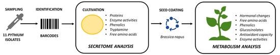
2.1. Isolation and Selection of Pythium Isolates

Approximately 20 g of soil sample was mixed thoroughly with 15 mL of sterilized (121 °C; 30 min) 15 g L−1 tap water agar (TWA) [9]. Then, it was poured into a Petri dish and left to solidify. After that, the surface of the Petri dish was covered with pure TWA, incubated in the dark at 24 °C, and observed daily.
After 5 days of incubation, the Petri dish with the soil sample was microscopically observed. In the case of ornamental oogonia detection, a small plug of agar medium with the oogonia was transferred to a Petri dish with TWA, followed by 24-h incubation at an elevated temperature (36–37 °C). After the incubation, a small plug of TWA with one separated growing hypha was transferred, using a sterile preparation needle and microscope, to a Petri dish with TWA and incubated for 24 h at 24 °C. After this last incubation period, a small plug of TWA with one separated hypha was transferred with a sterile needle to a Petri dish with malt extract agar (MEA, Merck, Darmstadt, Germany). After 7 days of incubation at 24 °C, the plugs of MEA with growing oomycete biomass were stored in paraffinic oil at 10 °C.
Only the strains with in vitro mycoparasitic abilities were selected for further study. The competition tests were conducted in triplicates on 9 cm diameter Petri dishes with MEA medium (25 °C, dark) using plant pathogenic fungi Alternaria alternata, A. brassicicola, Verticillium albo-atrum, and Phoma lingam. First, the plant pathogens were inoculated on one side of the plate and allowed to grow for 2–5 days, producing colonies 20–25 mm in size. Thereafter, the agar block with particular Pythium strain was placed onto the opposite side of the Petri dish, and the continuation of the growth of the tested mold and Pythium were evaluated every 2 days until 10 days. The ability of individual strain to parasitize the pathogens was assessed on the basis of visible overgrowth of the pathogen colony by Pythium hyphae, cessation of pathogen hyphae growth and loss or inability of the pathogen colony to change pigmentation compared to control growth without Pythium sp. Another characteristic feature of parasitism, which was evaluated using an optical microscope, was the ability of Pythium sp. hyphae to encircle the pathogen hyphae at sites of early contact between the two colonies.

2.2. Cultivation of Pythium Isolates

Eleven strains exhibiting spiny spores, a typical feature of P. oligandrum, and mycoparasitism ability were selected for the study. The composition of cultivation medium was 1000 g millet grain mixed with 500 mL of 2% (w/v) sucrose solution containing 30 μM cholesterol as a source of sterols. A quantity of 30 g of the solid cultivation medium was placed in a 250-mL Erlenmeyer flask, sterilized, inoculated by 5 small plugs of Pythium isolates, and incubated at 24 °C for 7 days. After the cultivation period, the biomass of growing mycelium and remnants of cultivation media were diluted 1:5 with sterile deionized water and centrifuged (8000× g, 15 min). The upper phase was used for the determination of secreted compounds. The biomass of Pythium was freeze-dried. The number of oospores was determined after lyophilization, and the inoculum concentration for further use (seed coating) was set to 12.0 × 10⁶ oospores.g−1 using sterile carrier silicon dioxide powder. Control (negative control) represents a sterile, non-inoculated cultivation medium cultivated under the same conditions. The M1 strain of P. oligandrum was provided by the company Biopreparáty, Ltd. (Únětice, Czech Republic) and corresponds to strain ATCC 38472. This oomycete was isolated from sugar beet [10].

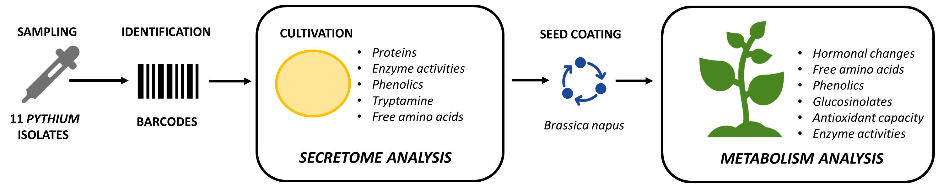
2.3. Identification of Pythium Isolates

Partial sequences of cytochrome c oxidase subunit I (COI), which is recommended as the main marker for oomycete identification [11,12], were studied in all strains. In addition, internal transcribed spacer (ITS) rDNA, another recommended barcode [12], was sequenced in the strains representing unique COI haplotypes. Genomic DNA was extracted using a DNeasy UltraClean Microbial Kit (Qiagen, Germantown, MD, USA). The ITS region was amplified with the primers ITS4 and ITS5 [13]. The COI gene was amplified with the primers Fm85mod and OomCoxI-Levup [12]. Amplification conditions were set as in [14]. The PCR products were purified and sequenced in Macrogen, Europe. The dataset for the phylogenetic analyses was assembled using reference sequences generated by [12,14] and supplemented with best Blast hits from NCBI GenBank. The other two P. oligandrum sequences were extracted from the whole genomic data of ATCC 38472 [15] and Po37 [16]. Sequence alignments was performed in MAFFT 6 using the G-INS-i strategy [17]. Maximum Likelihood phylogenetic analyses were done in PhyML 3.1. [18] using 500 bootstrap replicates and default settings. The COI gene alignment had 28 sequences and 680 characters, of which 91 were variable and 46 were parsimony informative. The ITS alignment had 29 sequences and 755 positions, of which 24 were variable and 12 parsimony informative. Barcode sequences were deposited in the EMBL sequence database (Table 1).

2.4. Plant Material

Brassica napus (rapeseed) seeds were treated with lyophilized Pythium spp. inoculum (oomycete biomass with medium inoculum) containing 12.0 × 10⁶ oospores.g−1. The inoculation of seeds was performed in a rotating seed machine at the concentration of 5 g of the inoculum on 1 kg of seeds. The mixture was moistened with deionized water and exposed to continuous rotation for 5 min. Then, it was left to dry and stored at room temperature.
Brassica napus treated and nontreated seeds were sown in pots with soil and grown for three weeks in growth chambers under 16/8 h light/dark period, ca. 150 µmol (photon) m−2 s−1 irradiance, at 20 °C, and 60% relative humidity. The samples of all mixed leaves from each group were immediately frozen in liquid N2 and kept at −80 °C.
Frozen leaf samples (0.5 g) were ground in liquid N2 with 1% polyvinylpyrrolidone. Then, 0.5 mL of extraction buffer (0.1 M Tris–HCl, pH 7.8, 1 mM EDTA, 10 mM DTT, 5 mM MgCl2; except for glycosidases and proteases—100 mM MOPS-NaOH, pH 5) was added to the ground tissue. The homogenate was centrifuged at 13,000× g for 10 min, and the resulting supernatant was used for measurements.

2.5. Protein Content

Protein measurements of all samples were performed spectrophotometrically at 595 nm using Protein Assay solution (Bio-Rad, Hercules, CA, USA) with bovine serum albumin as a standard [19].

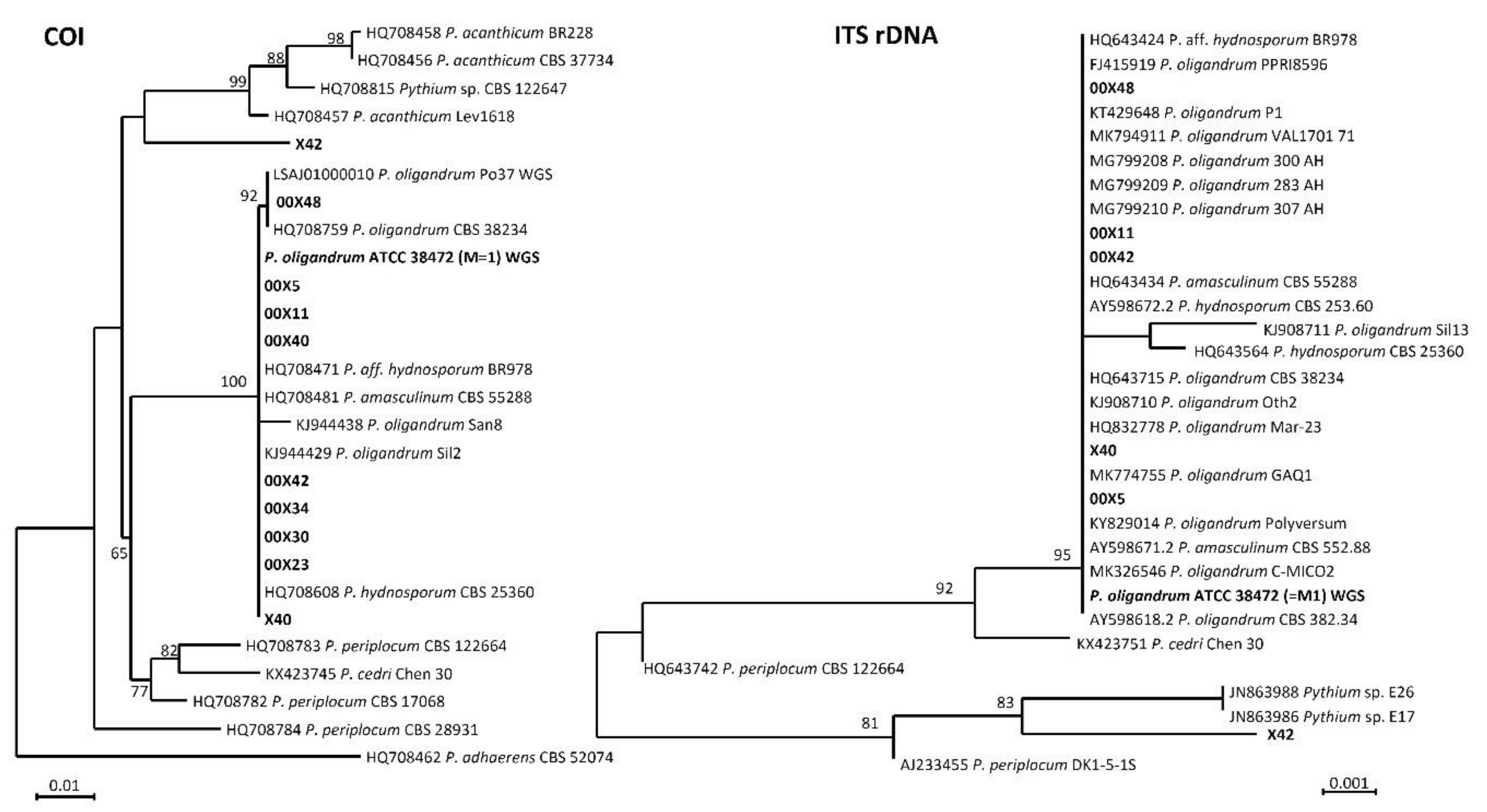
2.6. Electrophoretic Separation

Tricine-SDS-electrophoresis was performed to detect small molecules (such as oligandrins) secreted into the medium [20]. The gels consisted of 4% focusing and 16% separating layers. Medium samples were dissolved 1:1 in reducing sample buffer (6% (w/v) SDS; 3% (v/v) mercaptoethanol; 15% (w/v) glycerol; 0.025% (w/v) Coomassie Blue G-250; 150 mM Tris-HCl, pH 7.0). The proteins contained in the medium were separated by an SDS-PAGE run in 4% focusing and 15% separating gels [21].

2.7. Free Amino Acids and Tryptamine Content

Free proteinogenic amino acids and tryptamine were determined by capillary electrophoresis [22]. All electrophoretic experiments were conducted in a fused-silica capillary (Polymicro Technologies, Phoenix, AZ, USA) using a G7100A Capillary Electrophoresis Instrument (Agilent Technologies, Waldbronn, Germany) with a contactless conductivity detector. The detector consisted of two 4-mm-long cylindrical electrodes, with a 1-mm insulation gap. The inner diameter of the electrodes was 400 μm. The detection limits of all amino acids were in the range of 6 μg g−1 to 33 μg g−1. The limit of detection was calculated as 3-fold the standard deviation of the noise.

2.8. Phenolics and Antioxidant Capacity

The total phenolic content was determined using the standard Folin-Ciocâlteu colorimetric method [23,24]. Free Radical Scavenging, with the 2,2-diphenyl-1-picrylhydrazyl (DPPH) radical, and the Ferric Reducing Antioxidant Potential (FRAP) assays were used to determine the antioxidant capacity [23,24].

2.9. Phytohormone Analysis

In rapeseed leaves, indole-3-acetic acid (IAA), IAA-aspartate (IAA-Asp), indole-3-acetamide (IAM,), indole-3-acetonitrile (IAN), oxo-IAA (OxIAA), oxo-IAA-glucose ester (OxIAA-GE), phenylacetic acid (PAA), salicylic acid (SA), jasmonic acid (JA), JA-isoleucine (JA-Ile), and cytokinins (CKs) were determined. Frozen samples (ca. 50 mg fresh weight (FW)) were homogenized and extracted with cold (−20 °C) methanol/water/formic acid (15/4/1, v/v/v), as described previously [25,26,27]. The following isotope-labeled internal standards (10 pmol/sample) were added: 13C6-IAA (Cambridge Isotope Laboratories); 2H4-SA (Sigma-Aldrich); 2H3-PA, 2H3-DPA (NRC-PBI); 2H6-ABA, 2H5-JA, 2H5-transZ, 2H5-transZR, 2H5-transZ7G, 2H5-transZ9G, 2H5-transZOG, 2H5-transZROG, 2H5-transZRMP, 2H3-DZ, 2H3-DZR, 2H3-DZ9G, 2H6-iP, 2H6-iPR, 2H6-iP7G, 2H6-iP9G, 2H6-iPRMP (Olchemim). Phytohormones were separated with a reverse-phase cation exchange SPE column (Oasis-MCX, Waters) into the acid fraction by elution with methanol (auxins, ABA, SA, JA), and into the basic fraction by elution with 0.35 M NH4OH in 60% methanol (CKs and aminocyclopropane-1-carboxylic acid (ACC)). Fractions were analyzed using HPLC (Ultimate 3000, Dionex, Sunnyvale, CA, USA) coupled to a 3200 Q TRAP hybrid triple quadrupole/linear ion trap mass spectrometer (Applied Biosystems, Waltham, MA, USA). Hormone quantification was performed using the isotope dilution method with multilevel calibration curves (r2 > 0.99). Data processing was performed with the Analyst 1.5 software package (Applied Biosystems).

2.10. Extraction and Separation of Glucosinolates

Glucosinolates were extracted from 100 mg of lyophilized leaves ground in 1500 µL methanol/water (70/30, v/v). Then, the sample was shaken for 10 min (Vibramax 100 vibration platform shaker, Heidolph, Schwabach, Germany) and sonicated for 10 min (Elmasonic S 15, Elma, Germany). After centrifugation (10 min at 9000 rpm), 1200 µL of supernatant was evaporated and dissolved in 200 µL of background electrolyte (100 mM sodium tetraborate dissolved in water) and filtered through a 0.45-µm PVDF filter [28]. Again, the sample was shaken for 10 min and sonicated for 10 min. After centrifugation, 30 µL of supernatant was mixed with 5 µL of 1.8 mM sorbic acid as an internal standard.
Electrophoretic experiments were performed on an Agilent 7100 CE system (Agilent Technologies, Waldbronn, Germany) equipped with a UV/VIS detector. An unmodified fused-silica capillary of 50 µm ID and 375 µm OD (Polymicro Technologies, Phoenix, AZ, USA) was cut to 70.0 cm total length (effective length of 61.5 cm). Before the first measurement of the day, the capillary was flushed for 10 min with 1M NaOH and 10 min with deionized water. Between individual runs, the capillary was flushed with 1M NaOH for 2 min, and with background electrolyte for 2 min as well. Samples were injected using 5 kPa pressure for 5 s. Voltage of 20 kV (current approx. 76 µA) was applied during the 45-min analysis, and absorbance was detected at λ = 225 nm. The capillary temperature was 25 °C. The detection limits of glucosinolates were from 1 μg g−1 to 2 μg g−1. The limit of detection was calculated as 3-fold the standard deviation of the noise.

2.11. Enzyme Activities

Generally, enzyme activities were determined spectrophotometrically (Helios α, Thermo-Spectronics, Waltham, MA, USA) and expressed as activity in μmol of the respective product formed (substrate decreased) per min per mL of growth medium or per g of fresh plant weight (FW).
Catalase (CAT, EC 1.11.1.6) was detected at 240 nm as a H2O2 decomposition rate. Total ascorbate peroxidase (APOD, EC1.11.1.11) activity was determined as a decrease in the absorbance of ascorbate at 298 nm. Malic enzyme (NADP-ME, EC 1.1.1.40), glucose-6-phosphate dehydrogenase (EC 1.1.1.49), shikimate dehydrogenase (EC 1.1.1.25), and glutathione reductase (GR, EC 1.8.1.7) activities were assayed as NADPH production at 340 nm [29,30].
Proteolytic activity was determined using azocasein substrate or Nα-benzoyl-DL-arginine 4-nitroanilide [31,32]. Endo-β-1,3-glucanase (EC 3.2.1.6), cellulase (EC 3.2.1.4), and chitinase (EC 3.2.1.14) activities were determined based on the quantification of reducing sugars (products) with 3-methyl-2-benzothiazolinone hydrazone [33].
α-Glucosidase (EC 3.2.1.20), β-glucosidase (EC 3.2.1.21), α-galactosidase (EC 3.2.1.22), β-galactosidase (EC 3.2.1.23), α-mannosidase (EC 3.2.1.24), and β-N-acetylhexosaminidase (EC 3.2.1.52) activities were determined using the end-point method with specific synthetic p-nitrophenyl (p-NP-) substrates (p-NP-α,D-glucopyranoside, p-NP-β,D-glucopyranoside, p-NP-α,D-galactopyranoside, p-NP-β,D-galactopyranoside, p-NP-α,D-mannopyranoside, and p-NP-N-acetyl-β-D-glucosaminide, respectively). The reaction mixture contained a 50 mM citrate buffer, pH 4.5, 0.5 mM p-NP-substrate, and 10 µL of enzyme extract in total 40 µL volume. The reaction was stopped with 80 µL of 0.25 M borate buffer, pH 9.0. The hydrolyzed p-nitrophenol in the alkali region was quantified by measuring absorbance at 405 nm against a blank in which the enzyme was previously inactivated by the high pH of the borate buffer. Acid (EC 3.1.3.2) and alkaline phosphatase (EC 3.1.3.1) activities were determined with p-NP-phosphate at pH 5.5 and 9.0, respectively, modified from [34].
Superoxide dismutase (SOD, EC 1.15.1.1) isozyme patterns and activities were assessed after separation by 10% native PAGE [35,36].
Peroxidase (POD, EC 1.11.1.7) isozyme pattern was assessed after separation by 10% native PAGE. POD isozymes were detected in situ by staining gels in 1 M acetate buffer, pH 4.6, with 0.04% benzidine and 10 mM H2O2 [37].
Shikimate dehydrogenase (SDH, EC 1.1.1.25) isoenzyme pattern was detected after separation by 10% native PAGE by staining in 3 mM shikimate, 0.2 mM NADP+, 0.1 mg mL−1 nitroblue tetrazolium, and 5 µg mL−1 phenazine methosulfate.

2.12. Carbon, Nitrogen, and Sulfur Content Analysis

The lyophilized samples of leaves were used for elementary analysis of C, H, N, and S, which was performed on a Thermo Finningan Flash FA 1112 series CHNS/O analyzer.

2.13. Statistical Analysis

All measurements were performed at least in triplicate. Each plant experimental group consisted of three biological samples. Each sample represented mixed leaves of 30 plants. Three biological replications (i.e., independently cultured strains) were studied for each strain. Data were analyzed by one-way ANOVA (Bonferroni method) and t-test; differences were considered significant at p ≤ 0.05. The different letters in figures and tables denote significant differences (p ≤ 0.05).

3. Results and Discussion

3.1. Identification of Tested Pythium spp. Revealed not yet Described Pythium Species

In response to demands from many regulatory authorities and professional agricultural institutes, scientists have intensified the search for new and more effective biological control agents, which requires characterizing not only the specific organisms but also their effects on plant metabolism. As Pythium oligandrum is well-known for its beneficial features, we searched for other Pythium oomycetes with high potential for use as BCAs. For this purpose, we collected soil samples from different areas to identify new suitable organisms with unique properties (Table 1, Figure 1). We selected the strains that after cultivation showed ornamental oogonia and also behaved as mycoparasites. The mycoparasitism tests of the M1 strain as an example are shown in the Supplementary Materials (Figures S1–S4). Then, we characterized unknown Pythium strains by COI and ITS rDNA barcode genes.
Based on the COI gene, we determined that our study strains belonged to three unique haplotypes: (1) X42, (2) 00X48, and (3) others. Phylogenetically, the two latter haplotypes belonged to the well-supported clade consisting of sequences deposited as P. amasculinum (HQ708481, [12]); Pythium aff. hydnosporum (HQ708471, [12]); Pythium hydnosporum (HQ708608, [12]); and P. oligandrum (HQ708759, [12]; KJ944429, [14]) (genomic data from strains Po37 and ATCC 38,472 = M1) (Figure 1). The sequence of strain X42 showed a separate position, with the closest affinity to sequences deposited as Pythium acanthicum. Based on the ITS, all but one (X42) of the strains belonged to a single haplotype. The main haplotype belonged phylogenetically to the well-supported clade of P. oligandrum, P. hydnosporum, and P. amasculinum, whereas strain X42 showed a separate position with the closest affinity to Pythium sp. E26 JN863988 (97.50% sequence similarity).
Most of our isolates fit into the P. oligandrum clade consisting of P. oligandrum, P. hydnosporum, and P. amasculinum sequences. All three species have spiny oogonia and are morphologically related. For these reasons, P. oligandrum has been often misidentified as P. hydnosporum. P. amasculinum is considered a likely synonym of P. oligandrum [38].
There is a general consensus that the species inside this clade cannot be recognized based on the widely used barcode genes ITS and COI and should be formally synonymized pending more thorough investigations with multiple hypervariable genes [11,12,14]. Our strains morphologically fit into P. oligandrum, and accordingly we are designating them using this name. The strain of X42 has a separate position, sister to the P. oligandrum clade and P. acanthium. This strain is not ascribable to any sequenced species of Pythium. Morphologically, it is an ornamented oogonia, like P. oligandrum.

3.2. Secretomes Contained Proteins, Amino Acids, and Phenolic Compounds

The main goal of this study was to find out the biochemical background of the Pythium spp. life strategies, i.e., mycoparasitism and/or promoting plant growth. The oomycetes depend on their hosts in acquiring β-hydroxy sterols for sporulation [6] and thus produce extracellular protein elicitors (oligandrin and cell wall protein fractions) such as sterol carriers. A side effect of these elicitors is plant priming and activation of plant defense responses, which is advantageous during interaction with real pathogens [1]. In addition, Pythium spp. as mycoparasites secrete a large arsenal of effector proteins that allows the penetration of the host cell wall [39] (Figure 2, Figure 3, Figure 4 and Figure 5). Although the composition of the medium was identical among all studied Pythium strains, the contents of proteins, amino acids, and phenolic compounds in secretomes varied considerably (Figure 2). All Pythium strains secreted proteins into the medium; the highest content was in M1 and the lowest in 00X40 (Figure 2a). Differences were also found in the proportion of individual proteins analyzed by SDS-electrophoresis (Figure 3). Most proteins were of a molecular weight lower than 30 kDa, but proteins were also detected in the regions around 85 kDa and 55 kDa. Strains 00X40 and X40, and strains 00X23 and 00X42 showed similar protein profiles. However, the protein profile of X42 was quite unique; the relative molecular weights of its most abundant proteins differed from those of other strains analyzed in this study. By Tricine electrophoresis, suitable for separating small proteins and peptides, we identified molecules of approximately 10 kDa, which could correspond to oligandrins. In addition to oligandrins, X42 medium contained several peptides (<6.5 kDa), which could be either elicitors or degradation products (Figure 3).
One of the significant properties of P. oligandrum, which is responsible for plant growth promotion, is the production of tryptamine, the precursor of the phytohormone auxin. Thus, we determined the concentrations of tryptophan and tryptamine in the medium (Table 2, Figure 2b). Although we found low tryptamine concentrations and no free tryptophan in the control medium (without Pythium), after Pythium cultivation, tryptophan was present and tryptamine concentrations were significantly elevated, especially in X42 (5.5 times more than M1). The lowest tryptamine concentration was found in 00X23 medium, whose value was only 6% of that in X42 (Table 2, Figure 2b). Considering the amount of tryptophan and tryptamine provided in their secretomes, strains X42, 00X42, and 00X11 appear to have good predispositions to increase plant growth and fitness (Table 2, Figure 2b).
Due to differences in tryptophan content, we also determined other free amino acids and total amino acids (Table 2, Figure 2c). In the control medium, tyrosine, glutamine, and aspartate were absent, whereas histidine, lysine, and arginine were found in very low concentrations (Table 2). Key amino acids that could affect plant metabolism (tryptophan, methionine, phenylalanine, and partly tyrosine) were most abundant in secretomes of Pythium spp. X42, 00X42, and 00X11 (Table 2). The only absent amino acid in all media was cysteine, which has a reactive thiol group and therefore is likely bound in other compounds such as glutathione. The concentration of all free amino acids increased in the medium after Pythium cultivation; in X42, the increase was 14 times in comparison with the control (Figure 2c). Although the specific secondary metabolites that Pythium spp. produce remain somewhat unclear, we tested for the presence of phenolic compounds (Figure 2d). The highest value was determined in strain X42, 2.7 times exceeding M1. The presence of phenylpropanoid compounds is unlikely because we did not find the initial enzyme catalyzing the conversion of phenylalanine to cinnamic acid, phenylalanine ammonium lyase, in the published genome of the P. oligandrum strain ATTC 38,472 [15]. However, the synthesis of isoprenoids is probable because in the abovementioned genome, we found genes for enzymes capable of synthesizing mevalonate, isopenthenyldiphosphate, geranyldiphosphate, farnesyldiphosphate, and geranylgeranyldiphosphate from an acetyl-CoA precursor. This is in agreement with other oomycetes [40].

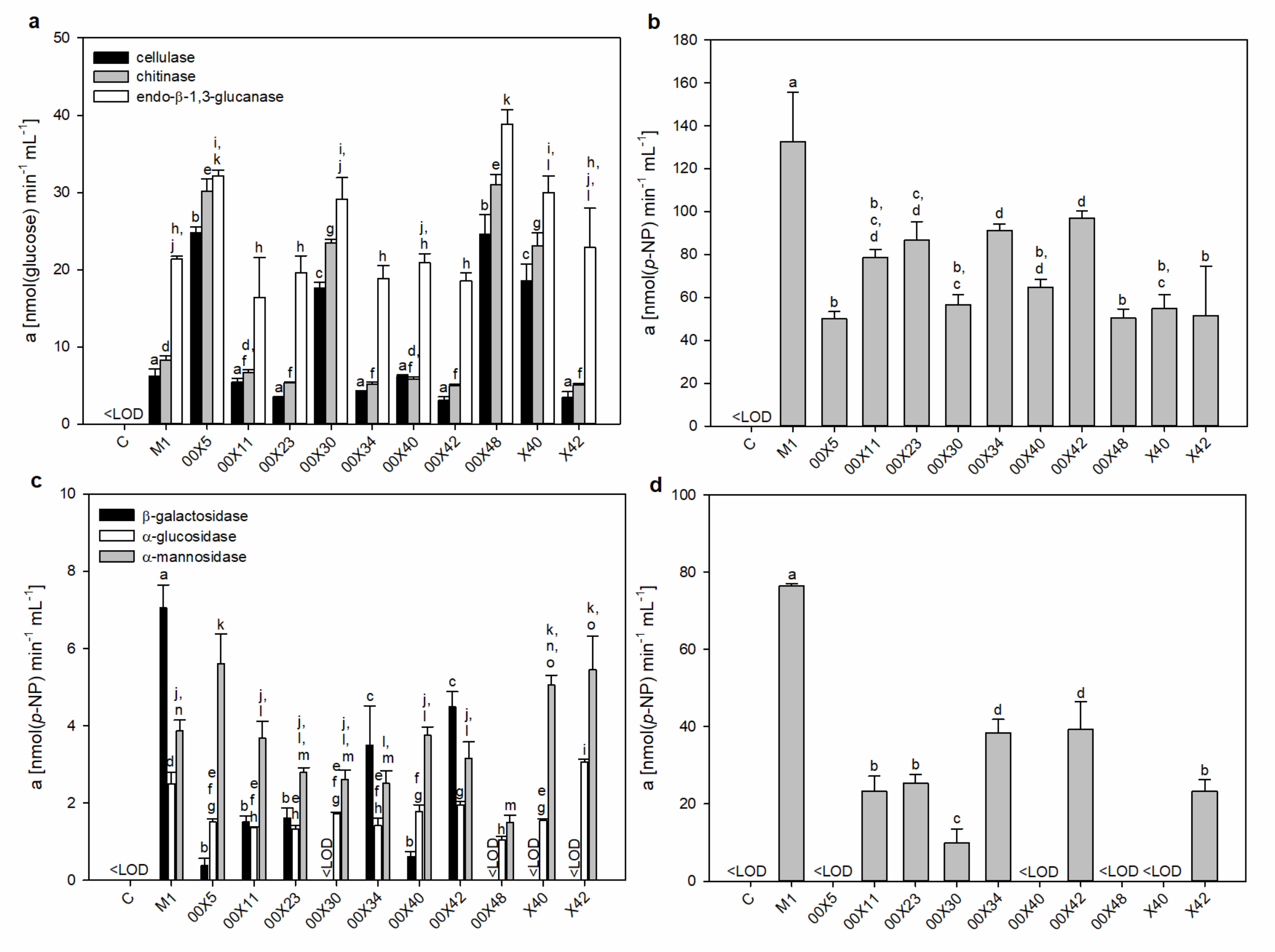
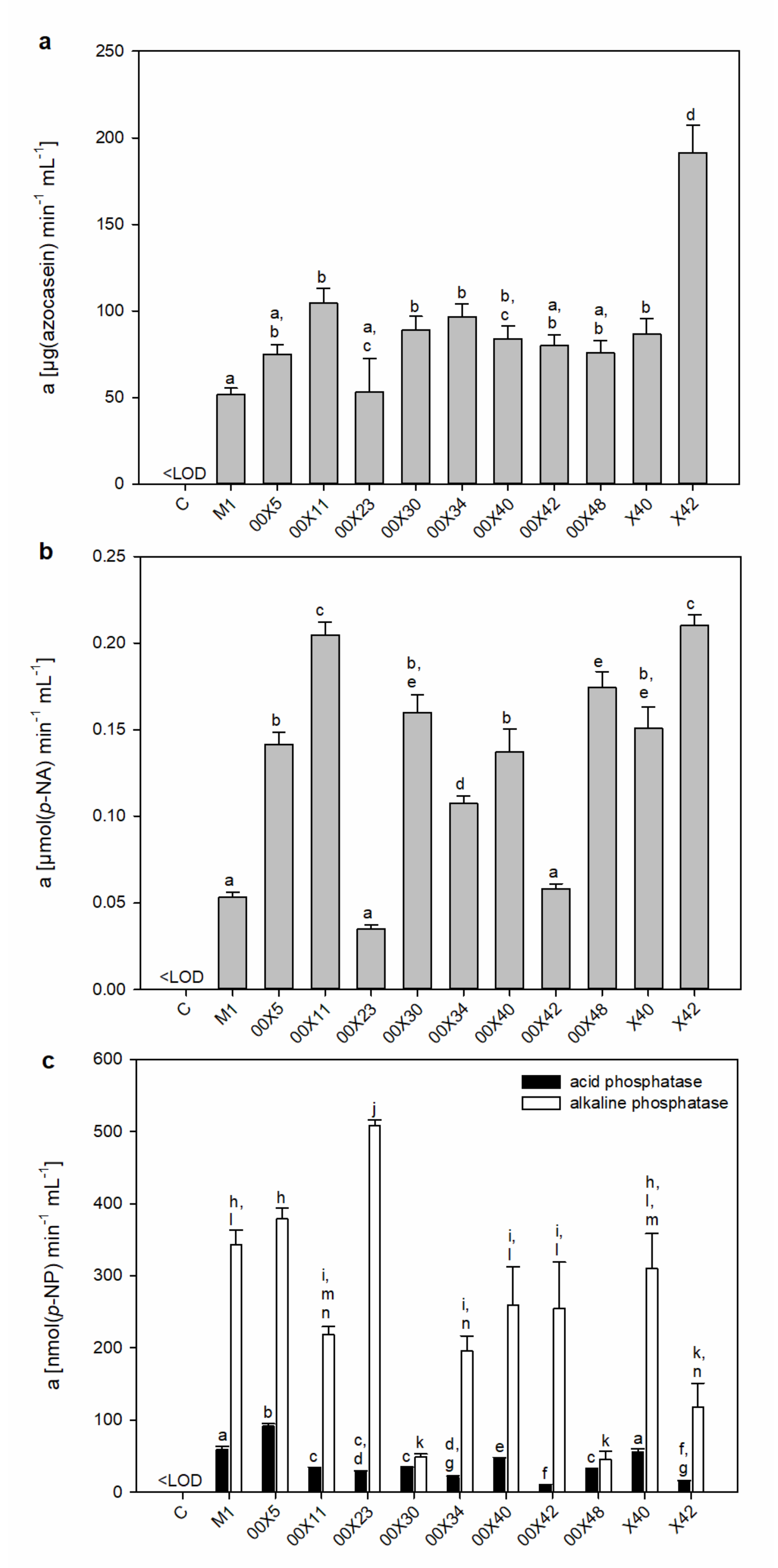
3.3. Pythium Secretes a Cocktail of Host Cell Wall-Degrading Enzymes

Hydrolytic enzymes facilitate cell wall penetration for host colonization. These enzymes include cutinases, glycosidases, pectinases, and proteases. A “cocktail” of different hydrolytic enzymes with overlapping substrate specificities apparently enables the formation of proper infection structures [41]. Most information about these enzymes in oomycetes derives from genomic analysis of Pythium spp. [15,39]. The glycoside hydrolases and proteases were represented in high numbers. Among oomycete effector proteins, glycoside hydrolases belong to the most abundantly secreted proteins (79 for P. oligandrum). Based on sequence analysis, glycoside hydrolases can be assigned to families [39], but their true substrate specificity and activity cannot be ascertained. This is the first study providing a comprehensive survey of enzyme activities of plant and fungal cell wall-degrading enzymes across the variety of Pythium strains. We showed that there is a high difference in the quantity and quality of the presented enzymes, even across strains of the same species (Figure 4 and Figure 5), and proper strain selection is crucial in searching for BCAs.
In comparison with M1, higher activities of 12ellulose (3–4-fold), chitinase (3-fold), and endo-β-1,3-glucanase (1.5-fold) were found in 00X5, 00X30, 00X48, and X40 media (Figure 4a).
In addition to endoglycosidases, exoglycosidases also complete the degradation of glycoconjugates. Thus, we measured the activity of α-glucosidase, β-glucosidase, β-galactosidase, α-mannosidase (Figure 4), α-galactosidase, and β-hexosaminidase. β-Glucosidase degrading β-oligoglucans into glucose monomers, used as a source of energy, showed the highest activity, especially in M1 (Figure 4b). The other strains’ media reached only 40–70% of M1 activity. α-Glucosidase activity was approx. 45 times lower than β-glucosidase activity, as expected because substrates such as α-glucans are a much less commonly found source in the natural environment. α-Mannosidase may degrade mannan structures, glycoproteins, or glycolipids in host cell walls or membranes. The activity of α-mannosidase in 00X5 and X42 media exceeded M1 by 40–45%; and X40 by 30%. (Figure 4c). β-Galactosidase activity at pH 4.5 was found only in some Pythium spp. (Figure 4c). Interestingly, even though exoglycosidases generally have an acidic optimum pH, β-galactosidase showed activity when measured under alkaline reaction conditions; the highest activity at pH 4.5 was found in M1, with no activity in 00X30, 00X48, X40, or X42 (Figure 4c). This enzyme may have a very broad optimum pH, but the 00X30 and X42 media showed activity only under alkaline conditions, reaching only 10% and 30% of M1, respectively. Therefore, we cannot rule out an alkaline β-galactosidase isoform (Figure 4d). Various glycoconjugates containing galactose, hemicelluloses, and pectins in plant cell walls, glycolipids, and glycoproteins could be substrates of this enzyme. No activity of α-galactosidase or β-hexosaminidase was detected, but the activity of these enzymes could be induced by the presence of an appropriate substrate. As far as we know, this is one of the first studies concerning the determination of exoglycosidase activity in Pythium spp.
Numerous pathogenic fungi and oomycetes produce saponin-detoxifying glycosidases, which are able to deglycosylate saponin molecules for their detoxification (degradation) [42,43,44]. Another role of exoglucosidases, namely β-glucosidase, could be the re-activation (deglycosylation) of O-glycosylated cytokinins, thus modulating the phytohormonal levels in planta [45]. Glycosylation is also the key regulating factor of the phenylpropanoid pathway by changing the solubility, stability, and toxicity of secondary metabolites. A vast array of glycoside hydrolases could help oomycetes to alter host metabolism, especially the level of defense compounds [46].
In addition to glycosidase activity, proteolytic activity is important for the ability of Pythium to penetrate the cell wall of the host. Accordingly, total proteolytic activity was assessed using azocasein as a substrate. All Pythium strains secreted proteases into the medium, and the highest activity was found for X42, whereas the lowest activity was found for M1. The activity was almost 4 times higher in X42 than in M1 (Figure 5a). In addition to X42, 00X11 strains also showed high activity of serine proteases in the media (both ca. 4-fold higher than M1) (Figure 5b), with significant differences in the proportion of individual classes of proteases in Pythium spp.
Phosphatases are enzymes with broad substrate specificities. Both alkaline and acidic phosphatases were present in all Pythium spp. media (Figure 5c). Alkaline phosphatase activity was much higher than acidic phosphatase activity. The highest differences between them were found for 00X23 (19-fold) and 00X42 (26-fold). Phosphatases remove phosphate groups from small organic molecules or polymers, which are then used to meet their own needs. This feature could also be useful for increasing nutrient availability for plants when phosphate is released near the roots.

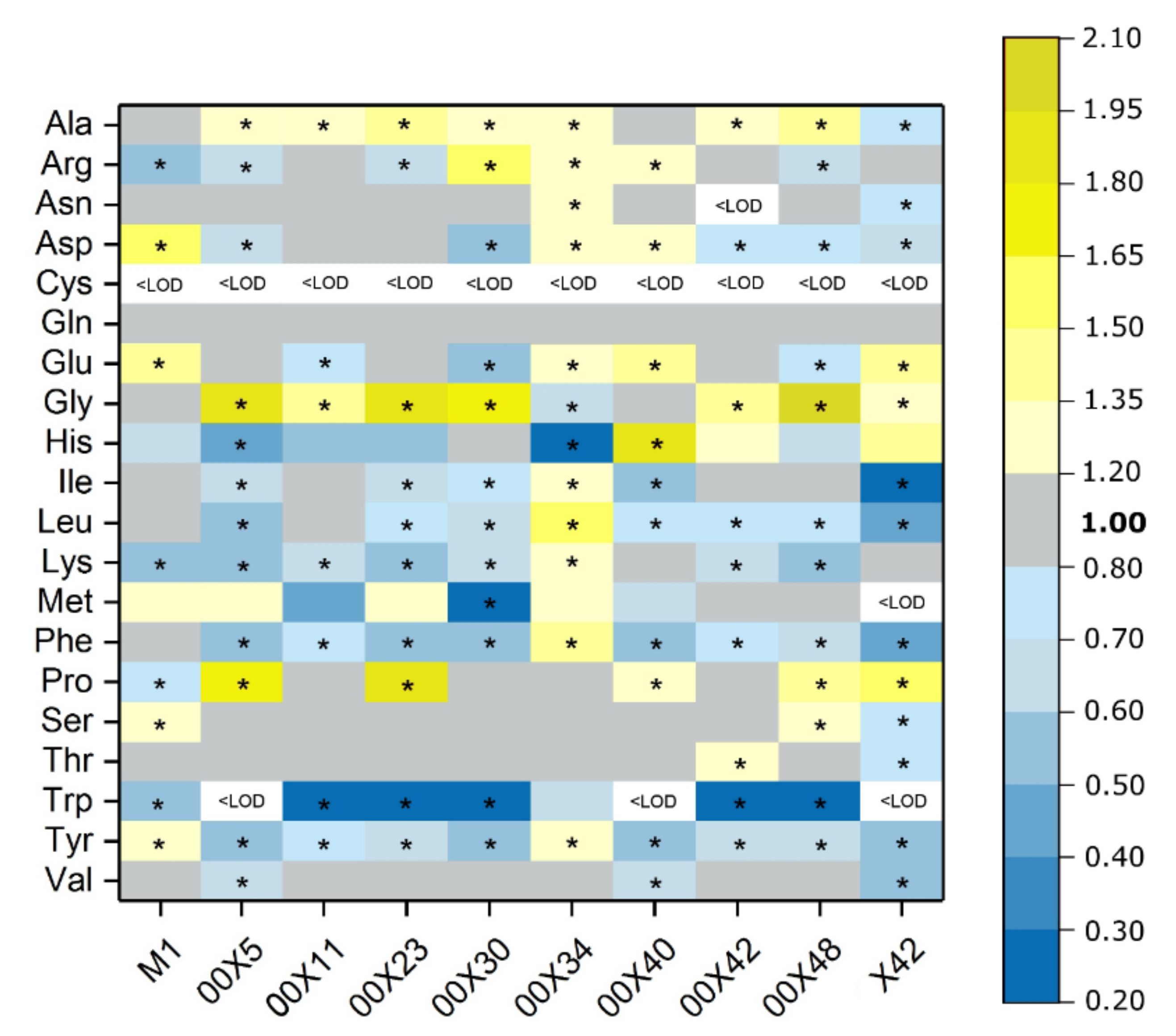
3.4. How Do Pythium spp. Affect Metabolism in Inoculated Rapeseed Plants?

The impact of inoculation of Brassica napus seeds with ten Pythium species on plant metabolism was followed in 3-week-old plants. Seed coating represents an efficient and convenient tool for introducing beneficial microorganisms into the soil and consequently into the rhizosphere or plant tissues [47]. Although most growth parameters were not affected in young plants, many metabolic pathways showed significant changes even within individual isolates. The inoculated plants showed no visible differences when compared to the control samples, or to each other. Similarly, the elementary analysis of dried leaf samples showed no significant differences in the content of carbon, nitrogen, or sulfur between plants treated with Pythium spp. and untreated control (Table S2). The carbon-to-nitrogen ratio, which characterizes the nitrogen supply to the plant, is shown in Figure S5. This ratio ranged from 6.2 to 8.2, and these differences were not significant, although the presence of various Pythium spp. may affect nitrogen use in plants. The content of photosynthetic pigments was also determined in leaf samples from experimental plants (Figure S6). The content of chlorophyll a was 11% and 28% higher in plants treated with M1 and 00X11 than in the control, respectively. The content of chlorophyll b was also higher in 00X11 and 00X42 than in other experimental groups. This corresponds to the previous assumption of positive influence of these strains on plant fitness.
In leaves, the soluble protein content varied, albeit not significantly (Figure 6a), and no differences in protein profile were detected by SDS-electrophoresis (Figure S7). However, some differences were found in the total amount of free amino acids. Plants inoculated with some Pythium strains showed increased total free amino acid content, whereas others showed lower content than the control (Figure 6b). More interestingly, the comparison of individual free amino acids (Figure 7) highlighted that the contribution of the first synthetized amino acid glutamine was equal in all plant groups. Conversely, the levels of some amino acids were decreased in all plant groups, e.g., aromatic amino acids, tryptophan, phenylalanine, and tyrosine. The decrease in tryptophan was particularly significant in plants treated with 00X40, 00X5, and X42 Pythium spp. Its concentration was under the detection limit; therefore, the tryptamine content was also measured (Figure 6c). However, tryptamine concentration was also low in plants treated with these strains, most likely because tryptamine was further metabolized to auxin. In turn, the plants inoculated with 00X30, 00X48, and 00X11 strains showed high tryptamine concentration, most likely due to slower conversion to auxin. The presence of tryptamine in control plants also documents its other uses and functions in plant metabolism. Alanine and glycine were among the more abundant free amino acids. The levels of proline, which is often synthetized in plants under stress conditions, were increased in some plant groups and unchanged or decreased in others (Figure 7).
Enhanced secondary metabolite synthesis, especially via phenylpropanoid and terpenoid pathways, has well-described effects on plant metabolism caused by P. oligandrum [3]. Surprisingly, no significant differences were found in phenolic content, although it was slightly elevated in some groups in comparison with the control (Figure 6d). The method detected only soluble phenolics and not compounds bound to the cell wall, so a significant part of polyphenols could have been incorporated as phenolic acids and especially lignin. Cell wall impregnation by lignin could significantly improve the defense against biotic stress. The soluble phenolic compounds contribute to the total antioxidative capacity; thus, this property was determined in plant extracts using two methods, DPPH (Figure 6e) and FRAP (Figure 6f). Considering the nonsignificant changes in phenolic content described above, the antioxidant capacity unsurprisingly showed no significant differences. Nevertheless, the FRAP method showed 20% enhancement of this value in plants treated with 00X5 and 00X34 strains.

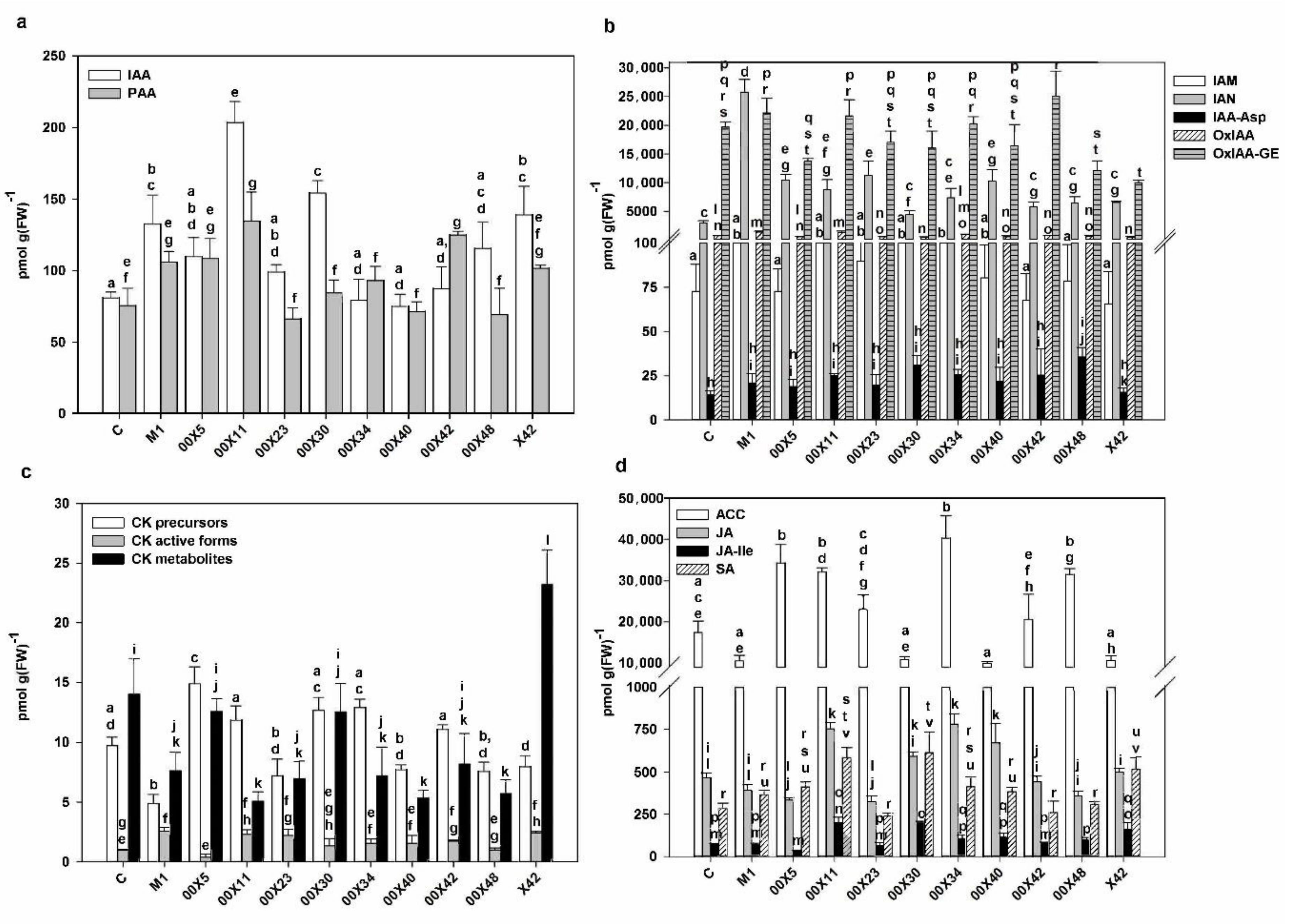
3.5. The Concentration of Phytohormones is Affected by Pythium spp. in the Rapeseed Plants

Plant growth and development as well as interactions with the environment are controlled by phytohormones. Accordingly, P. oligandrum may promote plant growth by increasing auxin concentrations. Indole-3-acetic acid (IAA), the most physiologically active auxin, may be synthetized from tryptamine provided by P. oligandrum [8]. Therefore, the auxin content of rapeseed plants treated with the Pythium strains was analyzed (Figure 8a). Considering the amount of tryptophan and tryptamine provided in their secretomes, the strains X42 and 00X11 appear to have good predispositions to increase plant growth and fitness (Table 2, Figure 2b). Indeed, the highest IAA content was found in plant leaves treated with 00X11, 00X30, X42, and M1 strains. Differences were also found in the contents of indole-3-acetamide (IAM) and indole-3-acetonitrile (IAN), which are precursors of IAA in different plant biosynthetic pathways (Figure 8b). In addition, differences were also found in the content of phenylacetic acid (PAA), a weak, evolutionarily ancient auxin. This compound was also present in higher amounts in some plants (e.g., treated with 00X11 or 00X42) than in the control (Figure 8a). These results suggest that Pythium likely affects auxin metabolism by multiple pathways other than just tryptamine delivery. The catabolic products of auxin metabolism were detected in all experimental groups (Figure 8b). Oxo-IAA and its glucosyl ester were the most abundant catabolites. Auxin inactivation is also performed by conjugation with amino acids, but this conjugation only occurred with aspartic acid in rapeseed plants. The highest contents of tryptamine (Figure 2c) and tryptophan (Table 2), determined in X42 medium, positively correlated with IAA level in leaves (Figure 8a). Negative correlation of tryptamine and tryptophan with the content of IAA degradation products (IAM, IAA-Asp, Ox-IAA, Ox-IAA GE) was not detected. The highest content of IAA and PAA was found in plants treated with 00X11 and 00X30 (Figure 8a).
The promotion of cell division and subsequently of plant growth requires cytokinins (CKs) as well as auxins. The active cytokinins, namely trans-zeatin and cis-zeatin, as well as their precursors, storage forms (CK O-glucosides), and degradation products (CK N-glucosides) were also determined in plants (Figure 8c). Higher concentrations of active cytokinins than in control (with the exceptions of 00X5 and 00X48) were found in all groups of rapeseed plants. High concentrations of precursors and degradation products in most experimental variants suggest intensive cytokinin metabolism.
In addition to auxins and cytokinins, hormones related to plant immunity were also monitored, particularly salicylic acid (SA) and jasmonate (JA) (Figure 8d). The treated rapeseed plants had higher SA content (except for 00X23 and 00X42) compared with the control. The highest SA concentrations were found in plants treated with 00X30 and 00X11, increasing to 216% and 205%, respectively. The contents of JA and especially of its active form JA-Ile were higher in plants treated with 00X11, 00X30, 00X34, and 00X40. SA and JA activate signaling pathways in response to biotroph or necrotroph/herbivore attack, respectively. Considering the possibility of attack by different pathogens, as well as the fact that some pathogens may change their lifestyle during infection, these hormones may enhance plant defense against a wide range of foes. The spatially separated activation of JA and SA defense pathways, which could increase the chance of successful elimination of greater pest diversity, has been described [48]. SA and JA functions are modulated by the presence of other phytohormones. Gaseous phytohormone ethylene (ET), the precursor of which is 1-aminocyclopropane-1-carboxylic acid (ACC), could modify the defense responses controlled by JA [49]. ACC was present in all leaf samples including untreated controls. Its concentration was in six groups higher than in the control; the highest increase was observed in plants treated with 00X34 (231%). The plants treated with the 00X11 strain contained higher concentrations of all aforementioned phytohormones (Figure 8, Table S1). The higher levels of JA, JA-Ile, and ACC in treated plants are in accordance with the reported induction of JA and ET signaling pathways by P. oligandrum elicitins [50,51]. In turn, SA signaling including PR1 and PR5 in tomato plants has been ruled out [50], but we found higher levels of SA in rapeseed plants inoculated with some Pythium spp. than in control (Figure 8d). However, it is possible to assume species specificity or simply only readiness to respond faster (and to a greater extent) in the case of infection. The concentrations of individual hormones and their metabolites are shown in Table S1.

3.6. Pythium Alters the Glucosinolate Composition of Inoculated Rapeseed Plants

Brassiacaceous plants contain glucosinolates, which protect their tissues from herbivores and likely also from pathogens. The profile of glucosinolates depends on the plant species, its genotype, and growth conditions including biotic and abiotic stresses [52]. Progoitrin and gluconapine were found to be the main glucosinolates in rapeseed meal [53]. In addition, glucoraphanin, glucobrassicanapin, and glucobrassicin were found in rapeseed plants [54]. In the leaves of our experimental plants, the most abundant glucosinolates were glucobrassicin and progoitrin. Furthermore, glucoraphanin was also identified, but only in plants treated with Pythium strains; in the control plants and in the plants grown with 00X34, the value was below the detection limit (Table 3). Pythium strains can alter glucosinolate content. In the Pythium-treated plants, glucobrassicin content was enhanced, whereas progoitrin was rather reduced. This could be advantageous, because progoitrin is metabolized to oxazolidine-2-thione, which causes reproductive and thyroid problems when consumed by animals [55,56]. In general, glucosinolates are of crucial importance in plant defense against herbivores [57] or plant pathogens [58,59,60]. Tryptophan is present in Pythium-treated plants at very low levels or under the detection limit (Figure 7); therefore, it could be a precursor not only of auxin but also of glucobrassicin (Table 3). Methionine as a precursor of glucoraphanin and progoitrin was secreted by Pythium strains into the medium, but its concentration in plants depended on the Pythium strain (Table 2, Figure 7). It is possible that methionine is provided by Pythium to the plant, thus stimulating metabolic pathways originating in methionine, e.g., biosynthesis of ethylene or S-adenosylmethionine, a methyl group donor [49]. It seems that seed coating could increase plant defense at a young growth stage when plants are most vulnerable and not ready for harvest. Each step towards higher natural plant resistance leads to a reduction in the use of pesticides.

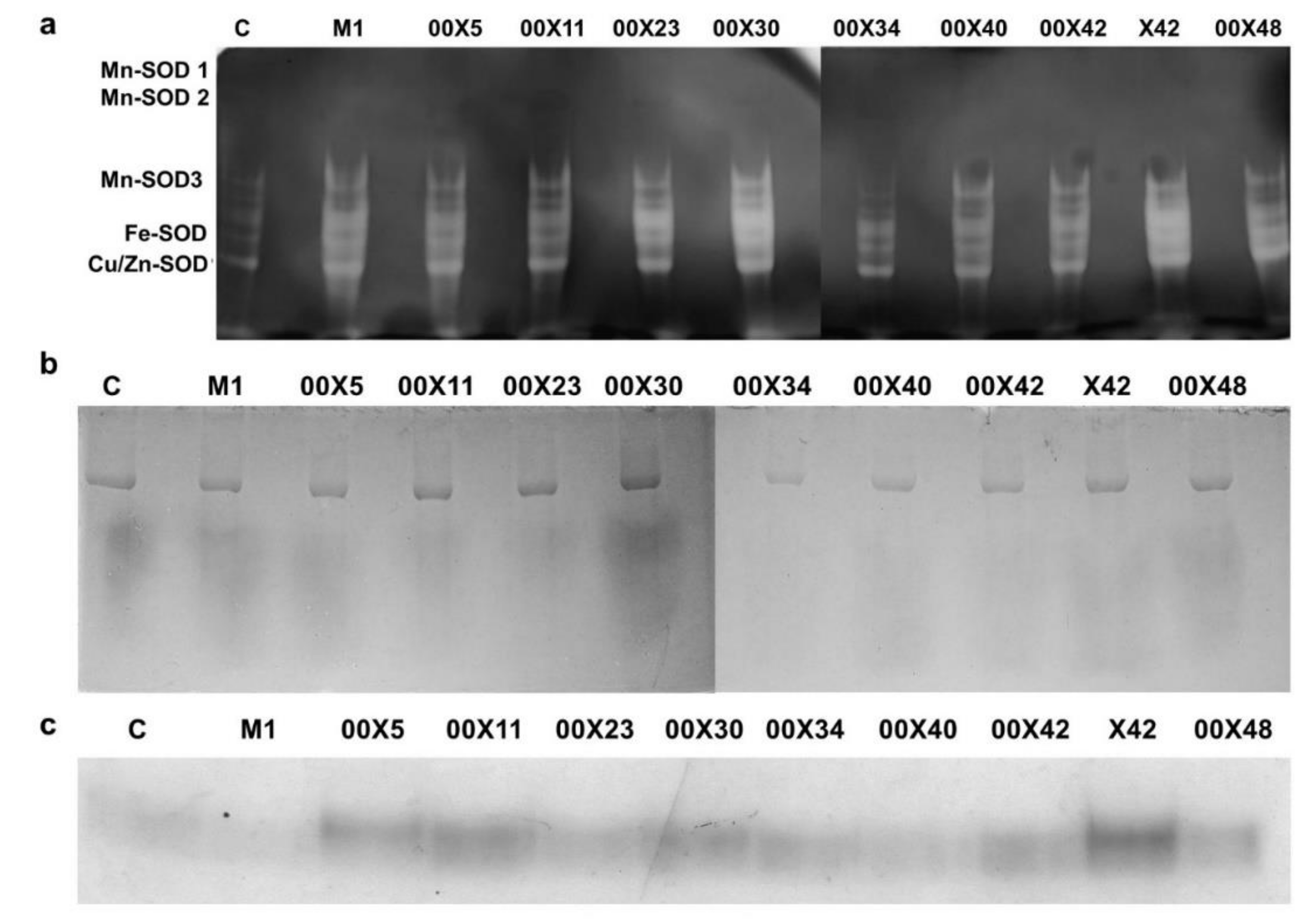
3.7. Could Pythium spp. Support the Activity of Antioxidant Enzymes in Rapeseed Plants?

The health status of a plant depends on the activity of antioxidant enzymes. The superoxide radical, one of the most dangerous reactive oxygen species, is detoxified by superoxide dismutase (SOD). SOD activity was detected in leaf extracts (Figure 9a). The individual SOD isoforms were more abundant in the plants inoculated with Pythium spp. than in the control plants; the highest intensity was found in 00X30 and X42 samples. The product of SOD reaction, H2O2, is also a reactive compound, which is degraded by catalases (CAT) (Figure S8a). CAT activity differed between plants; the highest activity was found in plants inoculated with X42, and the lowest in those treated with 00X11 and 00X42. H2O2 is also a substrate for peroxidases, which have broad substrate specificity. They participate in the synthesis of lignin, for example. At least two peroxidase isoforms were present. This microheterogeneity of the latter isoform could have been caused by glycosylation (Figure 9b). Another enzyme that uses H2O2 as a substrate is ascorbate peroxidase (Figure S8b). Its activity was higher in plants treated with Pythium spp. than in controls, except for 00X23 and 00X48.
Glutathione in its reduced form is important for the control of the reduced status of thiol groups in many proteins in plant cells. The change of glutathione from the oxidized form to the reduced one is catalyzed by glutathione reductase (GR), which uses NADPH as a coenzyme. However, the activity of GR in plants was not significantly affected by the Pythium spp. treatment (Figure S8c).
Plant redox state is also affected by the availability of NADPH, which is produced in chloroplasts during photosynthesis. The requirement for NADPH is enhanced under oxidative stress, and therefore other sources, such as NADPH-producing enzymes, could be used. However, the activity of the NADP-malic enzyme (NADP-ME) and of glucose-6-phosphate dehydrogenase (G6PDH) in experimental plants showed no significant effects from Pythium spp. The highest activity of NADP-ME and G6PDH was in plants treated with 00X48 and 00X34, respectively (Figure S8d,e).
NADPH is used also by enzymes of the shikimate pathway, which synthetize aromatic amino acids. The shikimate dehydrogenase (SDH) activity was higher in plants inoculated with Pythium spp. compared to the control, and the highest activity was found in plants treated with X42 (Figure 9c). The synthesis of aromatic amino acids, especially phenylalanine and partly tyrosine, is important for plants, not only for protein synthesis but also for the production of phenylpropanoid compounds including lignin, which fortifies cell walls and thus improves plant defense.

4. Conclusions

In this study, we assess a broad number of strains for traits related to mycoparasitism as well as plant-promoting activity. The secretomes of Pythium spp. varied significantly in terms of the activities of enzymes capable of acting on host cell walls. Even closely related P. oligandrum strains differed in their properties and effects on plants. The alterations in enzyme activities and tryptamine production may indicate advantages either for mycoparasitism or plant growth promotion. Some strains showed equal prerequisites for both effects. The preferred life strategy of Pythium spp. has a crucial importance in the selection of new biocontrol agents. This is the first study screening seed coating with Pythium spp. for effects on the metabolism of rapeseed plants. In addition to differences in free amino acids and antioxidant enzyme activities, major differences in phytohormone levels were also found, showing increased concentrations of auxin and jasmonates (and salicylic acid in some strains), indicating plant growth promotion as well as readiness for effective defense.

Supplementary Materials

The following are available online at https://www.mdpi.com/2076-2607/8/10/1472/s1. Figure S1. The in vitro interaction between Pythium oligandrum—strain M1 and Alternaria alternate, Figure S2. The in vitro interaction between Pythium oligandrum—strain M1 and Verticillium albo-atrum, Figure S3. The in vitro interaction between Pythium oligandrum—strain M1 and Alternaria brasicae, Figure S4. The in vitro interaction between Pythium oligandrum—strain M1 and Phoma lingam, Figure S5. C/N ratio in the rapeseed leaves, Figure S6. Chlorophyll and carotenoid content in the rapeseed leaves, Figure S7. Proteins of the rapeseed leaves separated by SDS-electrophoresis, Figure S8. Enzyme activities in the rapeseed leaves: catalase (a), ascorbate peroxidase (b), glutathione reductase (c), malic enzyme (d), glucose-6-phosphate dehydrogenase (e). The different letters above each bar denote significant differences (p ≤ 0.05) among the groups according to one way-ANOVA. Each code (00X5, 00X11, 00X23, 00X30, 00X34, 00X40, 00X42, 00X48, X42, and M1) represents rapeseed plants treated with Pythium strain. Control (C) was prepared as untreated rapeseed plants. Abreviations: FW, fresh weight, Table S1, Table S2. Carbon, nitrogen, sulfur, and hydrogen content in the rapeseed leaves, expressed as % of dry weight

Author Contributions

K.B. performed experiments, wrote the manuscript, and secured funding; K.V. performed experiments; T.V. took care of seed coating and plant growth; M.K. was responsible for bioinformatics; V.H. performed statistical analysis and edited the manuscript; A.Š., acquired Pythium isolates and performed microscopic analysis; R.V. and P.D. analyzed phytohormone content; T.K. and O.H. determined free amino acids; K.Č. and O.H. analyzed glucosinolate content; H.R. wrote the manuscript and designed and supervised the experiments. All authors have read and agreed to the published version of the manuscript.

Funding

This work was supported by Technology Agency of the Czech Republic TJ01000451 and Charles University projects GA UK No. 1304119 and SVV260427/2020.

Acknowledgments

This paper is dedicated to the memory of our inspirational colleague, teacher, and friend, Marie Stiborová, who recently passed away. We are grateful to Karel Černý, Markéta Hrabětová, and Marcela Mrázková from The Silva Tarouca Research Institute for Landscape and Ornamental Gardening (RILOG) in Průhonice (Czech Republic) for the molecular identification of the strains. We are also grateful to Martina Havelcová from the Institute of Rock Structure and Mechanics of the Czech Academy of Sciences (Czech Republic) for the elementary analysis. We would also like to thank Carlos Henrique Vieira Melo for English language editing.

Conflicts of Interest

Tomáš Vaněk and Adam Štípek are employees of the company Biopreparáty, which manufactures biological antifungal products based on Pythium oligandrum.

References

  1. Gerbore, J.; Benhamou, N.; Vallance, J.; Le Floch, G.; Grizard, D.; Regnault-Roger, C.; Rey, P. Biological control of plant pathogens: Advantages and limitations seen through the case study of Pythium oligandrum. Environ. Sci. Pollut. Res. Int. 2014, 21, 4847–4860. [Google Scholar] [CrossRef] [PubMed] [Green Version]
  2. Sivan, A.; Elad, Y.; Chet, I. Biological control effects of a new isolate of Trichoderma harzianum on Pythium aphanidermatum. Phytopathology 1984, 74, 498–501. [Google Scholar] [CrossRef]
  3. Benhamou, N.; le Floch, G.; Vallance, J.; Gerbore, J.; Grizard, D.; Rey, P. Pythium oligandrum: An example of opportunistic success. Microbiology 2012, 158, 2679–2694. [Google Scholar] [CrossRef] [PubMed]
  4. Gabrielova, A.; Mencl, K.; Suchanek, M.; Klimes, R.; Hubka, V.; Kolarik, M. The oomycete Pythium oligandrum can suppress and kill the causative agents of dermatophytoses. Mycopathologia 2018, 183, 751–764. [Google Scholar] [CrossRef] [Green Version]
  5. Kushwaha, S.K.; Vetukuri, R.R.; Grenville-Briggs, L.J. Draft genome sequence of the mycoparasitic oomycete Pythium periplocum strain CBS 532.74. Genome Announc. 2017, 5, e00057–17. [Google Scholar] [CrossRef] [Green Version]
  6. Ponchet, M.; Panabieres, F.; Milat, M.L.; Mikes, V.; Montillet, J.L.; Suty, L.; Triantaphylides, C.; Tirilly, Y.; Blein, J.P. Are elicitins cryptograms in plant-Oomycete communications? Cell Mol. Life Sci. 1999, 56, 1020–1047. [Google Scholar] [CrossRef]
  7. Masunaka, A.; Sekiguchi, H.; Takahashi, H.; Takenaka, S. Distribution and expression of elicitin-like protein genes of the biocontrol agent Pythium oligandrum. J. Phytopathol. 2010, 158, 417–426. [Google Scholar] [CrossRef]
  8. Le Floch, G.; Rey, P.; Benizri, E.; Benhamou, N.; Tirilly, Y. Impact of auxin-compounds produced by the antagonistic fungus Pythium oligandrum or the minor pathogen Pythium group F on plant growth. Plant Soil 2003, 257, 459–470. [Google Scholar] [CrossRef]
  9. Whipps, J.M. Effect of media on growth and interactions between a range of soil-born glasshouse pathogens and antagonistic fungi. New Phytol. 1987, 107, 127–142. [Google Scholar] [CrossRef]
  10. Vesely, D. Studies of the mycoparasitism in rhizosphere of emerging sugar-beet. Zentralbl. Bakteriol. Naturwiss. 1978, 133, 195–200. [Google Scholar] [CrossRef]
  11. Hyde, K.D.; Nilsson, R.H.; Alias, S.A.; Ariyawansa, H.A.; Blair, J.E.; Cai, L.; de Cock, A.W.; Dissanayake, A.J.; Glockling, S.L.; Goonasekara, I.D. One stop shop: Backbones trees for important phytopathogenic genera: I. Fungal Divers. 2014, 67, 21–125. [Google Scholar] [CrossRef] [Green Version]
  12. Robideau, G.P.; De Cock, A.W.; Coffey, M.D.; Voglmayr, H.; Brouwer, H.; Bala, K.; Chitty, D.W.; Desaulniers, N.; Eggertson, Q.A.; Gachon, C.M.; et al. DNA barcoding of oomycetes with cytochrome c oxidase subunit I and internal transcribed spacer. Mol. Ecol. Resour. 2011, 11, 1002–1011. [Google Scholar] [CrossRef]
  13. White, T.J.; Bruns, T.; Lee, S.; Taylor, J.W. Amplification and direct sequencing of fungal ribosomal RNA genes for phylogenetics. In PCR Protocols: A Guide to Methods and Applications; Innis, M.A., Gelfand, D.H., Sninsky, J.J., White, T.J., Eds.; Academic Press, Inc.: New York, NY, USA, 1990; pp. 315–322. [Google Scholar]
  14. Chen, J.; Lü, L.; Ye, W.; Wang, Y.-C.; Zheng, X.-B. Pythium cedri sp. nov. (Pythiaceae, Pythiales) from southern China based on morphological and molecular characters. Phytotaxa 2017, 309, 135–142. [Google Scholar] [CrossRef]
  15. Faure, C.; Veyssiere, M.; Boelle, B.; San Clemente, H.; Bouchez, O.; Lopez-Roques, C.; Chaubet, A.; Martinez, Y.; Bezouska, K.; Suchanek, M.; et al. Long-read genome sequence of the sugar beet rhizosphere mycoparasite Pythium oligandrum. G3 Genes Genom. Genet. 2020, 10, 431–436. [Google Scholar] [CrossRef] [Green Version]
  16. Berger, H.; Yacoub, A.; Gerbore, J.; Grizard, D.; Rey, P.; Sessitsch, A.; Compant, S. Draft genome sequence of biocontrol agent Pythium oligandrum strain Po37, an Oomycota. Genome Announc. 2016, 4, e00215–00216. [Google Scholar] [CrossRef] [PubMed] [Green Version]
  17. Katoh, K.; Standley, D.M. MAFFT multiple sequence alignment software version 7: Improvements in performance and usability. Mol. Biol. Evol. 2013, 30, 772–780. [Google Scholar] [CrossRef] [Green Version]
  18. Guindon, S.; Dufayard, J.F.; Lefort, V.; Anisimova, M.; Hordijk, W.; Gascuel, O. New algorithms and methods to estimate maximum-likelihood phylogenies: Assessing the performance of PhyML 3.0. Syst. Biol. 2010, 59, 307–321. [Google Scholar] [CrossRef] [PubMed] [Green Version]
  19. Bradford, M.M. A rapid and sensitive method for the quantitation of microgram quantities of protein utilizing the principle of protein-dye binding. Anal. Biochem. 1976, 72, 248–254. [Google Scholar] [CrossRef]
  20. Schagger, H. Tricine-SDS-PAGE. Nat. Protoc. 2006, 1, 16–22. [Google Scholar] [CrossRef] [PubMed]
  21. Laemmli, U.K. Cleavage of structural proteins during assembly of head of bacteriophage-T4. Nature 1970, 227, 680–685. [Google Scholar] [CrossRef]
  22. Hodek, O.; Krizek, T.; Coufal, P.; Ryslava, H. Design of experiments for amino acid extraction from tobacco leaves and their subsequent determination by capillary zone electrophoresis. Anal. Bional. Chem. 2017, 409, 2383–2391. [Google Scholar] [CrossRef] [PubMed]
  23. Dudonne, S.; Vitrac, X.; Coutiere, P.; Woillez, M.; Merillon, J.M. Comparative study of antioxidant properties and total phenolic content of 30 plant extracts of industrial interest using DPPH, ABTS, FRAP, SOD, and ORAC assays. J. Agric. Food Chem. 2009, 57, 1768–1774. [Google Scholar] [CrossRef] [PubMed]
  24. Tupec, M.; Hyskova, V.; Belonoznikova, K.; Hranicek, J.; Cerveny, V.; Ryslava, H. Characterization of some potential medicinal plants from Central Europe by their antioxidant capacity and the presence of metal elements. Food Biosci. 2017, 20, 43–50. [Google Scholar] [CrossRef]
  25. Dobrev, P.I.; Kaminek, M. Fast and efficient separation of cytokinins from auxin and abscisic acid and their purification using mixed-mode solid-phase extraction. J. Chromatogr. A 2002, 950, 21–29. [Google Scholar] [CrossRef]
  26. Dobrev, P.I.; Vankova, R. Quantification of abscisic acid, cytokinin, and auxin content in salt-stressed plant tissues. Methods Mol. Biol. 2012, 913, 251–261. [Google Scholar] [CrossRef]
  27. Prerostova, S.; Dobrev, P.I.; Konradyova, V.; Knirsch, V.; Gaudinova, A.; Kramna, B.; Kazda, J.; Ludwig-Muller, J.; Vankova, R. Hormonal responses to Plasmodiophora brassicae infection in Brassica napus cultivars differing in their pathogen resistance. Int. J. Mol. Sci. 2018, 19, 4024. [Google Scholar] [CrossRef] [Green Version]
  28. Lechtenberg, M.; Hensel, A. Determination of glucosinolates in broccoli-based dietary supplements by cyclodextrin-mediated capillary zone electrophoresis. J. Food Compos. Anal. 2019, 78, 138–149. [Google Scholar] [CrossRef]
  29. Hyskova, V.; Pliskova, V.; Cerveny, V.; Ryslava, H. NADP-dependent enzymes are involved in response to salt and hypoosmotic stress in cucumber plants. Gen. Physiol. Biophys. 2017, 36, 247–258. [Google Scholar] [CrossRef]
  30. Yannarelli, G.G.; Fernandez-Alvarez, A.J.; Santa-Cruz, D.M.; Tomaro, M.L. Glutathione reductase activity and isoforms in leaves and roots of wheat plants subjected to cadmium stress. Phytochemistry 2007, 68, 505–512. [Google Scholar] [CrossRef]
  31. Coelho, D.F.; Saturnino, T.P.; Fernandes, F.F.; Mazzola, P.G.; Silveira, E.; Tambourgi, E.B. Azocasein substrate for determination of proteolytic activity: Reexamining a traditional method using bromelain samples. BioMed Res. Int. 2016, 2016, ID8409183. [Google Scholar] [CrossRef] [Green Version]
  32. Nakata, H.; Ishii, S. Substrate activation of trypsin and acetyltrypsin caused by -N-benzoyl-L-arginine p-nitroanilide. J. Biochem. 1972, 72, 281–290. [Google Scholar] [CrossRef] [PubMed]
  33. Anthon, G.E.; Barrett, D.M. Determination of reducing sugars with 3-methyl-2-benzothiazolinonehydrazone. Anal. Biochem. 2002, 305, 287–289. [Google Scholar] [CrossRef] [PubMed]
  34. Maseko, S.T.; Dakora, F.D. Rhizosphere acid and alkaline phosphatase activity as a marker of P nutrition in nodulated Cyclopia and Aspalathus species in the Cape fynbos of South Africa. S. Afr. J. Bot. 2013, 89, 289–295. [Google Scholar] [CrossRef] [Green Version]
  35. Beauchamp, C.; Fridovich, I. Superoxide dismutase: Improved assays and an assay applicable to acrylamide gels. Anal. Biochem. 1971, 44, 276–287. [Google Scholar] [CrossRef]
  36. Synkova, H.; Semoradova, S.; Schnablova, R.; Muller, K.; Pospisilova, J.; Ryslava, H.; Malbeck, J.; Cerovska, N. Effects of biotic stress caused by Potato virus Y on photosynthesis in ipt transgenic and control Nicotiana tabacum L. Plant Sci. 2006, 171, 607–616. [Google Scholar] [CrossRef]
  37. Spoustova, P.; Hyskova, V.; Muller, K.; Schnablova, R.; Ryslava, H.; Cerovska, N.; Malbeck, J.; Cvikrova, M.; Synkova, H. Tobacco susceptibility to Potato virus Y-NTN infection is affected by grafting and endogenous cytokinin content. Plant Sci. 2015, 235, 25–36. [Google Scholar] [CrossRef]
  38. Plaats-Niterink, A.J.v.d. Monograph of the genus Pythium. Stud. Mycol. 1981, 21, 1–244. [Google Scholar]
  39. McGowan, J.; Fitzpatrick, D.A. Genomic, network, and phylogenetic analysis of the oomycete effector arsenal. MSphere 2017, 2, e00408–17. [Google Scholar] [CrossRef] [Green Version]
  40. Judelson, H.S. Metabolic diversity and novelties in the oomycetes. Annu. Rev. Microbiol. 2017, 71, 21–39. [Google Scholar] [CrossRef]
  41. Brunner, F.; Wirtz, W.; Rose, J.K.; Darvill, A.G.; Govers, F.; Scheel, D.; Nurnberger, T. A beta-glucosidase/xylosidase from the phytopathogenic oomycete, Phytophthora infestans. Phytochemistry 2002, 59, 689–696. [Google Scholar] [CrossRef]
  42. Bowyer, P.; Clarke, B.R.; Lunness, P.; Daniels, M.J.; Osbourn, A.E. Host range of a plant pathogenic fungus determined by a saponin detoxifying enzyme. Science 1995, 267, 371–374. [Google Scholar] [CrossRef]
  43. Crombie, W.M.L.; Crombie, L.; Green, J.B.; Lucas, J.A. Pathogenicity of ‘take-all’ fungus to oats: Its relationship to the concentration and detoxification of the four avenacins. Phytochemistry 1986, 25, 2075–2083. [Google Scholar] [CrossRef]
  44. Ivanov, D.A.; Bernards, M.A. Ginsenosidases and the pathogenicity of Pythium irregulare. Phytochemistry 2012, 78, 44–53. [Google Scholar] [CrossRef] [PubMed]
  45. Brzobohaty, B.; Moore, I.; Kristoffersen, P.; Bako, L.; Campos, N.; Schell, J.; Palme, K. Release of active cytokinin by a beta-glucosidase localized to the maize root meristem. Science 1993, 262, 1051–1054. [Google Scholar] [CrossRef]
  46. Le Roy, J.; Huss, B.; Creach, A.; Hawkins, S.; Neutelings, G. Glycosylation is a major regulator of phenylpropanoid availability and biological activity in plants. Front. Plant Sci. 2016, 7, 735. [Google Scholar] [CrossRef] [Green Version]
  47. Ma, Y. Seed coating with beneficial microorganisms for precision agriculture. Biotechnol. Adv. 2019, 37, 1–11. [Google Scholar] [CrossRef]
  48. Tsuda, K. Division of Tasks: Defense by the Spatial Separation of Antagonistic Hormone Activities. Plant Cell Physiol. 2017, 54, 3–4. [Google Scholar] [CrossRef]
  49. Pieterse, C.M.; Van der Does, D.; Zamioudis, C.; Leon-Reyes, A.; Van Wees, S.C. Hormonal modulation of plant immunity. Annu. Rev. Cell Dev. Biol. 2012, 28, 489–521. [Google Scholar] [CrossRef] [PubMed] [Green Version]
  50. Kawamura, Y.; Takenaka, S.; Hase, S.; Kubota, M.; Ichinose, Y.; Kanayama, Y.; Nakaho, K.; Klessig, D.F.; Takahashi, H. Enhanced defense responses in Arabidopsis induced by the cell wall protein fractions from Pythium oligandrum require SGT1, RAR1, NPR1 and JAR1. Plant Cell Physiol. 2009, 50, 924–934. [Google Scholar] [CrossRef] [Green Version]
  51. Takenaka, S.; Yamaguchi, K.; Masunaka, A.; Hase, S.; Inoue, T.; Takahashi, H. Implications of oligomeric forms of POD-1 and POD-2 proteins isolated from cell walls of the biocontrol agent Pythium oligandrum in relation to their ability to induce defense reactions in tomato. J. Plant Physiol. 2011, 168, 1972–1979. [Google Scholar] [CrossRef]
  52. Bjorkman, M.; Klingen, I.; Birch, A.N.; Bones, A.M.; Bruce, T.J.; Johansen, T.J.; Meadow, R.; Molmann, J.; Seljasen, R.; Smart, L.E.; et al. Phytochemicals of Brassicaceae in plant protection and human health-influences of climate, environment and agronomic practice. Phytochemistry 2011, 72, 538–556. [Google Scholar] [CrossRef] [PubMed]
  53. Ishikawa, S.; Maruyama, A.; Yamamoto, Y.; Hara, S. Extraction and characterization of glucosinolates and isothiocyanates from rape seed meal. J. Oleo Sci. 2014, 63, 303–308. [Google Scholar] [CrossRef] [PubMed] [Green Version]
  54. Schaefer, H.L.; Brandes, H.; Ulber, B.; Becker, H.C.; Vidal, S. Evaluation of nine genotypes of oilseed rape (Brassica napus L.) for larval infestation and performance of rape stem weevil (Ceutorhynchus napi Gyll.). PLoS ONE 2017, 12, e0180807. [Google Scholar] [CrossRef] [PubMed]
  55. Angelino, D.; Dosz, E.B.; Sun, J.; Hoeflinger, J.L.; Van Tassell, M.L.; Chen, P.; Harnly, J.M.; Miller, M.J.; Jeffery, E.H. Myrosinase-dependent and -independent formation and control of isothiocyanate products of glucosinolate hydrolysis. Front. Plant Sci. 2015, 6, 831. [Google Scholar] [CrossRef] [Green Version]
  56. Halkier, B.A.; Gershenzon, J. Biology and biochemistry of glucosinolates. Annu. Rev. Plant Biol. 2006, 57, 303–333. [Google Scholar] [CrossRef] [Green Version]
  57. Wittstock, U.; Kliebenstein, D.J.; Lambrix, V.; Reichelt, M.; Gershenzon, J. Chapter five Glucosinolate hydrolysis and its impact on generalist and specialist insect herbivores. Recent Adv. Phytochem. 2003, 37, 101–125. [Google Scholar] [CrossRef]
  58. Brader, G.; Mikkelsen, M.D.; Halkier, B.A.; Tapio Palva, E. Altering glucosinolate profiles modulates disease resistance in plants. Plant J. 2006, 46, 758–767. [Google Scholar] [CrossRef]
  59. Giamoustaris, A.; Mithen, R. Glucosinolates and disease resistance in oilseed rape (Brassica napus ssp. oleifera). Plant Pathol. 1997, 46, 271–275. [Google Scholar] [CrossRef]
  60. Schlaeppi, K.; Abou-Mansour, E.; Buchala, A.; Mauch, F. Disease resistance of Arabidopsis to Phytophthora brassicae is established by the sequential action of indole glucosinolates and camalexin. Plant J. 2010, 62, 840–851. [Google Scholar] [CrossRef] [Green Version]
Figure 1. Maximum likelihood (ML) tree of Pythium spp. based on COI (left) and ITS rDNA (right) barcode genes. The bootstrap values are indicated at the nodes. The strains used in this study are printed in bold. The tree was rooted with P. adherens HQ708462 and P. periplocum AJ233455, respectively. Each code (00X5, 00X11, 00X23, 00X30, 00X34, 00X42, 00X48, X40, X42, and M1) represents a Pythium strain.
Microorganisms 08 01472 g001
Figure 2. Characterization of compounds secreted into the medium by Pythium strains: protein content (a), tryptamine content (b), total free amino acids (c), total phenolics (d). Each code (00X5, 00X11, 00X23, 00X30, 00X34, 00X40, 00X42, 00X48, X40, X42, and M1) represents a Pythium strain. Control (C) is a sterile, non-inoculated cultivation medium. The different letters above each bar denote significant differences (p ≤ 0.05) between groups according to one-way ANOVA (Bonferroni method).
Microorganisms 08 01472 g002
Figure 3. Electrophoretic separation of proteins secreted into the medium by Pythium strains. The proteins were separated by 15% SDS-PAGE (a,b) and 16% Tricine-SDS-PAGE (c,d). ST1-Thermo Scientific PageRuler Plus Prestained Protein Ladder (kDa): 250; 130; 100; 70; 55; 35; 25; 15; 10. ST2-Polypeptide SDS-PAGE Bio-Rad (kDa): 26.6; 17; 14.4; 6.5; 3.5; 1.4. Each code (00X5, 00X11, 00X23, 00X30, 00X34, 00X40, 00X42, 00X48, X42, and M1) represents rapeseed plants treated with a Pythium strain. Control (C) was prepared as untreated rapeseed plants.
Microorganisms 08 01472 g003
Figure 4. Activities of endo- and exoglycosidases secreted into the medium by Pythium strains. Cellulase, chitinase, and endo-β-1,3-glucanase activities (a); β-glucosidase activity (b); β-galactosidase, α-glucosidase, and α-mannosidase activities (c); β-galactosidase activity in alkaline pH 9.0 (d). Each code (00X5, 00X11, 00X23, 00X30, 00X34, 00X40, 00X42, 00X48, X40, X42, and M1) represents a Pythium strain. Control (C) is a sterile, non-inoculated cultivation medium. The different letters above each bar denote significant differences (p ≤ 0.05) between groups according to one-way ANOVA (Bonferroni method).
Microorganisms 08 01472 g004
Figure 5. Activities of proteases and phosphatases secreted into the medium by Pythium strains. Total proteolytic activity (a), serine proteases (b), acid and alkaline phosphatases (c). Each code (00X5, 00X11, 00X23, 00X30, 00X34, 00X40, 00X42, 00X48, X40, X42, and M1) represents a Pythium strain. Control (C) is a sterile, non-inoculated cultivation medium. The different letters above each bar denote significant differences (p ≤ 0.05) between groups according to one-way ANOVA (Bonferroni method).
Microorganisms 08 01472 g005
Figure 6. Characterization of the rapeseed leaves. Protein content (a), total free amino acids (b), tryptamine content (c), total phenolics (d), antioxidant capacity—2,2-diphenyl-1-picrylhydrazyl (DPPH) method (e), antioxidant capacity—Ferric Reducing Antioxidant Potential (FRAP) method (f). Each code (00X5, 00X11, 00X23, 00X30, 00X34, 00X40, 00X42, 00X48, X42, and M1) represents rapeseed plants treated with a Pythium strain. Control (C) was prepared as untreated rapeseed plants. The different letters above each bar denote significant differences (p ≤ 0.05) between groups according to one-way ANOVA (Bonferroni method). Abbreviations: DW, dry weight; FW, fresh weight.
Microorganisms 08 01472 g006
Figure 7. Free amino acid content in the rapeseed leaves. The contents of individual amino acids of all samples are expressed in % of total amount (dry weight) and compared with those of the control (untreated rapeseed plants). Each code (00X5, 00X11, 00X23, 00X30, 00X34, 00X40, 00X42, 00X48, X42, and M1) represents rapeseed plants treated with a Pythium strain. Asterisks denote significant differences (p ≤ 0.05) between sample and control plants according to t-test. <LOD, under the limit of detection.
Microorganisms 08 01472 g007
Figure 8. Phytohormone contents in the rapeseed leaves. Active auxins (a), precursors and deactivation products of auxins (b), cytokinins (c), aminocyclopropane-carboxylic acid, jasmonic acid; jasmonate-isoleucine, salicylic acid (d). Each code (00X5, 00X11, 00X23, 00X30, 00X34, 00X40, 00X42, 00X48, X42, and M1) represents rapeseed plants treated with a Pythium strain. Control (C) was prepared as untreated rapeseed plants. The different letters above each bar denote significant differences (p ≤ 0.05) between groups according to one-way ANOVA (Bonferroni method). Abbreviations: FW, fresh weight; ACC, 1-aminocyclopropane-1-carboxylic acid; CK, cytokinin; IAA, indole-3-acetic acid; IAA-Asp, IAA-aspartate; IAM, indole-3-acetamide; IAN, indole-3-acetonitrile; JA, jasmonic acid; JA-Ile, JA-isoleucine; OxIAA, oxo-IAA; OxIAA-GE, oxo-IAA-glucose ester; PAA, phenylacetic acid; SA, salicylic acid.
Microorganisms 08 01472 g008
Figure 9. Detection of the activity of superoxide dismutase (SOD) isoenzymes (a), peroxidases (b), and shikimate dehydrogenase (c) after electrophoretic separation under native conditions. The individual SOD isoenzymes were determined in inhibition studies using H2O2 and KCN. Each code (00X5, 00X11, 00X23, 00X30, 00X34, 00X40, 00X42, 00X48, X42, and M1) represents rapeseed plants treated with a Pythium strain. Control (C) was prepared as untreated rapeseed plants.
Microorganisms 08 01472 g009
Table 1. Summary of Pythium spp. and their identification parameters (internal transcribed spacer (ITS) rDNA and cytochrome c oxidase subunit I (COI)).
IdentityStrainAccession No.
ITS rDNACOI
P. oligandrum00X5LR760206LR760205
P. oligandrum00X11MT249384LR760204
P. oligandrum00X23 LR760202
P. oligandrum00X30 LR760200
P. oligandrum00X34 LR760199
P. oligandrum00X40LR760209LR760201
P. oligandrum00X42LR760210LR760197
P. oligandrum00X48LR760208LR760198
P. oligandrumX40LR760207LR760196
Pythium sp.X42LR760211LR760203
Table 2. The free amino acid content in the medium. <LOD, under the limit of detection. Each code (00X5, 00X11, 00X23, 00X30, 00X34, 00X40, 00X42, 00X48, X40, X42, and M1) represents a Pythium strain. Control (C) is a sterile, non-inoculated cultivation medium.
C [µmol.L-1]
CM100X500X1100X2300X3000X3400X4000X4200X48X40X42
Ala993 ± 702578 ± 1341523 ± 1203055 ± 1997391 ± 1981951 ± 522984 ± 611625 ± 329734 ± 61832 ± 371130 ± 458088 ± 1769
Arg32 ± 8975 ± 501274 ± 421507 ± 66457 ± 251272 ± 211036 ± 41820 ± 8908 ± 592039 ± 891057 ± 831088 ± 89
Asn345 ± 39685 ± 97482 ± 1091167 ± 80784 ± 14812 ± 431056 ± 65794 ± 191171 ± 54623 ± 92442 ± 281462 ± 183
Asp<LOD426 ± 33181 ± 501077 ± 219709 ± 157353 ± 130854 ± 85455 ± 982027 ± 67295 ± 113225 ± 1312552 ± 120
Cys<LOD<LOD<LOD<LOD<LOD<LOD<LOD<LOD<LOD<LOD<LOD<LOD
Gln<LOD1207 ± 24771 ± 122555 ± 2412486 ± 651488 ± 1162410 ± 1022169 ± 1153609 ± 94777 ± 36797 ± 474169 ± 630
Glu63 ± 311731 ± 591208 ± 1122737 ± 1323332 ± 901934 ± 72806 ± 871415 ± 797537 ± 1061106 ± 30991 ± 308597 ± 937
Gly276 ± 311223 ± 60476 ± 691638 ± 1372445 ± 107861 ± 691726 ± 28806 ± 174364 ± 181778 ± 43417 ± 834338 ± 927
His1 ± 0251 ± 13204 ± 35383 ± 37128 ± 8249 ± 1337 ± 23199 ± 27351 ± 8250 ± 46163 ± 14360 ± 111
Ile106 ± 64650 ± 71471 ± 811038 ± 48705 ± 45645 ± 15901 ± 87517 ± 191365 ± 70533 ± 47371 ± 121662 ± 147
Leu659 ± 431237 ± 121068 ± 1611895 ± 671367 ± 1091311 ± 521602 ± 871014 ± 1322513 ± 661193 ± 92811 ± 1213099 ± 514
Lys26 ± 7900 ± 113750 ± 261327 ± 32779 ± 53935 ± 201096 ± 17679 ± 111408 ± 55896 ± 27546 ± 191669 ± 100
Met134 ± 28352 ± 10323 ± 45498 ± 147313 ± 47386 ± 31331 ± 84256 ± 27640 ± 55316 ± 29251 ± 14737 ± 129
Phe138 ± 67521 ± 47374 ± 84904 ± 182476 ± 127534 ± 43748 ± 69460 ± 116910 ± 82510 ± 38354 ± 191441 ± 240
Pro573 ± 1462030 ± 1552080 ± 2921970 ± 1634881 ± 892866 ± 1151494 ± 1051199 ± 1023888 ± 1083829 ± 1381677 ± 896118 ± 983
Ser228 ± 45879 ± 22558 ± 991331 ± 671865 ± 102809 ± 441249 ± 78734 ± 792859 ± 77715 ± 133470 ± 123113 ± 393
Thr170 ± 42846 ± 0520 ± 631211 ± 1021358 ± 47793 ± 351116 ± 125633 ± 282068 ± 42712 ± 43420 ± 222417 ± 452
Trp<LOD122 ± 1250 ± 15141 ± 5687 ± 5689 ± 17127 ± 2481 ± 14266 ± 4682 ± 2160 ± 20250 ± 61
Tyr<LOD419 ± 65378 ± 78676 ± 26339 ± 26440 ± 34555 ± 58351 ± 55842 ± 32376 ± 31277 ± 321118 ± 238
Val302 ± 311058 ± 158821 ± 351623 ± 471353 ± 881095 ± 111443 ± 102784 ± 1132461 ± 71884 ± 41656 ± 412806 ± 347
Table 3. The contents of glucosinolates in the rapeseed leaves. Each code (00X5, 00X11, 00X23, 00X30, 00X34, 00X40, 00X42, 00X48, X42, and M1) represents rapeseed plants treated with a Pythium strain. Control (C) was prepared as untreated rapeseed plants. The different letters in upper index denote significant differences (p ≤ 0.05) between groups according to one-way ANOVA (Bonferroni method). Abbreviations: DW, dry weight.
GlucobrassicinGlucoraphaninProgoitrin
[mg g(DW)−1][mg g(DW)−1][mg g(DW)−1]
C0.142 ± 0.002 a0 ± 0 i0.196 ± 0.004 l,n
M10.214 ± 0.004 b,c0.001 ± 0.000 i0.133 ± 0.008 m,p
00X50.200 ± 0.004 b0.002 ± 0.002 i,j0.166 ± 0.005 l,m
00X110.230 ± 0.008 c0.003 ± 0.000 i,j0.139 ± 0.004 m,p
00X230.180 ± 0.003 d0.009 ± 0.004 j,k0.210 ± 0.007 n,q
00X300.418 ± 0.013 e0.005 ± 0.004 i,j0.142 ± 0.026 m,p
00X340.049 ± 0.005 f0.014 ± 0.004 k0.086 ± 0.022 o
00X400.097 ± 0.002 g0.005 ± 0.001 i,j0.120 ± 0.005 o,p
00X420.318 ± 0.003 h0.003 ± 0.001 i,j0.226 ± 0.018 n
00X480.179 ± 0.002 d0.005 ± 0.002 i,j0.175 ± 0.008 l,m,q
X420.148 ± 0.003 a0 ± 0 i0.078 ± 0.017 o

