
Organellar Evolution: A Path from Benefit to Dependence


1
Institute of Parasitology, Biology Centre, Czech Academy of Sciences, 37005 České Budějovice, Czech Republic
2
Faculty of Science, University of South Bohemia, 37005 České Budějovice, Czech Republic
Microorganisms 2022, 10(1), 122; https://doi.org/10.3390/microorganisms10010122
Submission received: 15 November 2021
/
Revised: 4 January 2022
/
Accepted: 5 January 2022
/
Published: 7 January 2022
(This article belongs to the Section Systems Microbiology)
Abstract
:Eukaryotic organelles supposedly evolved from their bacterial ancestors because of their benefits to host cells. However, organelles are quite often retained, even when the beneficial metabolic pathway is lost, due to something other than the original beneficial function. The organellar function essential for cell survival is, in the end, the result of organellar evolution, particularly losses of redundant metabolic pathways present in both the host and endosymbiont, followed by a gradual distribution of metabolic functions between the organelle and host. Such biological division of metabolic labor leads to mutual dependence of the endosymbiont and host. Changing environmental conditions, such as the gradual shift of an organism from aerobic to anaerobic conditions or light to dark, can make the original benefit useless. Therefore, it can be challenging to deduce the original beneficial function, if there is any, underlying organellar acquisition. However, it is also possible that the organelle is retained because it simply resists being eliminated or digested untill it becomes indispensable.
1. Introduction
A eukaryotic cell is typical by hosting semiautonomous organelles, such as mitochondria and plastids. These organelles are deeply integrated into the host cell; however, they usually keep some level of independence by encoding a fraction of the organellar proteome and RNAs in their genomes [1,2,3,4], living to some extent like endosymbiotic bacteria [5,6]. Mitochondria and plastids are, with few exceptions, essential for the host cell survival; once the cell has captured an organelle, it can hardly get rid of it [1,2,3,4,6,7]. It is believed that mitochondria and plastids evolved in endosymbiotic events, involving an engulfment or invasion of a free-living organellar ancestor, followed by the endosymbiotic transfer of genes from the captured entity to the nucleus of the host cell, with a consequent import of nuclear-encoded proteins into the organelle [3,8,9]. Symbiosis is an intimate, long-time relationship of two dissimilar organisms living together [10]. Although it is often understood as mutualism, the state beneficial for both partners, symbiosis, in fact, involves a continuum of relationships ranging from mutualism to parasitism [11]. The evolutionary history of plastids by domesticating a cyanobacterium is apparent because they are evolutionarily younger, and a cyanobacterial ancestor was likely acquired by the regular eukaryotic cell capable of phagocytosis [3,8,9].
On the other hand, the origin of the evolutionary older mitochondrion is more elusive. It is not straightfoward as to what kind of cell engulfed the mitochondrial ancestor, what ancestor it was, what the original mitochondrial beneficial function was, if it had any, and what kind of a symbiotic relationship the endosymbiotic partners had [6,12,13,14]. The veil of time successfully obscures the evolutionary history of the mitochondrion.
2. The Plastid Benefit for the Host Is Photosynthesis
When talking about organelles, it is believed that benefits drive symbiotic relationships. However, it can be pretty challenging to trace the original benefit for which the endosymbiont is retained and integrated into the host cell. It is most likely that photosynthesis was the reason for adopting a cyanobacterial symbiont by a heterotrophic eukaryotic host (Figure 1) because there is no other way to get photosynthesis into the eukaryotic cell lacking it. Photosynthesis is managed by such complex molecular machinery that convergent evolution of the machinery appears highly improbable [6]. While the lateral gene transfer (LGT) likely stands behind the evolution of photosynthesis in prokaryotes [15,16,17], it played, except the endosymbiotic LGT, no role in the evolution of photosynthesis in eukaryotes. Instead, the evolutionary history of eukaryotic phototrophs is full of endosymbiotic events involving prokaryotic (at least twice) or eukaryotic phototrophs (many times) as donors of photosynthetic ability. Consequently, compartmentalization always physically separates photosynthesis from the host cell in eukaryotes (Figure 1). Photosynthesis has been transmitted throughout the tree of life for the apparent reason of the acquisition of a photoautotrophic lifestyle. Although it was supposed for a long time that plastid endosymbiotic events are rare in evolution [18,19], it recently appeared that at least two and six independent events were likely responsible for the appearance of primary and complex plastids, respectively, not counting complex plastids replacements. In addition to the primary endosymbiotic Archaeplastida, a relatively recent event involving heterotrophic amoebae and a cyanobacterium was proposed for the rhizarian Paulinella chromatophora, again with the apparent benefit of photoautotrophy [20,21,22,23,24]. Complex plastids have likely been independently acquired in Euglenophyta, Chorarachniophyta, Alveolata, Stramenopila, Haptophyta, and Cryptophyta [25,26,27,28,29,30,31] (Table 1).
Photosynthesis as a beneficial function is, however, not essential for the host cell‘s survival. Many photoautotrophic lineages became secondarily heterotrophic (Figure 1 and Table 1). Various eukaryotes have lost photosynthesis, being either forced by the lack of access to light or by chemical (e.g., antibiotic) disruption of the photosynthetic molecular machine [11,31,44,45,46,47,48,49,50]. For example, apicomplexan parasites (Sporozoa, Apicomplexa) likely became secondarily heterotrophic because of the easy availability of the organic carbon from the host or by the switch of the ancestral photoparasite from translucent to the opaque host [11]. Additionally, we have to consider that photosynthesis is not only beneficial for the primary producer, as it is quite costly and stands behind the outstanding production of reactive oxygen species (ROS), which can heavily damage the cell [45]. Additionally, a strictly phototrophic lifestyle forces the organisms to live in the access to light and thus limits their environmental distribution.
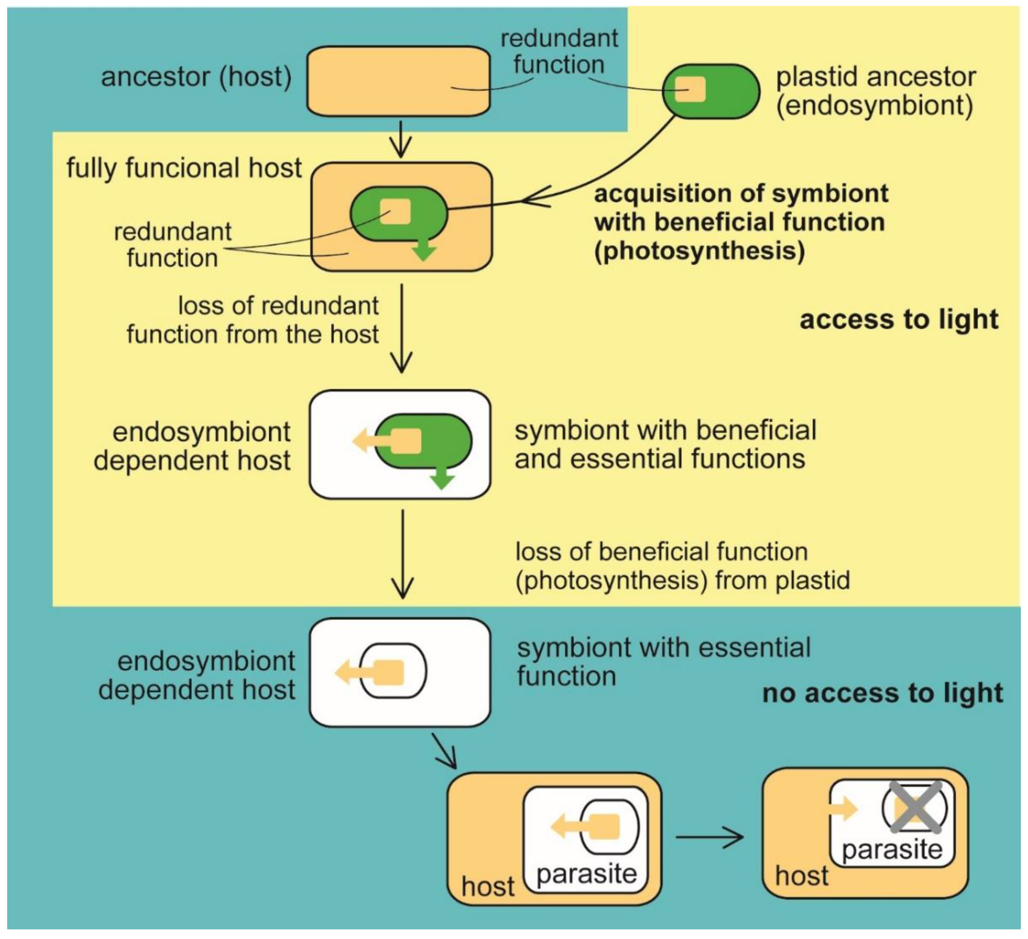
Figure 1.
Evolution of benefit and essential function in the plastid. The heterotrophic host acquired a photosynthetic endosymbiotic bacterium with the function (photosynthesis) beneficial for the host. The host cell lost the redundant function (e.g., synthesis of heme, fatty acids, and isoprenoids). At the same time, the delegation of the syntheses to the endosymbiont makes it essential for host survival (eukaryotic phototrophs, e.g., Archaeplastida and Paulinella sp., and algae with complex plastids such as Ochrophyta, Cryptophyta, Haptophyta, Dinophyta, Apicomplexa, Euglenopyhta, Chlorarachniophyta [1,2,3,11]). The endosymbiont retained its indispensability for the host even when it had lost photosynthesis, the original beneficial function (in nonphotosynthetic algae, e.g., Helicosporidium sp., Polytomella sp., Euglena longa, apicomplexan parasites, for example, Plasmodium falciparum, Toxoplasma gondii [1,2,3,7,11] Table 1). Switching to parasitism and scavenging the essential compounds from the host allows the complete loss of the plastid (apicomplexan parasite Cryptosporidium [42], parasitic dinoflagellate Hematodinium [40]).

Therefore, many phototrophs are, in fact, mixotrophs, which can still live heterotrophically. Such organisms may be prone to losing photosynthesis when getting to the nutrient-rich environment or finding a successful heterotrophic strategy, such as predation or parasitism. Moreover, many protists combine the phototrophic and heterotrophic lives to overcome a reduced availability of various compounds present in host or prey but rare in their environment, such as, for example, nitrogen, phosphorus, iron, and sulfur [11]. Therefore, photosynthesis was frequently lost from green parasitic algae [31,44], euglenophytes [46], apicomplexan parasites [11,31,47,48,49,50], and dinoflagellates [48]. Various secondarily heterotrophic strategies, including parasitism, have evolved repeatedly during the evolution of life among former phototrophs [11,38,39]. It is worth noting that such trophic switches are often found in the same taxonomic groups, such as, for example, in myozozans, group of alveolate protists, consisting of dinoflagellates, apicomplexan parasites, and apicomonads (chromerids and colpodellids). Apicomplexans and dinoflagellates are also the only known algae shown to lose their plastids completely. The plastid is absent from the apicomplexan parasitic genus Cryptosporidium [42] and gregarines [51], and the parasitic dinoflagellates of the genus Hematodinium [40]. The plastid losses have happened exclusively in parasites thanks to their ability to scavenge the essential compounds originally produced by plastids from the host (Table 2).
3. A Beneficial Function of the Mitochondrion
In contrast with plastids, the hypothetical benefit responsible for acquiring mitochondria is the subject of speculation. Frankly speaking, it is not even clear if the mitochondrion was indispensable for forming an early eukaryotic cell [12] or when it was acquired in the course of evolution (early versus late acquisition). The discovery of the eukaryote lacking a mitochondrion proved that the organelle is not essential for the eukaryotic cell as it exists now when the organism is a secondary anaerobic (Figure 2) [61]. Generally, mitochondria are great examples of reductive evolution. The diversity of this organelle involves mitochondria with large circular genomes (e.g., in jakobids [52,53,62]), mitochondria with highly reduced genomes (e.g., those in apicomplexans; [63]), mitochondria-related organelles (MRO), e.g., mitosomes and hydrogenosomes without genomes [60], and, in the end, completely lost mitochondrion (in oxymonads; Table 2 [61]). Consequently, various such organelles possess diverse molecular machinery: the complete respiratory chain of aerobic mitochondria or variously reduced respiratory chains lacking particular complexes: complex I (myzozans, e.g., apicomplexan parasites, and some fungi), complexes III and IV, while complexes I, II, and V retained [64], complexes I and III in chromerids [58,63] and the dinoflagellates of the genus Amoebophrya [59]), or complexes III, IV, and ATP synthase in (some) hydrogenosomes [60]. The missing complexes are substituted by alternative sources of electrons (e.g., alternative NADH dehydrogenases, L- and D- lactate cytochrome c oxidoreductases), or the electron transport chain was even completely lost (MRO).
Mitochondria host a wide range of metabolic functions, with oxidative phosphorylation being the most prominent (Figure 2), although it is found only in classical mitochondrial organelles, and lost from highly reduced MROs. In addition to that, mitochondria can be responsible for the metabolism of amino acids and nucleotides, steroid biosynthesis, heme synthesis, fatty acid catabolism, iron-cluster biogenesis, and many others [60,65,66,67,68]. Such metabolic complexity makes the search for the original beneficial function of the mitochondrion difficult. Various hypotheses have been formulated to explain the primary reason for acquiring a mitochondrion. It is further complicated by extensive genetic rearrangements of the organelle during organellogenesis because a substantial part of the mitochondrial proteome does not originate from the supposed mitochondrial ancestor [60].
The hydrogen hypothesis [69] proposed that the primary benefit of pre-mitochondrial symbiont was hydrogen production for the host, methanogenic Archaea. Some eukaryotes, such as Acanthamoeba castellanii (Amoebozoa), Brevimastigomonas motovehiculus (Rhizaria), Blastocystis spp. (Stramenopila), Nyctotherus ovalis (Alveolata) still contain hydrogen-producing mitochondria with complete (Acanthamoeba) or reduced respiratory chains [60,64,70]. Others host hydrogenosomes, organelles believed to represent modified hydrogen-producing mitochondria [13,60], which have lost the ATP generating part of the respiratory chain and retain just complex I (NADH hydrogenase), or complex II (succinate dehydrogenase) or both [56]. Martin et al. [13] also claim that the mitochondrial ancestor was a facultatively anaerobic bacterium.
Another possible beneficial function of a pre-mitochondrion was proposed by Thomas Cavalier-Smith [71], who speculated that the ancestor of mitochondrion was a photosynthetic purple bacterium, and the primary benefit was photoautotrophy, similar to plastids. This hypothesis assumes that both symbiont and host were facultative aerobes and that the host already has oxidative phosphorylation. A phototrophic symbiont would have an immediate intracellular synergy between a photosynthetic symbiont fixing CO2 and respiring and a phagotrophic host using oxygen and excreting CO2 [71].
Other hypotheses suppose that the primary benefit of the mitochondrion is related to dealing with free oxygen in the environment, preferring aerobic heterotrophic respiring bacterium as a mitochondrial ancestor [60]. Oxygen appeared in higher levels during the Great Oxidation Event between 2.4 and 2.1 Bya (billion years ago) [72]. If we take into account the possibility of early acquisition of mitochondrion, the oldest estimates of its appearance touch 2.1 Bya (1655–2094 Mya), while the youngest move around 1 Bya (943–1102 Mya) [73]. One of the earliest views on mitochondrial evolution supposes that the mitochondrion-free anaerobic eukaryotic ancestor acquired aerobic mitochondrion to detoxify oxygen accumulated in the environment after the Great Oxidation Event [74,75]. This scenario would even fit the timing of appearance of eukaryotes between 1 and 2 Bya. However, geochemical data indicate relatively low oxygen levels during the diversification of eukaryotes [72,76]. Moreover, Zimorski et al. [76] argued that those are metabolic processes in the mitochondrion, particularly electron transport chain generating reactive oxygen species, which may harm the cell and must be detoxicated. Therefore, oxidative phosphorylation can hardly be the primary benefit, at least in light of the oxygen detoxification hypothesis. However, we cannot ignore the fact that there is no eukaryote without classical mitochondrion with oxidative phosphorylation known that can permanently live in the presence of oxygen. The use of enzymes that can react with free oxygen does not allow obligate anaerobes to inhabit an oxygen environment. When looking at strictly anaerobic bacteria, they rely on low-potential flavoproteins used for anaerobic respiration. Exposure to oxygen likely causes superoxide and hydrogen peroxide production. They inactivate enzymes with these functional groups through the oxidation of dehydratase iron-sulfur clusters and sulphydryls. However, anaerobes utilize several classes of dioxygen-sensitive enzymes absent from aerobes, which maintain the redox balance during anaerobic fermentation. Their reaction mechanisms require exposure of the solvent of radicals or low-potential metal clusters to the oxygen that can react with it. Additionally, hydroxyl radicals damaging DNA and other biomolecules are generated because hydrogen peroxide oxidizes free iron [77]. Analogously, we can expect that oxygen could not be tolerated by anaerobes involved as the host cell in the early eukaryotic endosymbiotic events. A mitochondrial ancestor could eventually invade the host cell as an energy parasite. The proposed intracellularly parasitic ancestor of the mitochondrion was predicted to bear the ATP/ADP translocase importing ATP from the host. In such a case, there was no beneficial function behind a selection of an endosymbiont (parasite) in the event because the benefit was provided by the host cell [78].
4. Accumulation of Benefits
The origin of semiautonomous organelles cannot be seen in a static model. It is an evolutionary dynamic system in which an organelle is not in the cell forever; they can be replaced and lost, leaving leftovers in the genome and metabolism or can even disappear without a trace. Learning from the complex plastids, we see that the plastid replacing the old one partially uses the metabolic equipment of the former cell tenant [68,79]. This mechanism forms a highly evolutionary mosaic metabolic net, particularly in organisms passing through serial endosymbiotic events [7,80,81,82,83]. When applied to the origin of the mitochondrion (Figure 3), such hypothetical serial events can explain the evolutionary highly variable mitochondrial proteomes, where only a minor part of it fits, evolutionarily speaking, to the supposed mitochondrial ancestor [60].
Since every symbiont in the series could have a different beneficial function, it is rather challenging to specify the original benefit of current mitochondria. Even more, some of the symbionts (maybe most of them) may have been parasites [10], with no benefit for the host at the time of the invasion. There could also be theoretically more than a single symbiont present in the host cell at a time because multiple endosymbioses stimulate the evolution of what we see as a mutualistic partnership [6].
5. Benefit and Essential Function
Although the beneficial function such as photosynthesis is not necessarily essential for the host cell survival in general, it can be crucial in certain conditions or indispensable for life on a global scale. Thus, for example, an organism living in the absence of organic substances in the environment needs photosynthesis serving as the source of organic carbon and thus became essential for its survival. Although lithotrophic bacteria can also fix CO2, they likely play a minor role in the global carbon cycle [84]. On the global scale, photosynthesis is essential for life on Earth as we know it because it is the only way that the energy of sunlight can be transformed to the energy of chemical bond utilizable by all living organisms, including heterotrophs. There is no doubt about the fact that, without photosynthesis, life on Earth, if it could exist, would be present on an incomparably smaller scale than it is now. Therefore, the essentiality of the metabolic function always depends on the particular environmental conditions.
It is even possible that in eukaryotic organellogenesis, we face two fundamentally different endosymbiotic processes. The first type, presumably leading to the evolution of the mitochondrion, may not need the host cell capable of phagocytoses because it can be based on an active symbiont invasion into the host [6,74]. Since many (maybe most) symbiotic bacteria are or were parasitic in their evolutionary history [6,10], they were likely able to attack and penetrate the host cell and live as intracellular parasites. There was no benefit to the host in such a case, but the relationship was driven by an advantage to the symbiont. Such parasitic relationships could evolve to mutual dependence through the defense of the host cell against the parasite followed by organellogenesis. The second type represented by plastids is based on the phagocytic engulfment of a phototroph which is supposed to retain in the host (originally predatory) cell due to the benefit of photoautotrophy (Figure 4). It is even possible that the symbiotic relationship of a plastid was also not necessarily driven by the benefit of phototrophy but by the resistance of symbiont against digestion by the predatory host cell. Through the stage of kleptoplastidy, non-permanent symbionts, which are, in the end, digested by the host, evolved into permanent plastids [85]. They are, however, kind of “gradually digested” anyway by the host using primary metabolites exported from the symbiont; this process is analogous to the domestication of animals by humans, something like milking cows. Nevertheless, even the permanent plastids are in most eukaryotic phototrophs sometimes lyzed by the host cell, allowing mRNA release to the host cytosol, followed by reverse transcription and the endosymbiotic gene transfer to the host nucleus. Thus, the endosymbiotic gene transfer is an ongoing and never-ending process. Therefore, we can identify differences in the organellar gene repertoires even in groups with conserved plastid (plants) [86] or mitochondrial (animals) genomes [87].
Although endosymbiotic organelles are going through a reductive evolution, constructive evolution also plays a role in the formation of mitochondria and plastids. The protein composition of photosystems (PS) is highly reduced in complex plastids, with the highest level of reduction referred to in chromerids. Although protein subunits have been lost from their photosystems, new ones appeared. Thus, in the apicomonad alga Chromera velia, two superoxide dismutases are unprecedentedly permanently attached to the PSI. In addition, three novel subunits with no sequence homology in the databases and unknown functions were found in the PSI of C. velia [88].
While primary plastids of plants are pretty conserved in their structure and genomes [86], complex plastids in algae display quite diverse functions, and they also can be different in the ultrastructure. For example, while many amino acids s (e.g., glutamate/ glutamine, cysteine, lysine, branched-chain, and aromatic amino acids) are synthesized in the diatom plastid, similar to plants [89], chromerids locate their amino acid synthesis dominantly to the cytosol [90].
Mitochondria are somewhat evolutionarily frozen in their function and structure in some groups, such as, for example, animals. In protists, they are highly diverse, as shown in Table 2. Despite the dominant reductive evolution of mitochondria, we can also find, similar to plastids, constructively evolving characters in their genomes. For example, kinetoplastids (e.g., Trypanosoma and Leishmania) evolved an extremely complex mitochondrial RNA-editing system for the extensive posttranscriptional repair of mRNAs to the translatable form [91].
6. Organelle or Symbiotic Bacterium?
Two kinds of biological objects of endosymbiotic origins, mitochondria, and plastids, are currently classified as organelles. Despite their deep genetic, cellular, and metabolic integration into the host cell, they mostly still keep some aspects of individual biological entity: they encode part of the proteome in their own DNA, multiply by regular fission, and move inside the host cell. In my opinion, there is no strictly defined border between organelles and endosymbionts. It looks as though there is a continuum between endosymbionts and organelles, and the term “organelle” is just historically defined, with no solid biological relevance [6].
However, there have been many attempts to find the feature discriminating an organelle from a symbiont [4,6]. The revolutionary but widely accepted definition was proposed by Thomas Cavalier-Smith in 1985 [92]: the organelle exported, by endosymbiotic gene transfer (EGT), its genes to the host nucleus and imports their products from the host. The endosymbiotic gene transfer and nuclear-encoded proteins in the organellar metabolism seem to define organelles. Later, Archibald and Keeling expanded the definition of organelles by three aspects: genetic (EGT and targeting of proteins into the organelle), cellular (synchronization with the host’s cell cycle), and metabolic integration of endosymbiotic partners [4,93]. However, they clearly say that their criteria are soft and that the border between organelles and symbionts will “never be completely unambiguous.” Various symbiotic relationships are understandable as possible evolutionary intermediates from symbionts to organelles. Reyes-Prieto and colleagues [94] introduced the term “symbionelle” for symbionts of insects with highly reduced genomes and consequent metabolic integration to the host. Facultative plant-endosymbiotic nitrogen-fixing bacteria (Rhizobia) import peptides encoded in their plant’s symbiotic partners [95]. The aphid’s γ-proteobacterial symbiont Buchnera is enclosed in the host-derived membrane vesicles and imports nuclear-encoded proteins via the secretory pathway. However, no gene transfer from the endosymbiont to the insect cell nucleus has been reported [96]. Finally, it was shown by Husník and colleagues [97] that the mealybug’s endosymbiotic β-proteobacterium Tremblaya princeps hosting its own γ-proteobacterial endosymbiont Moranella endobia, is supported by 22 nuclear-encoded proteins originating from various bacteria, except for Tremblaya. Many of the above-mentioned symbiotic bacteria more or less meet the definition of organelles. However, some of them still keep a higher level of autonomy and do not integrate more with the host, likely because they would lose the benefit of possible transmission into new hosts or a new generation.
On the other hand, the photosynthetic dinoflagellates called “dinotoms” [68,98,99,100,101] host diatom tertiary endosymbionts responsible for their phototrophic lifestyle. These symbionts function as regular plastids; however, they are almost complete diatom cells containing the nucleus, mitochondrion, and the four membraned diatom chloroplasts. The symbiont is separated from the host cell just by a single membrane. Likely due to the single membrane bounding, the symbiont cannot import nuclear-encoded proteins, which may stand behind its unprecedented evolutionary conservation.
The diversity of endosymbiotic relationships in nature is marvelous. It comprises, for example, symbiotic assemblies from facultative nitrogen-fixing symbionts of diatoms and haptophytes [98,99], through nitrogen-fixing bacteria in plants, endosymbionts of insects, bacterial symbionts of various protists [94,95,96,97,98,99,100,101,102,103], and diverse forms of mitochondria [56], to cyanelles of Paulinella chromatophora [36,96], and primary and complex plastids of eukaryotic phototrophs and secondary heterotrophs [1,2,3,7,11,30].
7. Conclusions
It is believed that the evolution of endosymbiotic organelles is driven by a benefit to the endosymbiotic partners. While the benefit of photosynthesis seems to be obvious in the case of plastids, the original beneficial function of mitochondrion is the subject of discussion. The mosaic evolutionary origin of mitochondrial proteome opens a possibility of serial endosymbiotic events behind the current mitochondria, analogous to that of complex plastids. Plastids and mitochondria can represent two different types of endosymbiotic events: invasion of the alphaproteobacterial intracellular parasite into the pre-eukaryotic host and predation on the cyanobacterial ancestor of plastids. In the mitochondrial evolution, the host has adapted to the presence of the parasite, with a gradual transition of the parasitic symbiosis to a mutualism. Plastids evolved from cyanobacterial prey resistant to digestion by the host cell. In both these models, a benefit for the host was not a driving force for organellogenesis.
Funding
This research was funded by Czech Science Foundation, grant number 21-03224S and European Regional Development Fund ERDF/ESF, grant number CZ.02.1.01./0.0/0.0/16_019/0000759.
Acknowledgments
I thank the Institute of Parasitology BC CAS for administrative and technical support.
Conflicts of Interest
The authors declare no conflict of interest.
References
- Gould, S.B.; Waller, R.F.; McFadden, G.I. Plastid evolution. Annu. Rev. Plant. Biol. 2008, 59, 491–517. [Google Scholar] [CrossRef] [Green Version]
- Keeling, P.J. The number, speed, and impact of plastid endosymbioses in eukaryotic evolution. Ann. Rev. Plant. Biol. 2013, 64, 583–607. [Google Scholar] [CrossRef] [Green Version]
- Archibald, J.M. Endosymbiosis and eukaryotic cell evolution. Curr. Biol. 2015, 25, R911–R921. [Google Scholar] [CrossRef] [Green Version]
- Gruber, A. What’s a name? Why organelles of endosymbiotic origin are implicitly called by their eukaryotic species name and how they can be distinguished from endosymbionts. Microb. Cell 2019, 6, 123–133. [Google Scholar] [CrossRef]
- Pallen, M.J. Time to recognize that mitochondria are bacteria? Trends Microbiol. 2011, 19, 58–61. [Google Scholar] [CrossRef]
- Oborník, M. In the beginning was the word: How terminology drives our understanding of endosymbiotic organelles. Microb. Cell 2019, 6, 134–141. [Google Scholar] [CrossRef] [PubMed]
- Oborník, M. Endosymbiotic evolution of algae, secondary heterotrophy and parasitism. Biomolecules 2019, 9, 266. [Google Scholar] [CrossRef] [PubMed] [Green Version]
- Martin, W.; Rujan, T.; Richly, E.; Hansen, A.; Cornelsen, S.; Lins, T.; Leister, D.; Stoebe, B.; Hasegawa, M.; Penny, D. Evolutionary analysis of Arabidopsis, cyanobacterial, and chloroplast genomes reveals plastid phylogeny and thousands of cyanobacterial genes in the nucleus. Proc. Natl. Acad. Sci. USA 2002, 99, 12246–12251. [Google Scholar] [CrossRef] [PubMed] [Green Version]
- Timmis, J.N.; Ayliffe, M.A.; Huang, C.Y.; Martin, W. Endosymbiotic gene transfer: Organelle genomes forge eukaryotic chromosomes. Nat. Rev. Genet. 2004, 5, 123–135. [Google Scholar] [CrossRef]
- Keeling, P.J.; McCutcheon, J.P. Endosymbiosis: The feeling is not mutual. J. Theor. Biol. 2017, 434, 75–79. [Google Scholar] [CrossRef] [PubMed]
- Oborník, M. Photoparasitism as an intermediate state in the evolution of apicomplexan parasites. Trends Parasitol. 2020, 36, 727–734. [Google Scholar] [CrossRef]
- Hampl, V.; Čepička, I.; Eliáš, M. Was the mitochondrion necessary to start eukaryogenesis? Trends Microbiol. 2018, 27, 96–104. [Google Scholar] [CrossRef]
- Martin, W.F.; Garg, S.; Zimorski, V. Endosymbiotic theories for eukaryote origin. Philos. Trans. R. Soc. B 2015, 370, 20140330. [Google Scholar] [CrossRef] [PubMed] [Green Version]
- Muñoz-Gómez, S.A.; Wideman, J.G.; Roger, A.J.; Slamovits, C.H. The origin of mitochondrial cristae from alphaproteobacteria. Mol. Evol. Biol. 2017, 34, 943–956. [Google Scholar] [CrossRef] [PubMed]
- Raymond, J. The role of horizontal gene transfer in photosynthesis, oxygen production, and oxygen tolerance. Methods Mol. Biol. 2009, 532, 323–338. [Google Scholar]
- Koblížek, M.; Moulisová, V.; Muroňová, M.; Oborník, M. Horizontal transfers of two types of puf operons among phototrophic members of the Roseobacter clade. Folia Microbiol. 2015, 60, 37–43. [Google Scholar] [CrossRef]
- Fischer, W.W.; Hemp, J.; Johnson, J.E. Evolution of oxygenic photosynthesis. Annu. Rev. Earth Planet Sci. 2016, 44, 647–683. [Google Scholar] [CrossRef]
- Cavalier-Smith, T. Principles of protein and lipid targeting in secondary symbiogenesis: Euglenoid, dinoflagellate, and sporozoan plastid origins and the eukaryote family tree. J. Euk. Microbiol. 1999, 46, 347–366. [Google Scholar] [CrossRef]
- Cavalier-Smith, T. The phagotrophic origin of eukaryotes and phylogenetic classification of protozoa. Int. J. Syst. Evol. Microbiol. 2002, 52, 297–354. [Google Scholar] [CrossRef]
- Delaye, L.; Valadez-Cano, C.; Perez-Zamorano, B. How really ancient is Paulinella chromatophora? PLoS Curr. Tree Life 2016, 8. [Google Scholar] [CrossRef] [PubMed]
- Stephens, T.G.; Gabr, A.; Calatrava, V.; Grossman, A.R.; Bhattacharya, D. Why is primary endosymbiosis so rare? New Phytol. 2021, 231, 1693–1699. [Google Scholar] [CrossRef] [PubMed]
- Kies, L. Electron microscopical investigations on Paulinella chromatophora Lauterborn, a thecamoeba containing blue-green endosymbionts (cyanelles). Protoplasma 1974, 80, 69–89. [Google Scholar] [CrossRef] [PubMed]
- Kies, L.; Kremer, B.P. Function of cyanelles in the thecamoeba Paulinella chromatophora. Naturwissenschaften 1979, 66, 578–579. [Google Scholar] [CrossRef]
- Marin, B.; Nowack, E.C.; Melkonian, M. A plastid in the making: Evidence for a second primary endosymbiosis. Protist 2005, 156, 425–432. [Google Scholar] [CrossRef]
- Bodył, A.; Stiller, J.W.; Mackiewicz, P. Chromalveolate plastids: Direct sescent or multiple endosymbioses? Trend. Ecol. Evol. 2009, 24, 119–121. [Google Scholar] [CrossRef]
- Stiller, J.W.; Schreiber, J.; Yue, J.; Guo, H.; Ding, Q.; Huang, J. The evolution of photosynthesis in chromist algae through serial endosymbioses. Nat. Commun. 2014, 5, 5764. [Google Scholar] [CrossRef] [Green Version]
- Petersen, J.; Ludewig, A.K.; Michael, V.; Bunk, B.; Jarek, M.; Baurain, D.; Brinkman, H. Chromera velia, endosymbioses and the rhodoplex hypothesis—plastid evolution in cryptophytes, alveolates, stramenopiles, and haptophytes (CASH lineages). Genome Biol. Evol. 2014, 6, 666–684. [Google Scholar] [CrossRef] [Green Version]
- Dorrell, R.G.; Bowler, C. Secondary plastids of stramenopiles. Adv. Bot. Res. 2017, 84, 57–103. [Google Scholar]
- Strassert, J.F.H.; Irisarri, I.; Williams, T.A.; Burki, F. A molecular timescale of eukaryote evolution with implications for the origin of red algal-derived plastids. Nat. Commun. 2021, 12, 1879. [Google Scholar] [CrossRef]
- Oborník, M. The birth of red complex plastids: One, three, or four times? Trends Parasitol. 2018, 34, 923–925. [Google Scholar] [CrossRef] [PubMed]
- Hadariová, L.; Vesteg, M.; Hampl, V.; Krajčoviš, J. Reductive evolution of chloroplasts in non-photosynthetic plants, algae and protists. Curr. Genet. 2018, 64, 365–387. [Google Scholar] [CrossRef]
- Sato, S.; Nakamura, Y.; Kaneko, T.; Asamizu, E.; Tabata, S. Complete structure of the chloroplast genome of Arabidopsis thaliana. DNA Res. 1999, 29, 283–290. [Google Scholar] [CrossRef] [PubMed]
- Tajima, N.; Sato, S.; Maruyama, F.; Kurokawa, K.; Ohta, H.; Tabata, S.; Sekine, K.; Moriyama, T.; Sato, N. Analysis of the complete plastid genome of the unicellular red alga Porphyridium purpureum. J. Plant Res. 2014, 127, 389–397. [Google Scholar] [CrossRef] [PubMed]
- De Koning, A.P.; Keeling, P.J. The complete plastid genome sequence of the parasitic green alga Helicosporidium sp. is highly reduced and structured. BMC Biol. 2006, 4, 12. [Google Scholar] [CrossRef] [Green Version]
- Smith, D.R.; Lee, R.W. A Plastid without a Genome: Evidence from the Nonphotosynthetic Green Algal Genus Polytomella. Plant Physiol. 2014, 164, 1812–1819. [Google Scholar] [CrossRef] [PubMed] [Green Version]
- Nowack, E.C.M.; Melkonian, M.; Glockner, G. Chromatophore genome sequence of Paulinella sheds light on acquisition of photosynthesis by eukaryotes. Curr. Biol. 2008, 18, 410–418. [Google Scholar] [CrossRef] [Green Version]
- Hallick, R.B.; Hong, L.; Drager, R.G.; Favreau, M.R.; Monfort, A.; Orsat, B.; Spielmann, A.; Stutz, E. Complete sequence of Euglena gracilis chloroplast DNA. Nucleic Acids Res. 1993, 21, 3537–3544. [Google Scholar] [CrossRef] [PubMed] [Green Version]
- Gockel, G.; Hachtel, W. Complete gene map of the plastid genome of the nonphotosynthetic euglenoid flagellate Astasia longa. Protist 2000, 151, 347–351. [Google Scholar] [CrossRef]
- Zhang, Z.; Green, B.R.; Cavalier-Smith, T. Single gene circles in dinoflagellate chloroplast genomes. Nature 1999, 400, 155–159. [Google Scholar] [CrossRef]
- Gornik, S.G.; Cassin, A.M.; MacRae, J.I.; Ramprasad, A.; Rchiad, Z.; McConville, M.J.; Bacic, A.; McFadden, G.I.; Pain, A.; Waller, R.F. Endosymbiosis undone by stepwise elimination of the plastid in a parasitic dinoflagellate. Proc. Natl. Acad. Sci. USA 2015, 112, 5767–5772. [Google Scholar] [CrossRef] [Green Version]
- Oudot-Le Secq, M.-P.; Grimwood, J.; Shapiro, H.; Armbrust, E.V.; Bowler, C.; Green, B.R. Chloroplast genomes of the diatoms Phaeodactylum tricornutum and Thalassiosira pseudonana: Comparison with other plastid genomes of the red lineage. Mol. Genet. Genom. 2007, 277, 427–439. [Google Scholar] [CrossRef] [PubMed]
- Janouškovec, J.; Horák, A.; Oborník, M.; Lukeš, J.; Keeling, P.J. A common red algal origin of the apicomplexan, dinoflagellate, and heterokont plastids. Proc. Natl. Acad. Sci. USA 2010, 107, 10949–10950. [Google Scholar] [CrossRef] [Green Version]
- Zhu, G.; Marchewka, M.J.; Keithly, J.S. Cryptosoridium parvum appears to lack a plastid genome. Microbiology 2000, 146, 315–321. [Google Scholar] [CrossRef] [PubMed] [Green Version]
- Suzuki, S.; Endoh, R.; Manabe, R.; Ohkuma, M.; Hirakawa, Y. Multiple losses of photosynthesis and convergent reductive genome evolution in the colourless green algae Prototheca. Sci. Rep. 2018, 8, 940. [Google Scholar] [CrossRef] [Green Version]
- Khorobrykh, S.; Havurinne, V.; Mattila, H.; Tyystjärvi, E. Oxygen and ROSS in photosynthesis. Plants 2020, 9, 91. [Google Scholar] [CrossRef] [Green Version]
- Füssy, Z.; Záhonová, K.; Tomčala, A.; Krajčovič, J.; Yurchenko, V.; Oborník, M.; Eliáš, M. The cryptic plastid of Euglena longa defines a new type of nonphotosynthetic plastid organelle. mSphere 2020, 5, e00675-20. [Google Scholar] [CrossRef]
- Salomaki, E.D.; Kolisko, M. There is treasure everywhere: Reductive plastid evolution in Apicomplexa in light of their close relatives. Biomolecules 2019, 9, 378. [Google Scholar] [CrossRef] [Green Version]
- Waller, R.F.; Kořený, L. Plastid complexity in dinoflagellates: A picture of gains, losses, replacements and revisions. Adv. Bot. Res. 2017, 84, 105–143. [Google Scholar]
- Mathur, V.; Kolísko, M.; Hehenberger, E.; Irwin, N.A.T.; Leander, B.S.; Kristmundsson, Á.; Freeman, M.A.; Keeling, P.J. Multiple independent origins of apicomplexan-like parasites. Curr. Biol. 2019, 29, 1–6. [Google Scholar] [CrossRef]
- Janouškovec, J.; Paskerova, G.G.; Miroliubova, T.S.; Mikhailov, K.V.; Birley, T.; Aleoshin, V.V.; Simdyanov, T.G. Apicomplexan-like parasites are polyphyletic and widely but selectively dependent on cryptic plastid organelles. eLife 2019, 8, e49662. [Google Scholar] [CrossRef] [PubMed]
- Toso, M.A.; Omoto, C.K. Gregarina niphandrodes may lack both a plastid genome and organelle. J. Eukaryot. Microbiol. 2007, 54, 66–72. [Google Scholar] [CrossRef] [Green Version]
- Gray, W.; Burger, G.; Derelle, R.; Klimeš, V.; Leger, M.M.; Sarrasin, M.; Vlček, Č.; Roger, A.J.; Eliáš, M.; Lang, B.F. The draft nuclear genome sequence and predicted mitochondrial proteome of Andalucia godoi, a protist with the most gene-rich and bacteria-like mitochondrial genome. BMC Biol. 2020, 18, 22. [Google Scholar] [CrossRef]
- Lang, B.F.; Burger, G.; O’Kelly, C.J.; Cedergren, R.; Golding, G.B.; Lemieux, C.; Sankoff, D.; Turmel, M.; Gray, M.W. An ancestral mitochondrial DNA resembling a eubacterial genome in miniature. Nature 1997, 387, 493–497. [Google Scholar] [CrossRef]
- Anderson, S.; Bankier, A.T.; Barrell, B.G.; de Bruijn, M.H.L.; Coulson, A.R.; Drouin, J.; Eperon, I.C.; Nierlich, D.P.; Roe, B.A.; Sanger, F.; et al. Sequence and organization of the human mitochondrial genome. Nature 1981, 290, 457–465. [Google Scholar] [CrossRef]
- Dong, S.; Zhao, C.; Chen, F.; Liu, Y.; Zhang, S.; Wu, H.; Zhang, L.; Liu, Y. The complete mitochondrial genome of the early flowering plant Nymphaea colorata is highly repetitive with low recombination. BMC Genom. 2018, 19, 614. [Google Scholar] [CrossRef] [PubMed]
- De Graaf, R.M.; Ricard, G.; van Alen, T.A.; Duarte, I.; Dutilh, B.E.; Burgtorf, C.; Kuiper, J.W.P.; van der Staay, G.W.M.; Tielens, A.G.M.; Huynen, M.A.; et al. The Organellar Genome and Metabolic Potential of the Hydrogen-Producing Mitochondrion of Nyctotherus ovalis. Mol. Biol. Evol. 2011, 28, 2379–2391. [Google Scholar] [CrossRef] [PubMed] [Green Version]
- Gardner, M.J.; Bates, P.A.; Ling, I.T.; Moore, D.J.; McCready, S.; Gunasekera, M.B.R.; Wilson, R.J.M.; Williamson, D.H. Mitochondrial DNA of the human malarial parasite Plasmodium falciparum. Mol. Biochem. Prasitol. 1988, 31, 11–17. [Google Scholar] [CrossRef]
- Flegontov, P.; Michálek, J.; Janouškovec, J.; Lai, D.H.; Jirků, M.; Hajdušková, E.; Tomčala, A.; Otto, D.T.; Keeling, P.J.; Pain, A.; et al. Divergent mitochondrial respiratory chains in phototrophic relatives of apicomplexan parasites. Mol. Biol. Evol. 2015, 32, 1115–1131. [Google Scholar] [CrossRef]
- John, U.; Lu, Y.; Wohlrab, S.; Groth, M.; Janouškovec, J.; Kohli, G.S.; Mark, F.C.; Bickmeyer, U.; Frahat, S.; Felder, M.; et al. A aerobic eukaryotic parasite with functional mitochondria that likely lacks a mitochondrial genome. Sci. Adv. 2019, 5, 1110. [Google Scholar] [CrossRef] [PubMed] [Green Version]
- Roger, A.J.; Muñoz-Gómez, S.A.; Kamikawa, R. The origin and diversification of mitochondria. Curr. Biol. 2017, 27, R1177–R1192. [Google Scholar] [CrossRef] [Green Version]
- Karnkowska, A.; Vacek, V.; Zubacova, Z.; Treitli, S.C.; Petrzelková, R.; Eme, L.; Novaqk, L.; Zarsky, V.; Barlow, L.D.; Herman, E.K. A eukaryote without a mitochondrial organelle. Curr. Biol. 2016, 26, 1274–1284. [Google Scholar] [CrossRef]
- Burger, G.; Gray, M.W.; Forget, L.; Lang, B.F. Strikingly bacteria-like and gene-rich mitochondrial genomes throughout jakobid protists. Genom. Biol. Evol. 2013, 5, 418–438. [Google Scholar] [CrossRef] [Green Version]
- Oborník, M.; Lukeš, J. The organellar genomes of Chromera and Vitrella, the phototrophic relatives of apicomplexan parasites. Ann. Rev. Microbiol. 2015, 69, 129–144. [Google Scholar] [CrossRef]
- Müller, M.; Mentel, M.; van Hellemond, J.J.; Henze, K.; Woehle, C.; Gould, S.B.; Yu, R.Y.; van der Giezen, M.; Tielens, A.G.; Martin, W.F. Biochemistry and evolution of anaerobic energy metabolism in eukaryotes. Microbiol. Mol. Biol. Rev. 2012, 76, 444–495. [Google Scholar] [CrossRef] [Green Version]
- Calvo, S.E.; Mootha, V.K. The mitochondrial proteome and human disease. Annu. Rev. Genom. Hum. Genet. 2010, 11, 25–44. [Google Scholar] [CrossRef] [Green Version]
- Panigrahi, A.K.; Ogata, Y.; Zíková, A.; Anupama, A.; Dalley, R.A.; Acestor, N.; Myler, P.J.; Stuart, K.D. A comprehensive analysis of Trypanosoma brucei mitochondrial proteome. Proteomics 2009, 9, 434–450. [Google Scholar] [CrossRef] [Green Version]
- Gawryluk, R.M.R.; Chisholm, K.A.; Pinto, D.M.; Gray, M.W. Compositional complexity of the mitochondrial proteome of a unicellular eukaryote (Acanthamoeba castellanii, supergroup Amoebozoa) rivals that of animals, fungi, and plants. J. Proteom. 2014, 109, 400–416. [Google Scholar] [CrossRef] [PubMed]
- Cihlář, J.; Füssy, Z.; Horák, A.; Oborník, M. Evolution of the tetrapyrole biosynthetic pathway in secondary algae: Conservation, redundancy and replacement. PLoS ONE 2016, 11, e0166338. [Google Scholar] [CrossRef]
- Martin, W.; Müller, M. The hydrogen hypothesis for the first eukaryote. Nature 1998, 392, 37–41. [Google Scholar] [CrossRef] [PubMed]
- Boxma, B.; de Graaf, R.; van der Staay, G.; van Alen, T.A.; Ricard, G.; Gabaldón, T.; van Hoek, A.H.A.M.; van der Staay, S.Y.M.; Koopman, W.J.H.; van Hellemond, J.J.; et al. An anaerobic mitochondrion that produces hydrogen. Nature 2005, 434, 74–79. [Google Scholar] [CrossRef] [PubMed] [Green Version]
- Cavalier-Smith, T. Origin of mitochondria by intracellular enslavement of a photosynthetic purple bacterium. Proc. Biol. Sci. 2006, 273, 1943–1952. [Google Scholar] [CrossRef] [PubMed] [Green Version]
- Lyons, T.W.; Reinhard, C.T.; Planavsky, N.J. The rise of oxygen in Earth’s early ocean and atmosphere. Nature 2014, 506, 307–315. [Google Scholar] [CrossRef]
- Eme, L.; Sharpe, S.C.; Brown, M.W.; Roger, A.J. On the age of eukaryotes: Evaluating evidence from fossils and molecular clocks. Cold Spring Harb. Perspect. Biol. 2014, 6, a016139. [Google Scholar] [CrossRef] [PubMed] [Green Version]
- Vellai, T.; Takacs, K.; Vida, G. A new aspect to the origin and evolution of eukaryotes. J. Mol. Evol. 1998, 46, 499–507. [Google Scholar] [CrossRef] [PubMed]
- Kurland, C.G.; Andersson, S.G. Origin and evolution of the mitochondrial proteome. Microbiol. Mol. Biol. Rev. 2000, 64, 786–820. [Google Scholar] [CrossRef] [PubMed] [Green Version]
- Zimorski, V.; Mentel, M.; Tielens, A.G.M.; Martin, W.F. Energy metabolism in nanerobic eukaryotes and Earth’s late oxygenation. Free Radic. Biol. Med. 2019, 140, 279–294. [Google Scholar] [CrossRef] [PubMed]
- Imlay, J.A. How oxygen damages microbes: Oxygen tolerance and obligate anaerobiosis. Adv. Microb. Physiol. 2002, 46, 111–153. [Google Scholar]
- Wang, Z.; Wu, M. Phylogenomic reconstruction indicates mitochondrial ancestor was an energy parasite. PLoS ONE 2014, 9, e110685. [Google Scholar] [CrossRef]
- Larkum, A.W.D.; Lockhart, P.J.; Howe, C.J. Shopping for plastid. Trends Plant. Sci. 2007, 12, 189–195. [Google Scholar] [CrossRef]
- Sagan, L. On the origin of mitosing cells. J. Theor. Biol. 1967, 14, 225–274. [Google Scholar] [CrossRef]
- Goksøyr, J. Evolution of eukaryotic cells. Nature 1967, 214, 1161. [Google Scholar] [CrossRef] [PubMed]
- Margulis, L. Symbiosis in evolution. Sci. Am. 1971, 225, 48–57. [Google Scholar] [CrossRef]
- Lane, N. Serial endosymbiosis or singular event at the origin of eucaryotes? J. Theor. Biol. 2017, 434, 58–67. [Google Scholar] [CrossRef] [PubMed] [Green Version]
- Kelly, D.P. Autotrophy: Concepts of lithotrophic bacteria and their organic metabolism. Annu. Rev. Microbiol. 1971, 25, 177–210. [Google Scholar] [CrossRef]
- Bodył, A. Did some red alga-derived plastids evolved via kleptoplastidy? A hypothesis. Biol. Rev. 2018, 93, 201–222. [Google Scholar] [CrossRef]
- Logacheva, M.D.; Schelkunov, M.I.; Shtratnikova, V.Y.; Matveeva, M.V.; Penin, A.A. Comparative analysis of plastid genomes of non-photosynthetic Ericaceae and their photosynthetic relatives. Sci. Rep. 2016, 6, 30042. [Google Scholar] [CrossRef] [PubMed]
- Lavrov, D.V.; Pett, W. Animal Mitochondrial DNA as We Do Not Know It: Mt-Genome Organization and Evolution in Nonbilaterian Lineages. Genom. Biol. Evol. 2016, 8, 2896–2913. [Google Scholar] [CrossRef]
- Sobotka, R.; Esson, H.J.; Koník, P.; Trsková, E.; Moravcová, L.; Horák, A.; Dufková, P.; Oborník, M. Extensive gain and loss of photosystem I subunits in chromerid algae, photosynthetic relatives of apicomplexans. Sci. Rep. 2017, 7, 13214. [Google Scholar] [CrossRef] [PubMed] [Green Version]
- Nonoyama, T.; Kazamia, E.; Nawaly, H.; Gao, X.; Tsuji, Y.; Matsuda, Y.; Bowler, C.; Tanaka, T.G.; Dorrell, R. Metabolic Innovations Underpinning the Origin and Diversification of the Diatom hloroplast. Biomolecules 2019, 9, 322. [Google Scholar] [CrossRef]
- Füssy, Z.; Faitová, T.; Oborník, M. Subcellular Compartments Interplay for Carbon and Nitrogen Allocation in Chromera velia and Vitrella brassicaformis. Genom. Biol. Evol. 2019, 11, 1765–1779. [Google Scholar] [CrossRef] [Green Version]
- Gray, M.W.; Lukeš, J.; Archibald, J.M.; Keeling, P.J.; Doolittle, W.F. Irremediable complexity? Science 2000, 330, 920–921. [Google Scholar] [CrossRef] [PubMed]
- Cavalier-Smith, T.; Lee, J.J. Protozoa as Hosts for Endosymbioses and the Conversion of Symbionts into Organelles. J. Protozool. 1985, 32, 376–379. [Google Scholar] [CrossRef]
- Keeling, P.J.; Archibald, J.M. Organelle Evolution: What’s in a Name? Curr. Biol. 2008, 18, R345–R347. [Google Scholar] [CrossRef] [PubMed] [Green Version]
- Reyes-Prieto, M.; Latorre, A.; Moya, A. Scanty microbes, the ‘symbionelle’ concept. Environ. Microbiol. 2014, 16, 335–338. [Google Scholar] [CrossRef]
- van de Velde, W.; Zehirov, G.; Szatmari, A.; Debreczeny, M.; Ishihara, H.; Kevei, Z.; Farkas, A.; Mikulass, K.; Nagy, A.; Tiricz, H.; et al. Plant peptides govern terminal differentiation of bacteria in symbiosis. Science 2010, 327, 1122–1126. [Google Scholar] [CrossRef]
- Nowack, E.C.M. Paulinella chromatophora – rethinking the transition from endosymbiont to organelle. Acta Soc. Bot. Pol. 2014, 83, 387–397. [Google Scholar] [CrossRef] [Green Version]
- Husník, F.; Tashyreva, D.; Boscaro, V.; George, E.E.; Lukeš, J.; Keeling, P.J. Bacterial and archaeal symbiosis with prostists. Curr. Biol. 2021, 31, R1–R16. [Google Scholar] [CrossRef]
- Thompson, A.W.; Foster, R.A.; Krupke, A.; Carter, B.J.; Musat, N.; Vaulot, D.; Kuypers, M.M.M.; Zehr, J.P. Unicellular cyanobacterium symbiotic with a single-celled eukaryotic alga. Science 2012, 337, 1546–1550. [Google Scholar] [CrossRef]
- Nakayama, T.; Kamikawa, R.; Tanifuji, G.; Kashiyama, Y.; Ohkouchi, N.; Archibald, J.M.; Inagaki, Y. Complete genome of a nonphotosynthetic cyanobacterium in a diatom reveals recent adaptations to an intracellular lifestyle. Proc. Natl. Acad. Sci. USA 2014, 111, 11407–11412. [Google Scholar] [CrossRef] [Green Version]
- Nakabachi, A.; Ishida, K.; Hongoh, Y.; Ohkuma, M.; Miyagishima, S.Y. Aphid gene of bacterial origin encodes a protein transported to an obligate endosymbiont. Curr. Biol. 2014, 24, R640–R641. [Google Scholar] [CrossRef] [PubMed] [Green Version]
- Tomas, R.N.; Cox, E.R. Observations on symbiosis of Peridinium Balticum and its intracellular alga.1. Ultrastructure. J. Phycol. 1973, 9, 304–323. [Google Scholar]
- Jeffrey, S.W.; Vesk, M. Further evidence for a membrane bound endosymbiont within the dinoflagellate Peridinium foliaceum. J. Phycol. 1976, 12, 450–455. [Google Scholar]
- Imanian, B.; Pombert, J.-F.; Dorrell, R.G.; Burki, F.; Keeling, P.J. Tertiary Endosymbiosis in Two Dinotoms Has Generated Little Change in the Mitochondrial Genomes of Their Dinoflagellate Hosts and Diatom Endosymbionts. PLoS ONE 2012, 7, e43763. [Google Scholar] [CrossRef] [PubMed]
Figure 2.
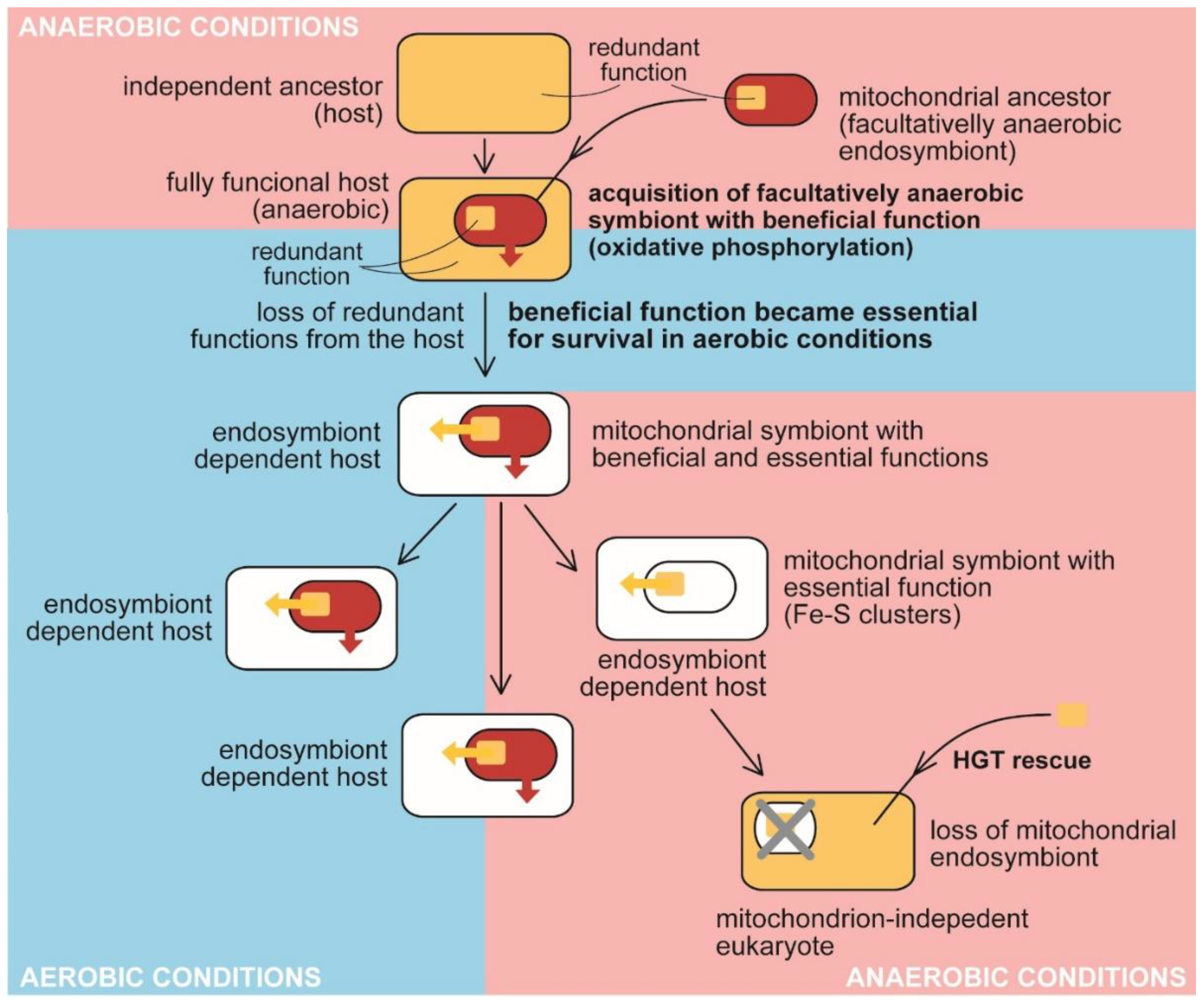
The traditional view on the evolution of benefit and essential function in the mitochondrion. Anaerobic host acquired a facultatively anaerobic endosymbiotic bacterium with the function beneficial for the host, presumably detoxifying oxygen. It became essential for the host in aerobic conditions. The redundant function was lost from the host (e.g., synthesis and assembly of Fe-S clusters). At the same time, the delegation of the synthesis to the endosymbiont makes it essential for host survival. The endosymbiont retained its indispensability for the host even when it had lost the original beneficial function by adaptating to anaerobic conditions. The acquisition of bacterial Fe-S clusters synthesis and assembly in the cytosol of oxymonads through HGT allowed the loss of the mitochondrion (MRO) [61].
Figure 2.
The traditional view on the evolution of benefit and essential function in the mitochondrion. Anaerobic host acquired a facultatively anaerobic endosymbiotic bacterium with the function beneficial for the host, presumably detoxifying oxygen. It became essential for the host in aerobic conditions. The redundant function was lost from the host (e.g., synthesis and assembly of Fe-S clusters). At the same time, the delegation of the synthesis to the endosymbiont makes it essential for host survival. The endosymbiont retained its indispensability for the host even when it had lost the original beneficial function by adaptating to anaerobic conditions. The acquisition of bacterial Fe-S clusters synthesis and assembly in the cytosol of oxymonads through HGT allowed the loss of the mitochondrion (MRO) [61].

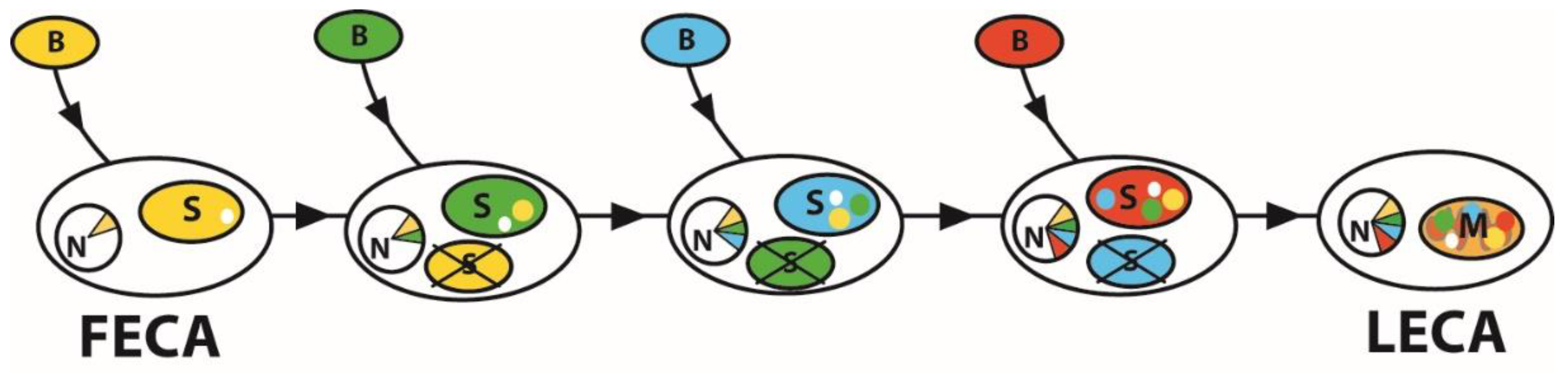
Figure 3.
Accumulation of benefits by serial primary endosymbiosis. Each endosymbiont in the evolutionary history from the First Eukaryotic Common Ancestor (FECA) to the Last Eukaryotic Common Ancestor (LECA) impacted the eukaryotic host genome and the proteome of an organelle. The transferred genes (EGT) are supposed to be expressed in the cytosol, and their products are targeted to the last survival, the mitochondrion. It led to the highly chimeric proteomes of the current mitochondria. Partners in the process are bacteria (B), transformed into symbionts (S). The symbiont genes migrate to the host nucleus (N). Such serial events could theoretically lead to the appearance of a mitochondrion (M) with a mosaic proteome.

Figure 4.
Two possible types of endosymbiotic events. In the first type, the endosymbiont (a–c) is a parasitic bacterium, which actively invades the host cell (a). The defense of the host ((b), blue arrows) against parasitic endosymbiont (red arrows) transformed the parasitic relationship to mutualism (c). Endosymbiotic gene transfer, together with gradual losses of redundant pathways, led to the mutual dependence of the host and symbiont. The second type of event (d–f) is based on hunting. The cyanobacterial prey is engulfed by phagocytosis (d). The bacterium resists (blue arrows) digestion (red arrows) (e). The photosynthetic endosymbiont provides primary metabolites (M) to the host cell in a mutualistic relationship. This type of organellogenesis resembles domestication. In both cases, the endosymbiont provides (non-essential) benefit to the host.


Table 1.
Selected plastids and their characteristics in various eukaryotes. It demonstrates the reductive evolution of plastids in eukaryotes.
Table 1.
Selected plastids and their characteristics in various eukaryotes. It demonstrates the reductive evolution of plastids in eukaryotes.
| Organism | Supergroup | Type of the Plastid | Genes (Cds) | Genome | Reference |
|---|---|---|---|---|---|
| Arabidopsis thaliana | Archaeplastida | primary | 85 | circular | [32] |
| Porphiridium purpureum | Archaeplastida | primary | 224 | circular | [33] |
| Helicosporidium sp. | Archaeplastida | primary | 26 | circular | [34] |
| Polytomella sp. | Archaeplastida | primary | 0 | circular | [35] |
| Paulinella chromatohpora | Cercozoa (SAR) | primary (cyanelle) | 867 | circular | [36] |
| Euglena gracilis | Eugenophyta | complex (secondary) | 67 | circular | [37] |
| Euglena longa | Eugenophyta | complex (secondary) | 46 | circular | [38] |
| Heterocapsa triquetra | Dinophyta (SAR) | complex | 14 | Circular (minicircles) | [39] |
| Hematodinium sp. | Dinophyta (SAR) | - | - | - | [40] |
| Thalassiosira pseudonana | Bacillariophyta (SAR) | complex | 141 | circular | [41] |
| Chromera velia | Apicomonada (SAR) | complex | 78 | linear | [42] |
| Vitrella brassicaformis | Apicomonada (SAR) | complex | 94 | circular | [42] |
| Toxoplasma gondii | Sporozoa (SAR) | complex | 29 | circular | NCBI |
| Cryptosoridium muris | Sporozoa (SAR) | - | - | - | [43] |
Table 2.
Examples of mitochondria and MROs and their characteristics in various eukaryotes. It demonstrates the reductive evolution of mitochondria, from mitochondrial organelles with large genomes to hydrogenosomes and mitosomes lacking genome and eukaryotic cells without mitochondrion.
Table 2.
Examples of mitochondria and MROs and their characteristics in various eukaryotes. It demonstrates the reductive evolution of mitochondria, from mitochondrial organelles with large genomes to hydrogenosomes and mitosomes lacking genome and eukaryotic cells without mitochondrion.
| Species | Taxonomy | Type of Mitochondrion | Genes (Cds) | Genome | Reference |
|---|---|---|---|---|---|
| Andalucia godoyi | Jakobida | Aerobic/OXPHOS | 67 | circular | [52] |
| Reclinomonas americana | Jakobida | Aerobic/OXPHOS | 66 | circular | [53] |
| Homo sapiens | Metazoa (Obazoa) | Aerobic/OXPHOS | 13 | circular | [54] |
| Nymphaea colorata | Archaeplastida | Aerobic/OXPHOS | 42 | circular | [55] |
| Nyctotherus ovalis | Ciliophora (SAR) | Anaerobic/H-producing | 16 | linear | [56] |
| Plasmodium falciparum | Sporozoa (SAR) | Aerobic/OXPHOS | 3 | linear | [57] |
| Chromera velia | Apicomonada (SAR) | Aerobic/OXPHOS | 2 | linear | [58] |
| Amebophrya ceratii | Dinophyta (SAR) | Aerobic/OXPHOS | 0 | - | [59] |
| Neocallimastix sp. | Chytridiomycota (Obazoa) | Hydrogenosome | - | - | [60] |
| Giardia intestinalis | Metamonada | Mitosome (Fe-S clusters) | - | - | [60] |
| Monocercomonoides sp. | Oxymonadida | - | - | - | [61] |
Publisher’s Note: MDPI stays neutral with regard to jurisdictional claims in published maps and institutional affiliations. |
© 2022 by the author. Licensee MDPI, Basel, Switzerland. This article is an open access article distributed under the terms and conditions of the Creative Commons Attribution (CC BY) license (https://creativecommons.org/licenses/by/4.0/).
Share and Cite
MDPI and ACS Style
Oborník, M. Organellar Evolution: A Path from Benefit to Dependence. Microorganisms 2022, 10, 122. https://doi.org/10.3390/microorganisms10010122
AMA Style
Oborník M. Organellar Evolution: A Path from Benefit to Dependence. Microorganisms. 2022; 10(1):122. https://doi.org/10.3390/microorganisms10010122
Chicago/Turabian StyleOborník, Miroslav. 2022. "Organellar Evolution: A Path from Benefit to Dependence" Microorganisms 10, no. 1: 122. https://doi.org/10.3390/microorganisms10010122
Note that from the first issue of 2016, this journal uses article numbers instead of page numbers. See further details here.