Review

Development in the Mammalian Auditory System Depends on Transcription Factors

by
Karen L. Elliott
1,
Gabriela Pavlínková
2,
Victor V. Chizhikov
3,
Ebenezer N. Yamoah
4 and
Bernd Fritzsch
1,*
1
Department of Biology, University of Iowa, Iowa City, IA 52242, USA
2
Institute of Biotechnology of the Czech Academy of Sciences, 25250 Vestec, Czechia
3
Department of Anatomy and Neurobiology, The University of Tennessee Health Science Center, Memphis, TN 38163, USA
4
Department of Physiology and Cell Biology, School of Medicine, University of Nevada, Reno, NV 89557, USA
*
Author to whom correspondence should be addressed.
Int. J. Mol. Sci. 2021, 22(8), 4189; https://doi.org/10.3390/ijms22084189
Submission received: 13 February 2021 / Revised: 15 April 2021 / Accepted: 16 April 2021 / Published: 18 April 2021
(This article belongs to the Special Issue Neurons of the Auditory Pathways)

Abstract

:
We review the molecular basis of several transcription factors (Eya1, Sox2), including the three related genes coding basic helix–loop–helix (bHLH; see abbreviations) proteins (Neurog1, Neurod1, Atoh1) during the development of spiral ganglia, cochlear nuclei, and cochlear hair cells. Neuronal development requires Neurog1, followed by its downstream target Neurod1, to cross-regulate Atoh1 expression. In contrast, hair cells and cochlear nuclei critically depend on Atoh1 and require Neurod1 expression for interactions with Atoh1. Upregulation of Atoh1 following Neurod1 loss changes some vestibular neurons’ fate into “hair cells”, highlighting the significant interplay between the bHLH genes. Further work showed that replacing Atoh1 by Neurog1 rescues some hair cells from complete absence observed in Atoh1 null mutants, suggesting that bHLH genes can partially replace one another. The inhibition of Atoh1 by Neurod1 is essential for proper neuronal cell fate, and in the absence of Neurod1, Atoh1 is upregulated, resulting in the formation of “intraganglionic” HCs. Additional genes, such as Eya1/Six1, Sox2, Pax2, Gata3, Fgfr2b, Foxg1, and Lmx1a/b, play a role in the auditory system. Finally, both Lmx1a and Lmx1b genes are essential for the cochlear organ of Corti, spiral ganglion neuron, and cochlear nuclei formation. We integrate the mammalian auditory system development to provide comprehensive insights beyond the limited perception driven by singular investigations of cochlear neurons, cochlear hair cells, and cochlear nuclei. A detailed analysis of gene expression is needed to understand better how upstream regulators facilitate gene interactions and mammalian auditory system development.

1. Introduction

The mammalian auditory system evolved in tetrapods out of the vestibular system [1,2] that diversifies into distinct hair cells with a unique innervation of spiral ganglion neurons (SGNs) which reach the cochlear nuclei for information processing [3,4]. The development of the auditory system depends on sequential activation of various genes. Once early development is completed, age-dependent loss progresses to reduce auditory hair cells that cannot be replaced [5,6]. Loss of hair cells may result in a delayed loss of SGNs with age [7,8], which will affect the cochlear nuclei [9]. We aim to provide an overview of transcription factors to allow a framework to develop the auditory system’s earliest steps, from neurons to hair cells and cochlear nuclei.
The mammalian auditory system consists of SGNs, two types of cochlear hair cells (inner and outer hair cells; IHC, OHC), and the cochlear nuclei subdivided into three nuclei: the anteroventral, posteroventral, and dorsal cochlear nucleus. Insights into these interlinked neurosensory components will be detailed beyond the initial morphogenetic early steps [9,10]. The proliferation pattern within each auditory system component affects the temporal and spatial differentiation and interconnects with various neurosensory cells. SGNs are the first cells of the auditory system to exit the cell cycle, progressing from base-to-apex from ~E10.5–12.5, followed by cochlear HCs, which exit the cell cycle opposite direction, from apex-to-base between ~E12.5–14.5 [11,12]. The temporal pattern of proliferation progression in the cochlea raises questions: why do developing neurons proliferate from the apex-to-base, and the cochlear HCs proliferate from base-to-apex (Figure 1)? The cochlear nuclei exit the cell cycle between E10.5–14.5 [13], partially overlapping with spiral ganglion neurons. Granular cells exit the cell cycle between E12-18, which is delayed relative to spiral ganglion proliferation progression [14]. We want to follow the formation of SGNs, hair cells, and cochlear nuclei and relate them to the three interlinked connections.
Spiral ganglion neurons: SGNs depend upon Eya1 and Sox2, followed by Neurog1 [15] and Neurod1 [16]. Neurog1 was shown to be regulated by Neurod1 [17]. In contrast to the complete loss of neurons in Neurog1 null mice [15], a few SGNs remain following the loss of Neurod1 [16,18]. A degree of interaction and crosstalk between Neurog1, Neurod1, and Atoh1 and various additional transcription factors remain incompletely understood and will be discussed in this review.
Figure 1. A composite set of development of the auditory system is revealed—Base-to-apex progression of α9-AChr expression in differentiating hair cells along the cochlea (A,B). The two different types of spiral ganglion neurons connect the cochlear hair cells with the cochlear nuclei (anteroventral (AVCN), posteroventral (PVCN), dorsal (DCN)), maintaining basal to apical tonotopic representation (C). The PVCN, located in rhombomere 4, expresses Hoxb1. An apex-to-base proliferation (E12.5–14.5) for cochlear hair cells is followed by a base-to-apex differentiation initiated by Atoh1 (E13.5–P0). The organ of Corti contains two different types of hair cells, inner and outer hair cells (IHC, OHC) arranged from high-frequency detection at the base (thick basilar membrane) to low-frequency detection at the apex (thinner basilar membrane). Approximately 15 type I fibers converge on a single IHC, whereas a single type II fiber expands along several OHCs. Central projections of type I and type II fibers are mostly parallel in the AVCN, maintaining a topographic map, though a slightly different pattern of organization exists in the PVCN. Modified after [9,19,20,21].
Ijms 22 04189 g001
Cochlear hair cells: The unusual progression of cell cycle exit from apex to base and subsequent differentiation from base to apex in cochlear HCs has been studied by gene expressions, including Eya1, Sox2, and Atoh1. The gene, Cdkn1b (p27(kip)), showed a distinct progression from the apex to the base [22,23] and is a marker of cell cycle exit followed by differentiation (Figure 1). About the same time, it was shown that cochlear HCs depend on the bHLH gene Atoh1 (Math1), as Atoh1 null mice lacked all HC differentiation [24]. Atoh1-eGFP mice showed a clear base to apex progression of Atoh1 expression [25], showing a different progression compared to cell cycle exit. Another bHLH null mutant, Neurog1, showed an altered pattern of cochlear HCs: the presence of up to 2 rows of IHCs and 4 to 5 rows of OHCs near the apex, a complete absence of neurons, and a reduced cochlear length of ~50% [15] suggesting that Neurog1 is upstream of Atoh1 expression [26,27]. A similar effect of cochlear length reduction was observed following the loss of another bHLH gene, Neurod1. In these mice, the length of the cochlea was reduced by about 1/3 and showed alterations of HCs: some OHCs showed an “IHC”-like phenotype [18,28]. Neurod1, like Atoh1, is also downstream of Neurog1 [17], suggesting an interaction of these bHLH genes with each other [27,29]. Finally, HC differentiation from the base to the apex was first described using Chrna9 (α9-acetylcholine)-eGFP expression [19]. It showed that HCs began differentiating first near the base around E16 and progressed over time toward the apex (Figure 1). Furthermore, expression begins in IHC before its expression in OHC, consistent with Atoh1 expression that contrasts with the proliferation of cell cycle exit from apex to base [30,31], including Nmyc2 [32].
Cochlear nuclei: Eya1 and Sox2 are expressed in the cochlear nuclei that requires the upregulation of Atoh1 for cochlear nuclei development [33]. Using Gata3 LacZ expression in delaminating and differentiating SGNs, it was established that these neurons reach the cochlear nuclei starting at E12.5 [34,35]. No cochlear projections ever form in Neurog1 null mice, given the loss of all SGNs [15]. The claim of central projection loss in Neurod1 null mice [18] was overstated as it was subsequently shown that the central projections of SGNs were formed and reached the cochlear nuclei [16,36]. In contrast to the possible transient expression of Neurog1 in cochlear nucleus neurons, there was a massive expression of Neurod1 [37], overlapping with Atoh1 expression [33] in the cochlear nuclei. In the cerebellum and the cochlear nucleus, Neurod1 negatively regulates Atoh1 expression [38]. Later work showed discrete expression of Atoh1 and Ptf1a in cochlear nuclei progenitors: Atoh1 is expressed in the progenitors of the excitatory cochlear nuclei neurons Ptf1a in the progenitors of the inhibitory cochlear nuclei neurons [39]. Simultaneously, it has been shown that expression of Atoh1 in progenitors that predominantly populate dorsal cochlear nuclei depends on secreted signals regulated by Lmx1a and Lmx1b in the hindbrain roof plate [40].
This review seeks to compare various aspects of the three different neurosensory components: the spiral ganglion neurons (SGN), the cochlear hair cells (HCs), and connect them to cochlear nuclei. We provide a comprehensive overview to interlink the spiral ganglia, cochlear HCs, and cochlear nuclei in the context of regulation by bHLH (Neurog1, Atoh1, and Neurod1) and other transcription factors (Eya1/Six1, Sox2, Pax2, Gata3, Lmx1a/b) to understand the development and aging processes of related neurosensory cells and their central projections to HCs and cochlear nuclei. Gene regulation in the SGNs, HCs, and cochlear nuclei is critical for long-term viability and potential regeneration of lost neurosensory elements [5,6,41]. What remains to be demonstrated is how the different genes interact within the mammalian auditory system’s various components.

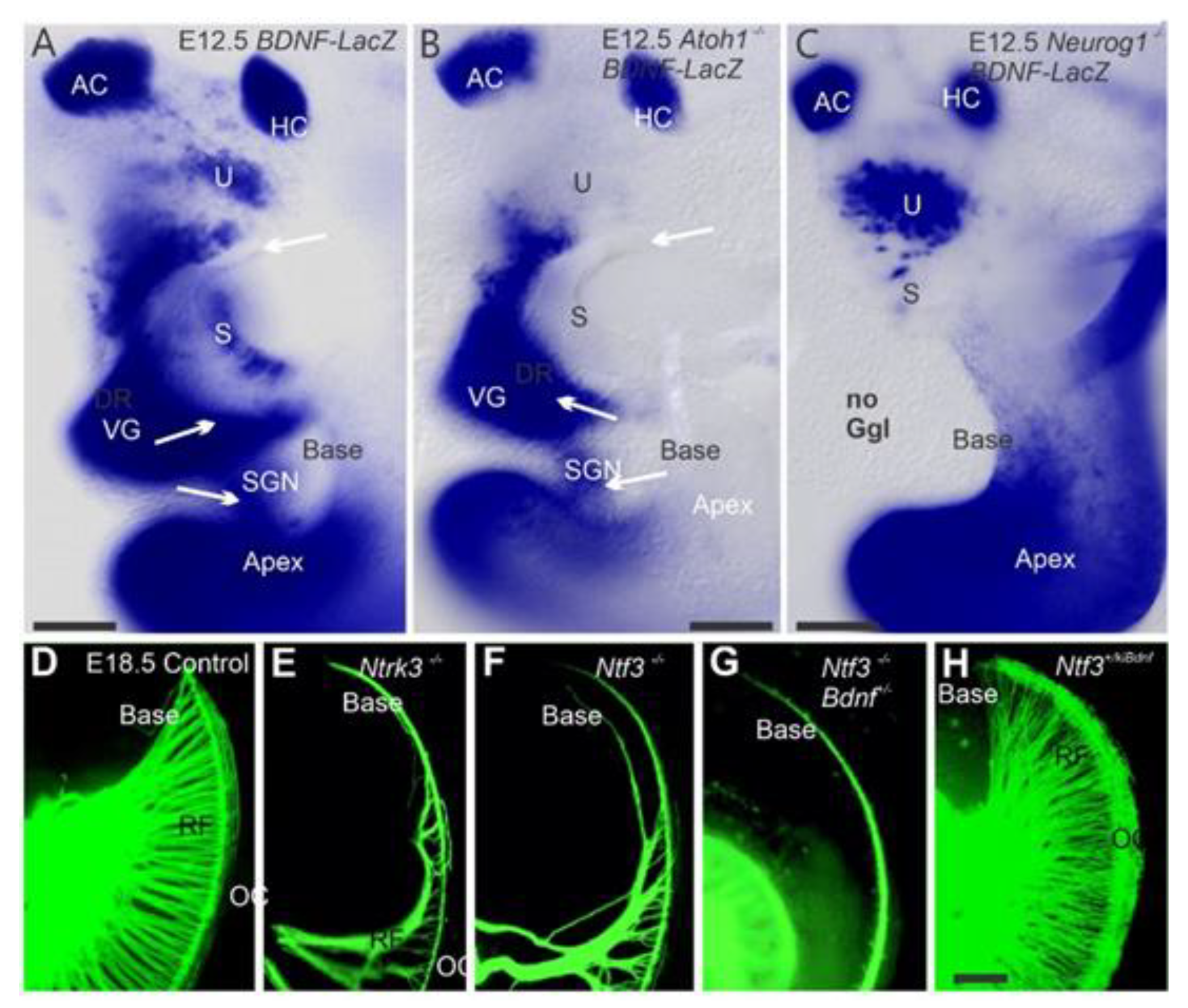
2. Neurog1 Regulates Neurod1 and Atoh1 Expression and Is Essential for Spiral Ganglion Neurons

The absence of Neurog1 resulted in a reduced length of the organ of Corti and prevented the development of all sensory neurons (Figure 2A–C), as they never initiated neuronal differentiation [12,17]. In contrast, Neurod1 null mice did not eliminate all sensory neurons [16]. Loss of Neurod1 resulted in a prolonged Atoh1 expression in sensory neurons and differentiation of some of these Atoh1-positive cells into ectopic “intraganglionic” HCs [28]. Furthermore, Neurod1 elimination in differentiating SGNs resulted in the formation of an abnormal spiral-vestibular ganglion [42,43], including the formation of vesicles containing several Atoh1-positive “HCs” [28,44]. These neurons overlap with the transient Atoh1-positive cells, suggesting Atoh1 is typically suppressed by Neurod1, consistent with the regulation of Atoh1 in the cerebellum [38] and the intestine [45]. Mice null for both Atoh1 and Neurod1 do not form “intraganglionic” HCs as was observed in mice lacking only Neurod1 [43].
SGNs are dependent on neurotrophins (Figure 2D–F). Loss of Ntf3 results in a complete loss of neurons innervating the cochlea base, whereas loss of Bdnf results in fewer neurons innervating the apex [48,49]. The loss of both copies of Ntf3 and one copy of Bdnf further reduces innervation to the base (Figure 2G). However, misexpression of Bdnf from the Ntf3 promoter causes an exuberant projection of neurons to the basal turn (Figure 2H), resulting in redirecting vestibular fibers to innervate the cochlea broadly [47]. Loss of both neurotrophin receptors (Ntrk2 and Ntrk3; [50]) or both neurotrophins (Bdnf/Ntf3) results in the complete loss of all neurons that leads to a subsequent loss of HCs [8].
SGNs can be classified as type I neurons, which innervate the IHCs, and type II neurons that innervate OHCs (Figure 1 and Figure 3). The latter is a much smaller proportion, constituting only 5-7% of the total spiral ganglion neurons. Currently, we do not fully understand the molecular mechanisms regulating the development, segregation, and neuronal projections of SGNs, although several genes involved in these processes are known [51,52,53,54,55]. For example, in Prox1 mutants, type II fibers turn at random and merge into a single bundle compared to the three parallel fibers that turn toward the base in controls (Figure 3A,B; [30,56]). Furthermore, the loss of Schwann cells following Sox10 deletion results in fibers projecting beyond the target HCs (Figure 3C; [54,57,58]). Additional genes, such as the Wnt/planar cell polarity (PCP) genes, Vangl2, Fzd3, and Fzd6, have been shown to affect type II fiber guidance to OHCs [58].
Mice in which Atoh1 was replaced by Neurog1 (Atoh1kiNeurog1) showed a different inner ear innervation pattern. In this model, there is an increased density of innervation to undifferentiated HCs [59,60]. Conditional deletion of Neurod1 results in incomplete neuronal loss, and the remaining fibers formed aberrant central and peripheral projections [36,42]. Dense neuronal projections to the flat epithelia were found in Atoh1/Neurod1 null mice in the absence of HCs [43], suggesting an uncoupling of innervation and HC differentiation.
In summary, the interrupted development of HCs in double Atoh1/Neurod1 null mutants uncouples HC development and projections growth. Expanded innervation of the remaining neurons in mouse mutants [43] could help hearing loss restoration [61], as well as the formation of neurons upstream of the proliferation of otic progenitors [62]. Regeneration of SGNs is also an essential task for future studies [62].

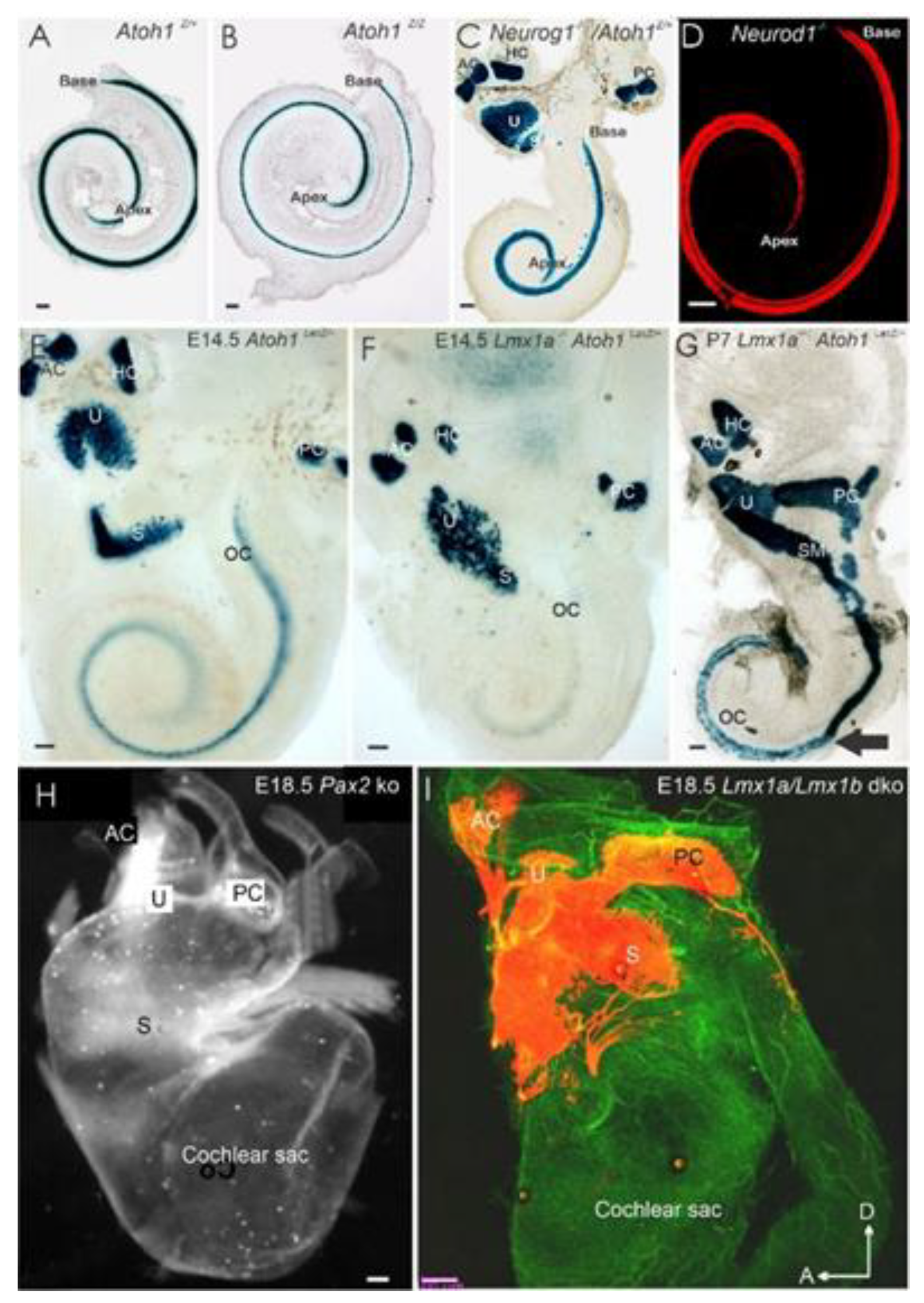
2.1. Is Atoh1 Playing a Role in Spiral Ganglion Neurons

Analysis of Atoh1-eGFP transgenic mice or Atoh1-cre allele showed transient expression of Atoh1 in some SGNs [12]; some of these neurons retained long-term expression Atoh1 [12]. These data suggest that some precursors may give rise to both HCs and sensory neurons. More substantial support of this hypothesis comes from recent data using multicolor labeling, which indicates a clonal relationship between several different inner ear cell types, including some sensory neurons [63]. It was later shown that the ordinarily transient expression of Atoh1 in inner ear sensory neurons could be sustained following the loss of Neurod1 [28], suggesting that Neurod1 inhibits Atoh1 expression, consistent with crosstalk of these two bHLH genes. In Neurod1 null mice, Atoh1 becomes upregulated in a different pattern (apex to base), also allowing “neurons” in the vestibular and cochlear ganglions to differentiate as ectopic “HCs” that were often grouped around intraganglionic vesicles [28,42,43]. The cochlear-derived ectopic HCs follow transient expression of Atoh1 that interacts with ErbB2, and Sox10 derived Schwann cells [57,64]. Without neuron formation, we see a delay of Atoh1 expression (Figure 4A,B), including different disorganized central projections in near-complete absence of IHCs, using exuberant type II projections [44,65].
Hair cells depend on neurons for their long-term maintenance [8], and conversely, neurons depend upon HCs and supporting cells [9,47]. Logically, one would assume that the absence of HCs will cause degeneration of eventually all neurons due to missing neurotrophic support. While the initial targeted growth of afferents and efferents to the undifferentiated “HCs” was not affected in Atoh1-null mice, there was indeed a later loss of innervation to certain inner ear areas [68]. Interestingly, the areas of sustained innervation by neurotrophins correlated with areas that still expressed Bdnf and Ntf3 in the absence of Atoh1 protein, suggesting that fiber retention is possible in the absence of Atoh1 differentiated HCs. Only immature HCs are formed in Pou4f3 null mice, and gradually HCs disappear in these null mice with time, and yet, some of the afferents are retained for a long time in these mice with limited expression of Bdnf and Ntf3 [70,71]. Furthermore, various Atoh1 conditional deletion mutations show a residual partial formation of HCs and proportional loss of neurons [43,72,73]. Finally, conditional deletion of Neurod1 results in a loss of many SGNs, yet near-normal cochlear HCs developed [43,68], suggesting that HCs’ survival depends on SGNs that require neuronal connections for their neurons.
In summary, neurons depend on HCs and vice versa. Understanding how the expansion of neuronal projections happens in the absence of HCs could restore lost innervation [61] and allow the flat epithelial to go beyond the expansion in the absent HCs [43,74]. Transforming SGNs to develop as HCs would provide a new population of HCs that adds to a possible source of HCs [5,28].

2.2. Spiral Ganglion Neurons Depend Upon Eya1 and Sox2 and Other Genes

Eya1 is expressed before Sox2, and both play a critical role in the proliferation and neurogenesis of neurosensory cells, and their loss will eliminate all sensory neurons [75,76,77]. Conditional deletion of Sox2 by Foxg1-cre likewise showed the absence of spiral ganglia but permitted a transient development of vestibular neurons, including developing peripheral and central processes [78]. A similar lack of SGNs is demonstrated in deleting Eya1/Six1 following the loss of Brg1 expression, indicating a major role for SWI/SNF chromatin remodeling [77]. Furthermore, a delayed deletion of Sox2 using Isl1-cre resulted in a complete loss of HCs and sensory neurons in the apex but had a transient expansion of disorganized HCs, with an unusual base innervation [79]. The similarities and differences between the different Sox2 deletions (Lcc, Ysb, Isl1-cre; Foxg1-cre) remain incompletely analyzed [78,80].
Eya1 elimination results in all neurons’ obliteration in the inner ear [63,77,81]. Also, Pax2-null mice resulted in a near absence of SGNs except for a small network of neurons innervating the cochlear duct [66], comparable to the partial innervation Isl1-cre; Sox2f/f mice [79]. Deleting Pax2 also results in the cochlear sack’s prolapse into the braincase beneath the brainstem (Figure 4H; [66]). Various other genes either entirely or partially derail the process of innervation [30,51,58,82]. For example, deleting even a single neurotrophin (Bdnf or Ntf3) results in some neuronal loss and aberrant innervation of the remaining neurons’ sensory epithelia [46,47]. Other genes, such as Gata3 and Dicer, affect inner ear neurosensory development [83,84]. Conditional deletion of either Gata3 or Dicer by Atoh1-cre results in a partial loss of neurons and HCs [83,84]; however, conditional deletion of Gata3 or Dicer by Foxg1-cre results in the complete or near-complete absence of neurons and HCs in a cochlear duct [83,85].
Similarly, in Lmx1a/b double knockout (DKO) mice, the cochlea exists as a simple sac devoid of HCs and SGNs, whereas the vestibular neurons and vestibular HCs do form [67]. Finally, even if there is no loss of neurons, Schwann cells’ absence significantly affects neuronal guidance. Wnt1-cre; Sox10f/f mice lack Schwann cells and had unusual SGN migration and projections to the cochlea’s lateral wall, bypassing the organ of Corti (Figure 3C; [57]), suggesting that Schwann cells provide a stop signal for SGNs.
In summary, the deletion of specific genes results in either complete neurosensory loss or some residual and transient sensory neuron formation. At least two and possibly five bHLH genes [29] interact to facilitate regular Atoh1 expression (Figure 2, Figure 3 and Figure 4). Among these genes are Neurog1 and Neurod1, which interact before Atoh1 upregulation occurs [27].

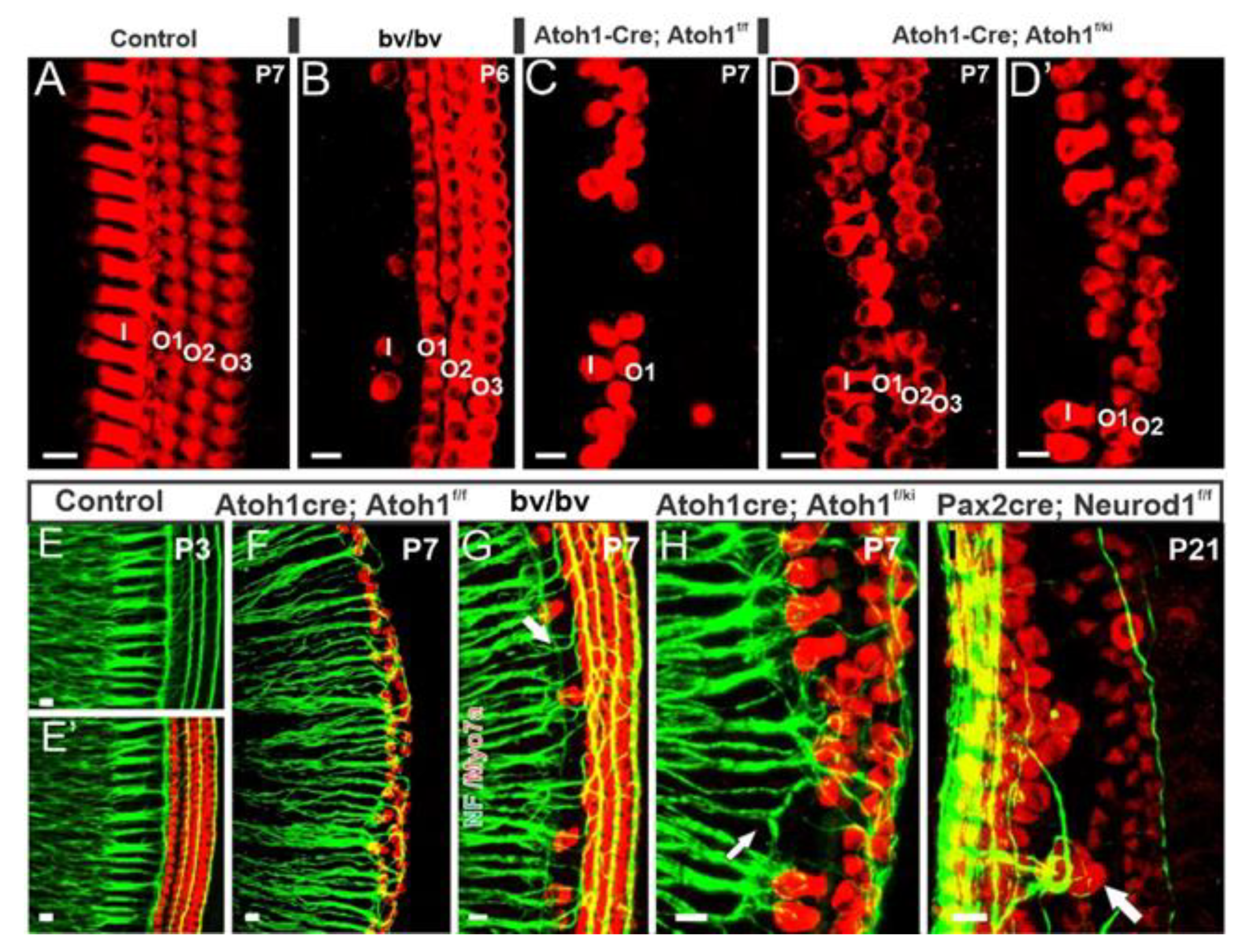
3. Cochlear Hair Cells Require Atoh1

The original study of Atoh1 in the inner ear showed that all HCs critically depend upon Atoh1 expression: without Atoh1 expression, all HCs are absent [24]. A follow-up study showed that the specific role of Atoh1 shows undifferentiated Atoh1-LacZ-positive HCs [25]. Atoh1 expression progresses around E13.5 from the upper-middle turn bilaterally toward the base and apex and finishes in the apex between E17–E18 [25,68]. Labeling with Atoh1-LacZ shows the latest forming “undifferentiated” HCs in the apex (Figure 4B) [24,68]. Expression of ”undifferentiated” cochlear HCs was near the cochlear IHCs positive cells expressed Atoh1-LacZ and demonstrated the loss of HCs at E13.5–E18.5 [25,68]. Furthermore, in situ hybridization showed expression of Atoh1 in the middle turn at E13.5 [12,28].
Using a short-term BrdU exposure, HCs were shown to exit the cell cycle at E12.5 in the apex and progress toward the base (Figure 1 and Figure 2) [12,86]. In addition, cell cycle exit progresses radially from IHCs to OHCs [28,31,44,87,88]. Questions remain regarding HC maturation progression from base to apex [19]. How the coordination of different cell cycle exits and cell fate determination occurs remains unclear, particularly in the Atoh1 expressing non-HCs, such as inner pillar cells [12,89]. Specifically, the cellular processes driving remodeling of the prosensory domain during cochlear development suggest combinations of cellular growth and cochlear extension from base-to-apex, allowing a different OHC progression for interpretation [31,44,87,90].
In parallel, various ways of analyzing the cellular progression focusing on Atoh1 deletions showed an increase in late forming apical HCs. They demonstrated loss of Atoh1 throughout the organ of Corti [68] correlated with cell apoptosis in identifiable, undifferentiated IHCs and OHCs. Likewise, conditional deletion of Atoh1 using Pax2-cre showed that most HCs were lost during late embryonic development; however, some undifferentiated cells expressed Myo7a, a marker for differentiated hair cells postnatal stages, and were innervated by neurons [72]. A ‘self-terminating system (Atoh1-cre; Atoh1f/f), in which there is a transient expression of Atoh1 resulting in some initial HC development, demonstrated progressive loss of IHC and most OHCs shortly after birth [73]. These data suggest that a brief upregulation of Atoh1 suffices to induce differentiation of IHCs (Figure 5C,F) and mostly the first row of OHCs. Atoh1 deletion at different HC differentiation stages showed different effects on HC survival and stereocilia development depending on the deletion time [91,92], suggesting that Atoh1 is necessary throughout HC differentiation. Using an ingenious expression system, in which induction of Atoh1 is under the control of a tetracycline-response element, generated viable ectopic “HCs” adjacent to the organ of Corti in early postnatal mice, with characteristics consistent with endogenous HCs [93], and in line with an upper limit of the proliferation of later HCs [94], consistent with a delay formation of HCs that remain Myo7a-positive in undifferentiated HCs.
Finally, loss of IHCs in Srrm4 mutants (a serine/arginine repetitive matrix 4 genes referred to as bv/bv mutations) shows that most type I afferent fibers reroute to OHCs (Figure 5B,G; [44,65]). Notably, the expression of dominant-negative REST in double Srrm3/4 mutants ablates all IHCs [95]. Previous work showed that delayed HCs loss, such as in Bdnf and Ntf3 double CKO mutants, results in delayed innervation [8]. The latter findings are consistent with reducing neurotrophins in Pou4f3 mutants, resulting in delayed neurosensory loss [70,71].
In mammals, HCs do not regenerate as they do in other vertebrates; however, supporting cells can be induced to transdifferentiate into HCs [5,94,96,97,98,99,100]. Despite this tremendous initial success of HC induction in vitro [41,101,102], reliable generation of new cochlear HCs has not yet been achieved to replace lost HCs, in particular, in older adults [6,103]. Attempts are underway to induce new hair cells by combinations of several transcription factors [104]. Given that Eya1 regulates Sox2 that depends on Atoh1, no attempt has been made to combine these three transcription factors [77,78].

3.1. Neurog1 and Neurod1 Regulate Atoh1 in Cochlear Hair Cells

Among the several bHLH genes known to participate in neurosensory development (Neurog1, Neurod1, Nscl1, Nscl2, bHLHb5), we selected a set of genes that have an apparent effect on the cochlea when deleted [15,28,105]. Loss of Neurog1 in mice results in a premature upregulation of Atoh1 in an apex-to-base progression and early differentiation of HCs [12,15]. In these mice, HCs exit the cell cycle in an apex to base progression about two days earlier than controls [12], reducing the cochlear length (Figure 4C). Deletion of Neurod1 also reduces cochlear length (Figure 4A,B) but is less reduced compared to Neurog1 null mice [18,36,42]. In contrast, Atoh1 deletion has minimal effect on cochlear length, either by itself or when deleted together with Neurog1 (Table 1). However, Neurod1/Atoh1 double null showed an additional reduction relative to that observed in Neurod1 null mice [43], suggesting a unique interaction of Atoh1 and Neurod1. Similar to Neurog1 null mice, premature differentiation HCs were found in Neurod1 mutants (Figure 4C). In these mice, there was an early upregulation of several genes, such as Atoh1 and Fgf8, in the apex where ectopic IHC-like cells in the region of OHCs were found, suggesting these gene proteins interact with each other to specify HC subtype as well [18,28,42,73]. Consistent with Neurod1 and Insm1 in the pancreas [106], we suggest the same interactions with OHC to transform into IHCs [28,42,107,108].
Replacing Atoh1 with Neurog1 (Atoh1kiNeurog1/kiNeurog1; Figure 5D,H) resulted in a few patches of undifferentiated “HCs” beyond the flat epithelia of Atoh1 null mice [59], suggesting that Neurog1 can partially rescue the Atoh1 phenotype. The co-expression of both Neurog1 and Atoh1 in Atoh1+/kiNeurog1 heterozygotes resulted in disorganization of HC polarity and stereocilia. It was not observed in Atoh1 heterozygote mice, suggesting that Neurog1 expression, and not Atoh1 haploinsufficiency, disrupts HC organization [59]. Combining the novel Atoh1 self-terminating mouse with the Atoh1kiNeruog1 mouse (Atoh1-cre; Atoh1kiNeurog1/Atoh1f/f) showed significantly more differentiated HCs and a more prolonged rescue than in the Atoh1 “self-terminating” littermates [60]. It is essential instead of transiently rescuing a few apical HCs as in the Atoh1 “self-terminating” mutant (Figure 5C,F; [73]), significantly more HCs differentiated and persisted for up to nine months in the Atoh1-cre; Atoh1kiNeurog1/Atoh1f/f mutant mouse model [60]. Together, these results suggest that, while Neurog1 can partially replace Atoh1, it cannot fully compensate for it. Thus, although replacing Atoh1 with fly ortholog atonal rescued the HC differentiation [109], the subsequent duplication and diversification of atonal family bHLH genes [110] no longer allow for the substitution of one for another to ultimately rescue hair cell loss.
These data suggest that the crosstalk of Neurog1, Neurod1, and Atoh1 affects cochlear extension and HC morphology and patterning (Figure 5I; [44]). Fully understanding the various mutations and putting them into the context of different cell fates requires additional work [111,112,113].

3.2. Eya1 and Sox2 and Other Transcription Factors Are Essential for HC Development

Eya1 is regulated by Sox2, which governs the Atoh1 gene [80,114], vital for HC formation [80]. Interestingly, two independent approaches used a delayed deletion of Sox2 [79,114] but showed different results. In one study, a delayed loss of Sox2 demonstrated effects in the apex only [114]. In the other study, conditional deletion of Sox2 resulted in the loss of HCs in the apex and a delayed loss in the base, showing unusual basal turn HCs/supporting cells and inner pillar cells [79]. Timing of Sox2 expression was demonstrated to be important for sensory development [76,115]. Furthermore, a complete deletion of Sox2 using Foxg1-cre demonstrated the overall cochlear reduction while showing no HC development [78]. These works provide the essential roles of Sox2, though the interaction of Sox2 with Atoh1 is not fully understood [87,90,99,100].
Other genes are also essential for cochlear development. For example, Eya1/Six1 are critical for early ear development and are needed to form the cochlea [77,81,116,117]. Another gene, Pax2, is necessary for the organ of Corti formation [66] and cooperates with Sox2 to activate Atoh1 expression [118]. Conditional deletion of Gata3 using Pax2-cre showed an incomplete loss of HCs compared to the complete absence of HCs using Foxg1-cre [34,83]. In Foxg1-cre; Gata3f/f mice, levels of Atoh1 expression were significantly reduced, and genes downstream of Atoh1 were not detected. Mice mutant for Lmx1a shows a delayed Atoh1 expression (Figure 4E–G) followed by transforming some organ of Corti HCs into differentiated vestibular hair cells [69,119,120,121]. In addition, Lmx1a deletion results in a shortened cochlea (Figure 4). Similarly, Foxg1 null mice also show a reduced cochlear length and a disorganized apex containing multiple rows of HCs with disoriented polarities [122]. Furthermore, HC survival is shortened in Foxg1 null mice [123]. A somewhat similar phenotype is reported for Nmyc2 null mutants, accompanied by apical cell fate changes [1,32].
The partial deletion of some HCs, but not others, following various gene mutations is an exciting perspective that needs to be explored. For example, the elimination of Fgfr1 by Foxg1-cre in the inner ear epithelium leads to a drastic reduction in the number of auditory HCs found in sensory patches [124]. Likewise, Sox2 deletion shows a similar partial loss of HCs in the Yellow submarine (Ysb) mutant [80]. Using Pax2-cre to delete Dicer [84] conditionally or Gata3 [83] resulted in incomplete HC loss compared to Foxg1-cre conditional deletion of these genes [83,85]. Like the latter conditional mutants, Lmx1a/b null mice show a complete loss of all cochlear HC development [67]. These data indicate that cochlear HCs are affected by single deletions and complex interactions of several genes. To date, the effects of compound deletions remain mostly unexplored [101,125]. Atoh1 may be the dominant gene in HC development [24], but interactions with other genes need to be better understood [99,117].
In summary, various genes upstream of Atoh1 are essential for HC development, including Eya1, Sox2, Pax2, Foxg1, Gata3, Lmx1a, and Fgfr1, as their deletion results in aberrant HC formation. Focusing on the effects of early gene deletions with massive HC loss or complete depletion of Atoh1 will help understand normal HC development [5], possibly using combinations of Eya1, Sox2, and Atoh1 to induce new hair cells [77,78].

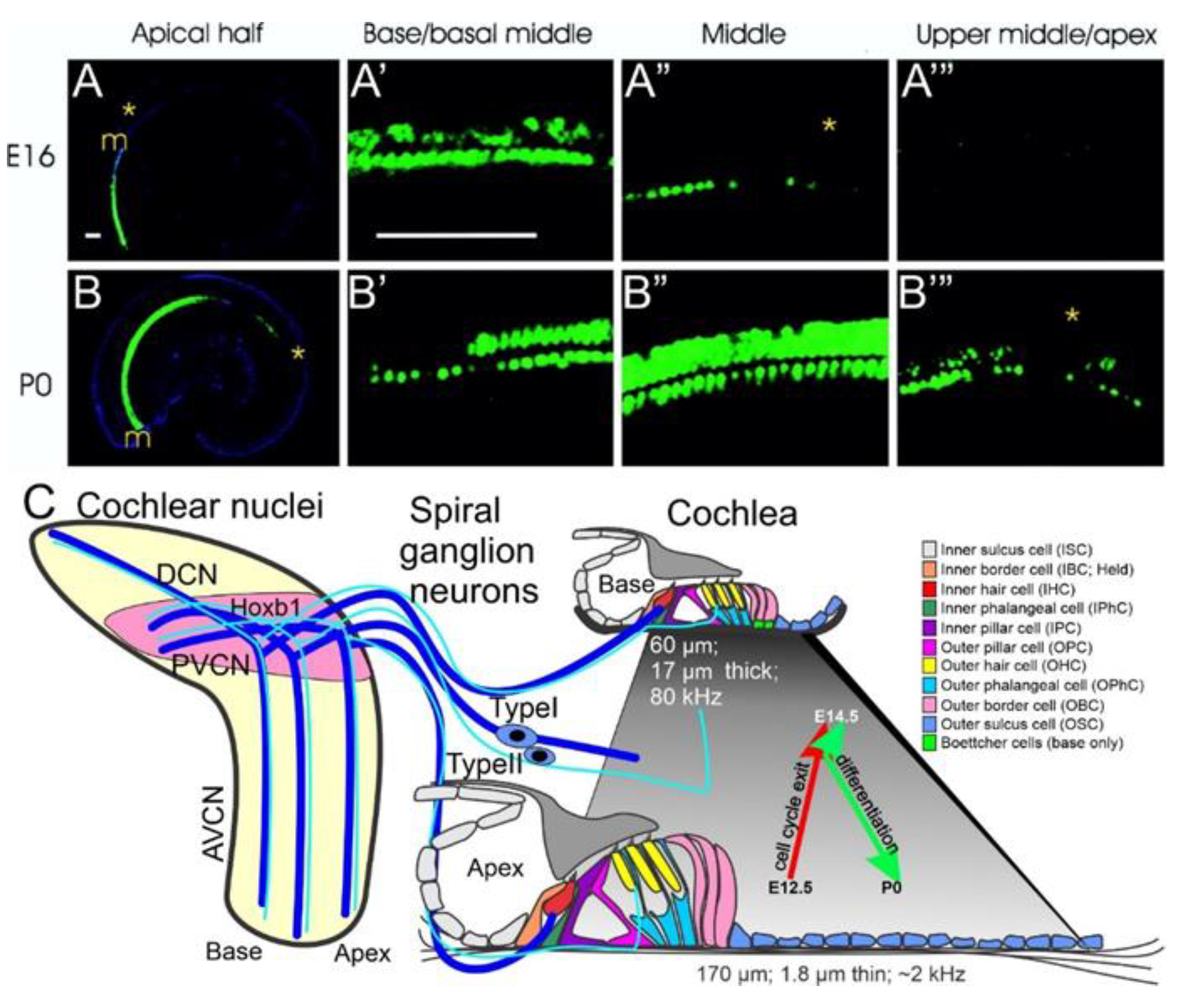
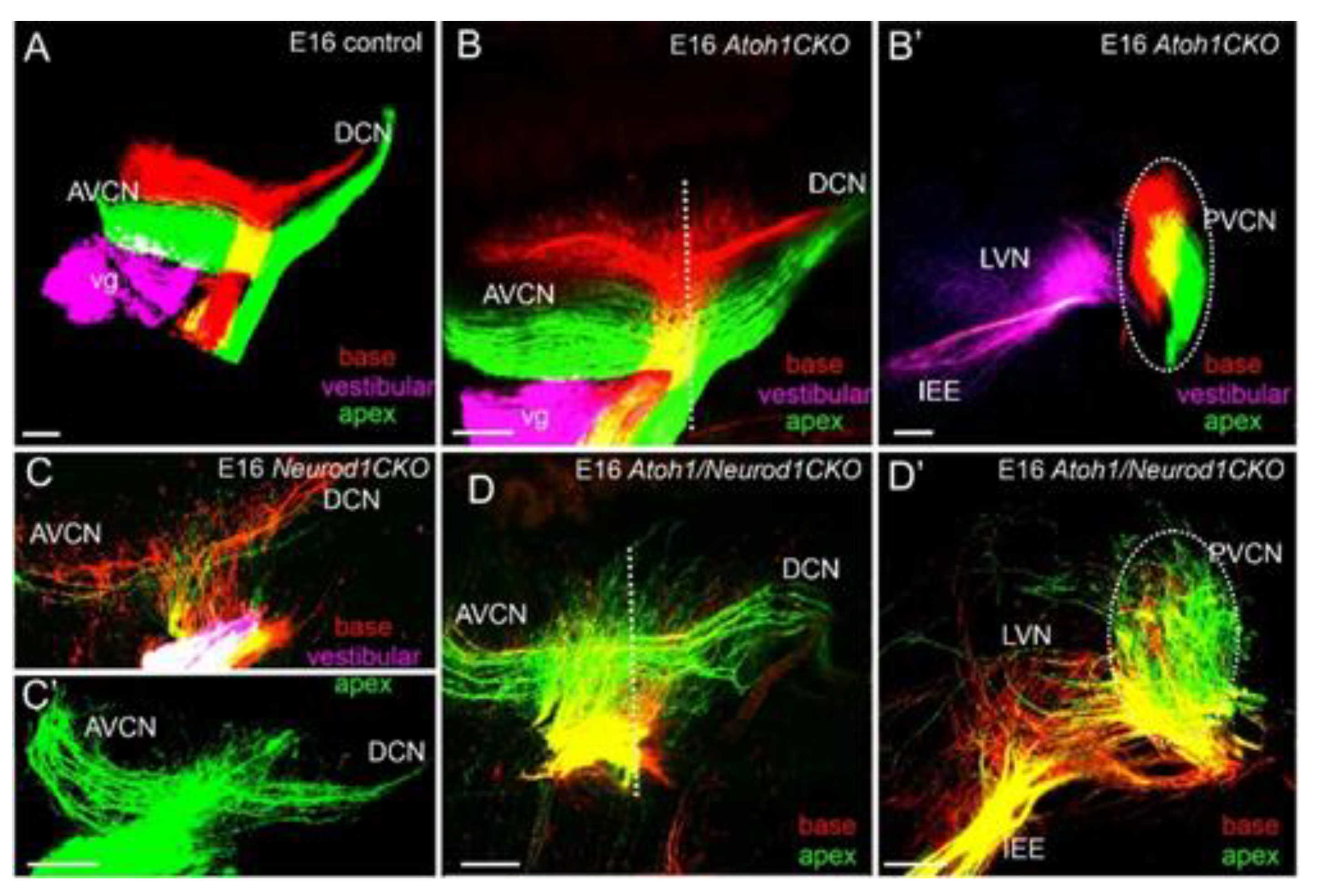
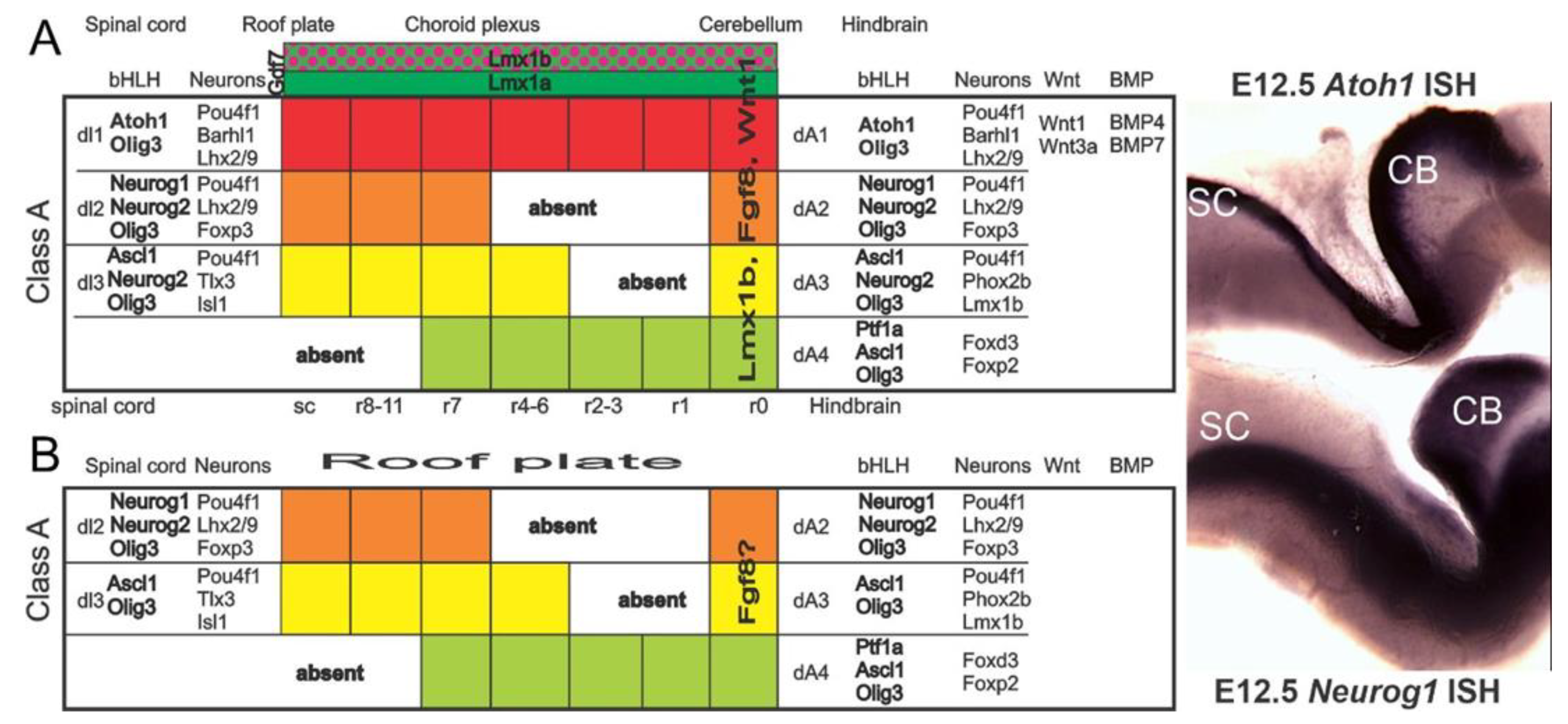
4. Cochlear Nuclei Depend on Atoh1

Atoh1 deletion disrupted the spinal cord, brainstem, and cerebellum development [126,127] and resulted in loss of all cochlear HCs [33,37,128,129]. Rhombomere-specific deletion of Atoh1 demonstrated that the cochlear nucleus is formed from cells in rhombomeres 3–5 [129,130]. Furthermore, loss of Atoh1 or another bHLH gene, Ptf1a, resulted in a loss of excitatory or inhibitory cochlear nucleus neurons, respectively, signifying their importance for regulating cell fate determination [39,131]. Moreover, Lmx1a and Lmx1b LIM-homeodomain transcription factors expressed in the hindbrain roof plate [40] together regulate Atoh1 expression [67]. Lmx1a/b double null mice lack cochlear development and excitatory neurons in the auditory nuclei, most likely due to the lack of Atoh1 in these mice [67]. In Lmx1a/b double mutant mice, roof plate does not differentiate into the choroid plexus, which is associated with lack of expression of roof plate/choroid plexus-derived secreted molecules, such as Gdf7 and Bmps, known to induce Atoh1 expression in adjacent neuroepithelium [132,133,134].
Central projections of inner ear afferents of Atoh1 null mice show near-normal projections, despite the absence of differentiated HCs and cochlear nucleus neurons [135]. Moreover, conditional deletion of Atoh1 in the ear, but retaining Atoh1 expression in cochlear nuclei, show near-normal segregation of central projections [43], expanding the critical independence of Atoh1 in neuronal pathfinding (Figure 6). In contrast, central projections are highly disorganized in Neurod1 mutants [36,42]. These data suggest that inner ear afferents rely more on molecular cues in the region of the targets than on direct interactions with target cells.
In summary, Atoh1 regulates cochlear nuclei formation [9,33,37], but has no apparent role in central projections’ guidance [43,135]. More work is needed to understand the role of Atoh1 to regulate central projections fully.

4.1. Neurod1 Interacts with Atoh1 in the Cochlea

Beyond a transient and limited expression of Neurog1 in vestibular nuclei [37,136], other bHLH genes are expressed in cochlear nuclei (Figure 7 [110,137]). In addition to Atoh1, Neurod1 and Neurod4 are also expressed in cochlear nuclei [37,136]. Neurod1 and Atoh1 negatively interact in the cerebellum [38], in the cochlear HCs and neurons [28], and the intestine [45]. Also, differential levels and expression patterns of Atoh1 and Neurod1 are found in the cochlear nucleus [38,138]. For example, Atoh1 is highly expressed in the ventral cochlear neurons, whereas Neurod1 is expressed more prominent in the dorsal cochlear nucleus (Figure 8; [18]). An additional bHLH transcription factor, bHLHb5 [130], is also required to form the dorsal cochlear nucleus properly. Its expression overlaps with yet another bHLH gene, Ptf1a, in the dorsal cochlear nucleus [131]. These data implicate the interaction of different bHLH genes in forming the cochlear nuclei (Atoh1, Neurod1, Ptf1a, bHLHb5) to crosstalk with each other in the spinal cord [139].
Loss of Neurod1, and likely Neurod4 [136], negatively affects the central targeting and branch pattern of fibers, leading to highly aberrant central pathfinding of both auditory and vestibular afferents [36,42]. Likewise, the inferior colliculi projections are disorganized [42], expanding previous work with Hoxb2 mutants [140]. Not surprisingly, Atoh1/Neurod1 double null mice had more severely disorganized projection of cochlear afferents [43] beyond that observed in Neurod1 single null mice (Figure 6) and Atoh1 null mice that lack a similar phenotype [135].
In summary, intricate interactions of different bHLH genes (Figure 8) to each other and with Atoh1 drive the formation of central projections [127,128,134] and, thus, require additional detailed analysis.

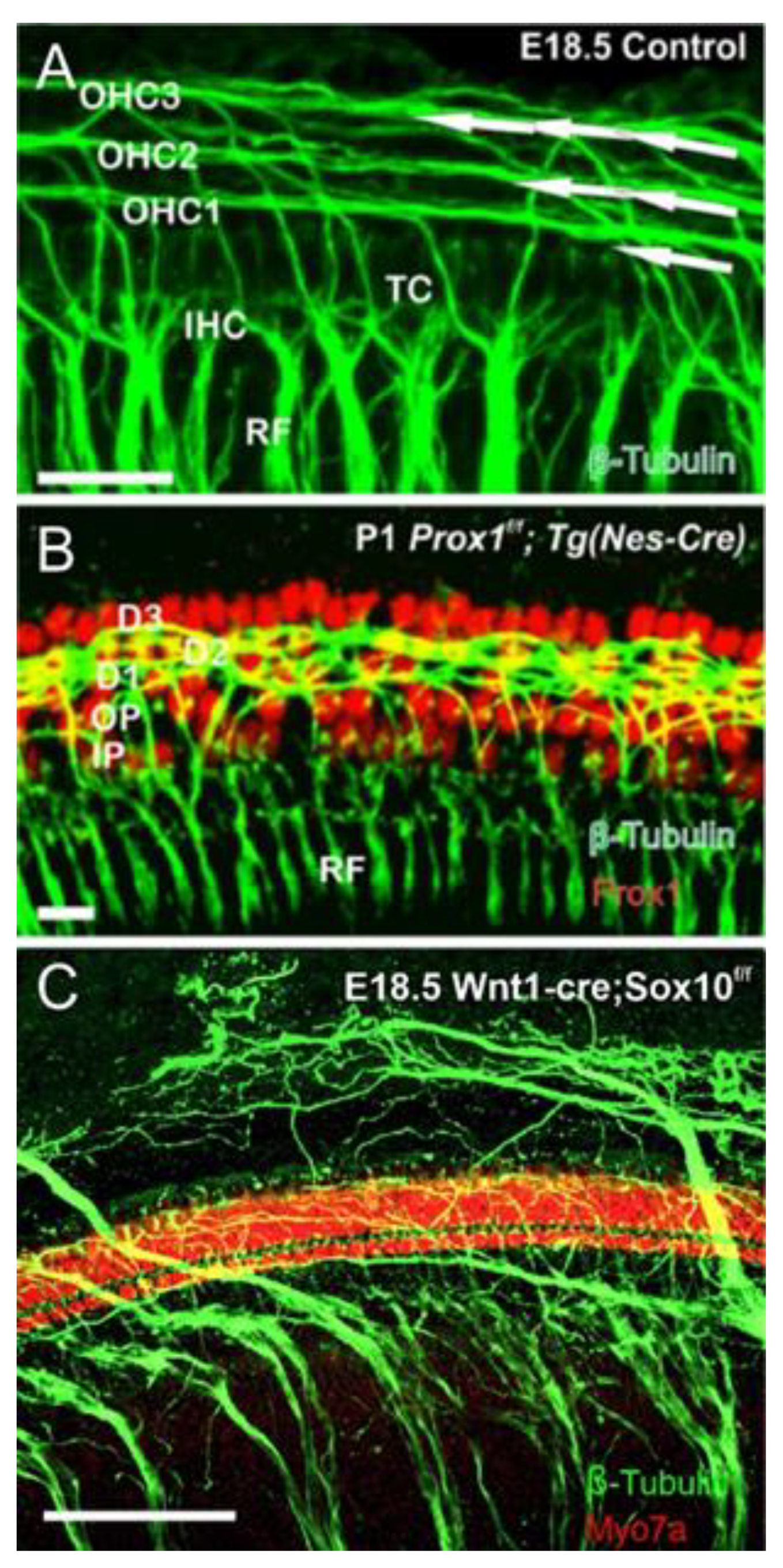
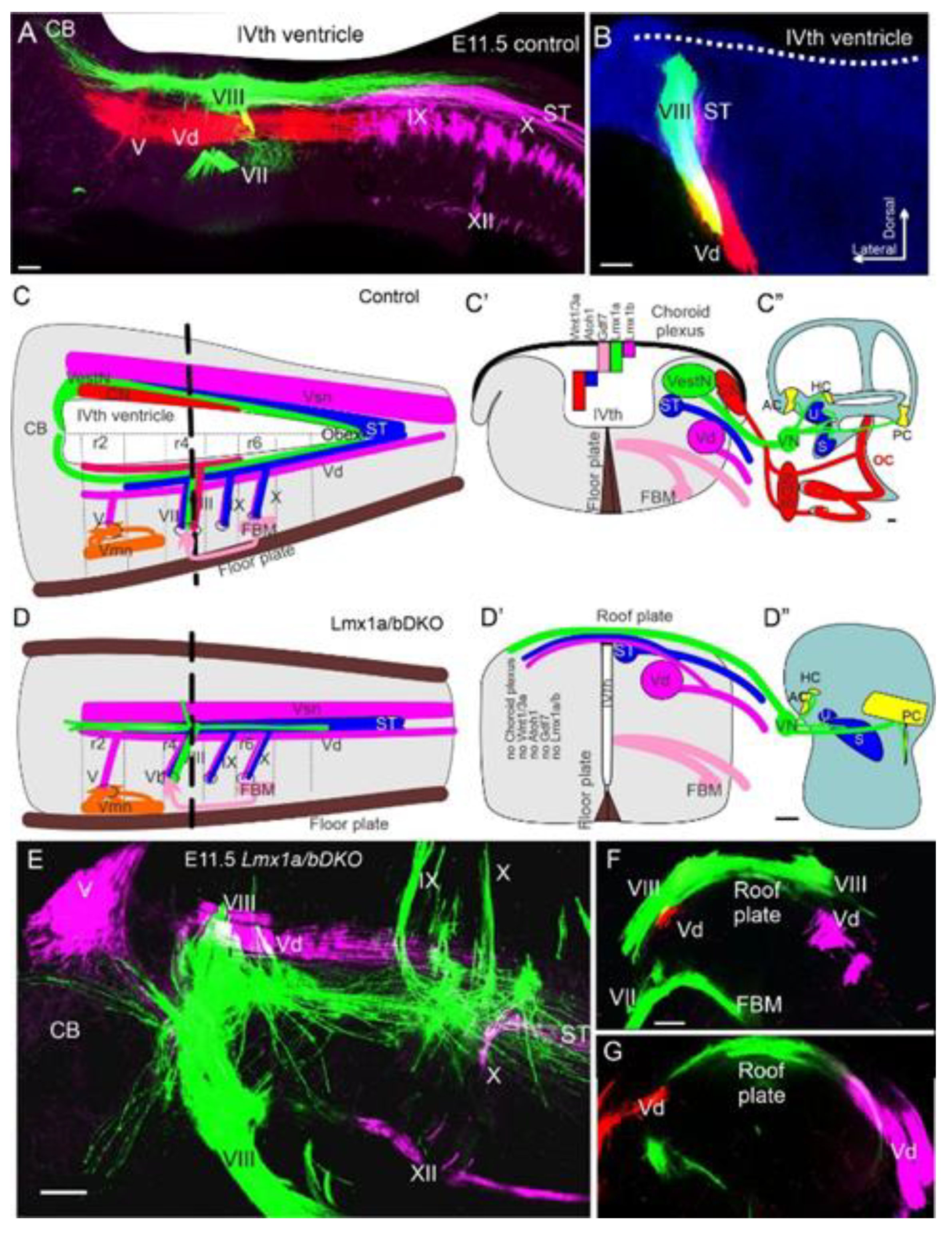
4.2. Sox2 Function Is Unclear, but Lmx1a/b Double Null Mice Eliminate All Cochlear Nuclei

Sox2 is essential for proneuronal regulation in the entire brain [77,141,142,143] and is broadly expressed in cochlear nuclei; however, the effects of selective Sox2 deletion in cochlear nuclei have not yet been analyzed. Eya1 is expressed in the rhombomere [144] but has not been analyzed selectively; targeted deletion of Eya1 could clarify its role for the cochlear nuclei. Using Foxg1-cre to eliminate Sox2 in the inner ear results in a complete loss of spiral ganglion formation but does not initially affect vestibular ganglion development [78].
As mentioned above, Lmx1a/b double null mutants lack cochlear nuclei [67]. While Lmx1a mutants show near-normal central projections [67], spiral ganglion projections of Lmx1a/b double null mutants are lost, and vestibular fibers project bilaterally to the dorsal hindbrain and interdigitate with contralateral vestibular fibers (Figure 7 and Figure 8) [67]. The presence of bilateral vestibular projections correlated with other genes’ expression, such as Wnt3a, which in Lmx1a/b double mutant becomes ectopically expressed at the dorsal midline than in the rhombic lip [67]. The suggested Wnt3a attraction expands on previous data showing that loss of the Wnt receptor, Fzd3 [145], or downstream Wnt signaling component, Prickle1 [82], affects central projections. Recent work suggests that another gene, Npr2, affects central projections, showing the gain and loss of afferents to different cochlear nuclei [146]. Central projections in mutants for other genes (Eya1/Six1, Pax2, Foxg1, Fgfr1, Gata3) are necessary for cochlear development to have not been investigated in great detail, aside from some data showing aberrant central projections in Pax2-cre; Gata3f/f mice [83].
In summary, the expression of Lmx1a/b is essential for proper hindbrain development, and deletion of these genes causes loss of the cochlear nucleus and projections to them. In contrast to the detailed description of Lmx1a/b loss, there is minimal information on the role of Sox2. Other central genes for cochlear nucleus development and central projections could replace lost neurons and block any auditory cochlear nucleus in Lmx1a/b null mice that act upstream of Atoh1 [33,67] to repopulate lost cochlear neurons that is an age-related loss of specific nuclei.

5. Summary and Conclusions

The auditory system requires developing the spiral ganglion neurons that develop before hair cells and partially overlapping with auditory nuclei. SGNs depend on Eya1 and Sox2 to be followed by Neurog1 and Neurod1, resulting in a shortening of the cochlea and smaller cochlear nuclei. Both hair cells and cochlear nuclei depend on Eya1, Sox2, and Atoh1 but develop central and peripheral neuron projections to reach hair cells and cochlear nuclei in either connection. A unique dependency on Lmx1a/b double null mutants and the loss of SGNs, cochlear hair cells, and cochlear nuclei followed the roof plate’s loss. All transcription factors are early and require jump-start forming all three components of the auditory system that will lose hair cells and neurons. Following the Eya1 > Sox2 > Atoh1 sequence of gene expression suggests the co-expression of at least three interlinked transcription factors may be needed to lead to the new formation of hair cells.

Author Contributions

Conceptualization, K.L.E., G.P., V.V.C. and B.F.; writing—original draft preparation, K.L.E., V.V.C., E.N.Y. and B.F.; writing—review and editing, G.P. and E.N.Y.; funding acquisition, E.N.Y., G.P., B.F. All authors have read and agreed to the published version of the manuscript.

Funding

This work was supported by a grant from the NIH/NIA (R01 AG060504, P01, AG051443, 1R01 DC015135, DC016099, 1R01 DC015252-01) to E.N.Y., by a grant from the NIH/NIA (R01 AG060504) to B.F. and by a grant from the Czech Science Foundation (20-06927S) and the Czech Academy of Sciences (RVO 86652036) to G.P.

Acknowledgments

We wish to thank several collaborators, in particular, Ma, Q.; Zoghbi, H.; Pirvola, U.; Matei, V.; Nichols, D.; Pauley, S.; Beisel, K.; He, D.; Soukup, G.; Jahan, I.; Kopecky, B.; Yang, T.; Duncan, J.; Kersigo, J. and Pan, N.

Conflicts of Interest

The author declares no conflict of interest.

Abbreviations

Ascl1Achete-scute family bHLH transcription factor 1
Atoh1Atonal bHLH transcription factor 1
Barhl1BarH-like homeobox 1
BdnfBrain-derived neurotrophic factor
bHLHe22 (bHLHb5)Basic helix–loop–helix family member e22
Chrna9Cholinergic receptor, nicotinic, alpha polypeptide 9 (α9-acetylcholine)
Cdkn1b (p27Kip)Cyclin-dependent kinase inhibitor 1b
Dicer1Dicer 1, ribonuclease type III
Eya1EYA transcriptional coactivator, and phosphatase 1 (eyes absent homolog 1)
Gata3GATA-binding protein 3
Gdf7Growth differentiation factor 7
Gfi1Growth factor independent 1 transcription repressor
Fgf8Fibroblast growth factor 8
Fgfr1Fibroblast growth factor receptor 1
Foxd3Forkhead box D3
Foxg1Forkhead box G1
Foxp2Forkhead box P2
Fzd3/6Frizzled class receptor 3/6
Insm1Insulinoma-associated 1
Isl1ISL1 transcription factor, LIM/homeodomain
Lhx2/9LIM homeobox protein 2/9
Lmx1a/bLIM homeobox transcription factor 1 alpha/beta
Myo7aMyosin VIIA
Neurod1Neurogenic differentiation 1
Neurog1/2Neurogenin 1/2
Nhlh1Nescient helix loop helix 1 (Nscl1)
Nhlh2Nescient helix loop helix 2 (Nscl2)
Nmyc2Neuroblastoma myc-related oncogene 2
Npr2Natriuretic peptide receptor 2
Ntf3Neurotrophin 3 (Nt3)
Ntrk2Neurotrophic tyrosine kinase, receptor, type 2 (TrkB)
Ntrk3Neurotrophic tyrosine kinase, receptor, type 3 (TrkC)
Olig3Oligodendrocyte transcription factor 3
Pax2/8Paired box 2/8
Phox2bPaired-like homeobox 2b
Pou4f1,3POU domain, class 4 transcription factor 1/3
Prickle1Prickle planar cell polarity protein 1
Ptf1aPancreas-specific transcription factor, 1a
RestRE1-silencing transcription factor (REST)
Six1Sine oculis-related homeobox 1
Smarca4SWI/SNF related, matrix associated, actin-dependent regulator of chromatin, subfamily a, member 4 (Brg1)
Sox2SRY (sex-determining region Y)-box 2
Srrm4Serine/arginine repetitive matrix 4
Tbr1T-box brain transcription factor 1
Tlx3T cell leukemia, homeobox 3
Vangl2VANGL planar cell polarity 2
Wnt1/3aWingless-type MMTV integration site family, member 1/3a

References

  1. Fritzsch, B.; Pan, N.; Jahan, I.; Duncan, J.S.; Kopecky, B.J.; Elliott, K.L.; Kersigo, J.; Yang, T. Evolution and development of the tetrapod auditory system: An organ of Corti-centric perspective. Evol. Dev. 2013, 15, 63–79. [Google Scholar] [CrossRef] [Green Version]
  2. Luo, Z.-X.; Manley, G.A. Origins and early evolution of mammalian ears and hearing function. In The Senses; Fritzsch, B., Ed.; Elsevier: Iowa City, NA, USA, 2021; Volume 2, pp. 207–252. [Google Scholar]
  3. Grothe, B.; Carr, C.E.; Casseday, J.H.; Fritzsch, B.; Köppl, C. The Evolution of Central Pathways and Their Neural Processing Patterns. In Evolution of the Vertebrate Auditory System; Springer Science & Business Media: Berlin, Germany, 2004; pp. 289–359. [Google Scholar]
  4. Grothe, B. The Auditory System Function—An Integrative Perspective. In The Senses; Fritzsch, B., Ed.; Elsevier: Iowa City, NA, USA, 2021; Volume 2, pp. 1–17. [Google Scholar]
  5. Zine, A.; Messat, Y.; Fritzsch, B. A human induced pluripotent stem cell-based modular platform to challenge sensorineural hearing loss. Stem Cells 2021. [Google Scholar] [CrossRef]
  6. Yamoah, E.N.; Li, M.; Shah, A.; Elliott, K.L.; Cheah, K.; Xu, P.-X.; Phillips, S.; Young, S.M., Jr.; Eberl, D.F.; Fritzsch, B. Using Sox2 to alleviate the hallmarks of age-related hearing loss. Ageing Res. Rev. 2020, 59, 101042. [Google Scholar] [CrossRef]
  7. Resnik, J.; Polley, D.B. Cochlear neural degeneration disrupts hearing in background noise by increasing auditory cortex internal noise. Neuron 2021, 109, 984–996.e4. [Google Scholar] [CrossRef]
  8. Kersigo, J.; Fritzsch, B. Inner ear hair cells deteriorate in mice engineered to have no or diminished innervation. Front. Aging Neurosci. 2015, 7, 33. [Google Scholar] [CrossRef] [Green Version]
  9. Rubel, E.W.; Fritzsch, B. Auditory system development: Primary auditory neurons and their targets. Annu. Rev. Neurosci. 2002, 25, 51–101. [Google Scholar] [CrossRef] [Green Version]
  10. De No, R.L. The Primary Acoustic Nuclei; Raven Press: New York, NY, USA, 1981. [Google Scholar]
  11. Ruben, R.J. Development of the inner ear of the mouse: A radioautographic study of terminal mitoses. Acta Otolaryngol. 1967, 220, 1–44. [Google Scholar]
  12. Matei, V.; Pauley, S.; Kaing, S.; Rowitch, D.; Beisel, K.; Morris, K.; Feng, F.; Jones, K.; Lee, J.; Fritzsch, B. Smaller inner ear sensory epithelia in Neurog1 null mice are related to earlier hair cell cycle exit. Dev. Dyn. 2005, 234, 633–650. [Google Scholar] [CrossRef] [Green Version]
  13. Pierce, E.T. Histogenesis of the dorsal and ventral cochlear nuclei in the mouse. An autoradiographic study. J. Comp. Neurol. 1967, 131, 27–53. [Google Scholar] [CrossRef]
  14. Fritzsch, B.; Elliott, K.L.; Pavlinkova, G. Primary sensory map formations reflect unique needs and molecular cues specific to each sensory system. F1000Reseach 2019, 8. [Google Scholar] [CrossRef]
  15. Ma, Q.; Anderson, D.J.; Fritzsch, B. Neurogenin 1 null mutant ears develop fewer, morphologically normal hair cells in smaller sensory epithelia devoid of innervation. J. Assoc. Res. Otolaryngol. 2000, 1, 129–143. [Google Scholar] [CrossRef] [Green Version]
  16. Kim, W.-Y.; Fritzsch, B.; Serls, A.; Bakel, L.A.; Huang, E.J.; Reichardt, L.F.; Barth, D.S.; Lee, J.E. NeuroD-null mice are deaf due to a severe loss of the inner ear sensory neurons during development. Development 2001, 128, 417–426. [Google Scholar]
  17. Ma, Q.; Chen, Z.; del Barco Barrantes, I.; de la Pompa, J.L.; Anderson, D.J. neurogenin1 is essential for the determination of neuronal precursors for proximal cranial sensory ganglia. Neuron 1998, 20, 469–482. [Google Scholar] [CrossRef] [Green Version]
  18. Liu, M.; Pereira, F.A.; Price, S.D.; Chu, M.-j.; Shope, C.; Himes, D.; Eatock, R.A.; Brownell, W.E.; Lysakowski, A.; Tsai, M.-J. Essential role of BETA2/NeuroD1 in development of the vestibular and auditory systems. Genes Dev. 2000, 14, 2839–2854. [Google Scholar] [CrossRef] [Green Version]
  19. Zuo, J.; Treadaway, J.; Buckner, T.W.; Fritzsch, B. Visualization of α9 acetylcholine receptor expression in hair cells of transgenic mice containing a modified bacterial artificial chromosome. Proc. Natl. Acad. Sci.USA 1999, 96, 14100–14105. [Google Scholar] [CrossRef] [Green Version]
  20. Booth, K.T.; Azaiez, H.; Jahan, I.; Smith, R.J.; Fritzsch, B. Intracellular Regulome Variability Along the Organ of Corti: Evidence, Approaches, Challenges, and Perspective. Front. Genet. 2018, 9, 156. [Google Scholar] [CrossRef]
  21. Muniak, M.A.; Connelly, C.J.; Suthakar, K.; Milinkeviciute, G.; Ayeni, F.E.; Ryugo, D.K. Central Projections of Spiral Ganglion Neurons. In The Primary Auditory Neurons of the Mammalian Cochlea; Springer: New York, NY, USA, 2016; pp. 157–190. [Google Scholar]
  22. Lowenheim, H.; Furness, D.N.; Kil, J.; Zinn, C.; Gultig, K.; Fero, M.L.; Frost, D.; Gummer, A.W.; Roberts, J.M.; Rubel, E.W.; et al. Gene disruption of p27(Kip1) allows cell proliferation in the postnatal and adult organ of corti. Proc. Natl. Acad. Sci.USA 1999, 96, 4084–4088. [Google Scholar] [CrossRef] [Green Version]
  23. Chen, P.; Segil, N. p27(Kip1) links cell proliferation to morphogenesis in the developing organ of Corti. Development 1999, 126, 1581–1590. [Google Scholar]
  24. Bermingham, N.A.; Hassan, B.A.; Price, S.D.; Vollrath, M.A.; Ben-Arie, N.; Eatock, R.A.; Bellen, H.J.; Lysakowski, A.; Zoghbi, H.Y. Math1: An essential gene for the generation of inner ear hair cells. Science 1999, 284, 1837–1841. [Google Scholar] [CrossRef] [Green Version]
  25. Chen, P.; Johnson, J.E.; Zoghbi, H.Y.; Segil, N. The role of Math1 in inner ear development: Uncoupling the establishment of the sensory primordium from hair cell fate determination. Development 2002, 129, 2495–2505. [Google Scholar]
  26. Fritzsch, B.; Beisel, K.W.; Bermingham, N.A. Developmental evolutionary biology of the vertebrate ear: Conserving mechanoelectric transduction and developmental pathways in diverging morphologies. Neuroreport 2000, 11, R35–R44. [Google Scholar] [CrossRef] [PubMed]
  27. Fritzsch, B.; Beisel, K.W.; Hansen, L.A. The molecular basis of neurosensory cell formation in ear development: A blueprint for hair cell and sensory neuron regeneration? Bioessays 2006, 28, 1181–1193. [Google Scholar] [CrossRef] [PubMed] [Green Version]
  28. Jahan, I.; Pan, N.; Kersigo, J.; Fritzsch, B. Neurod1 suppresses hair cell differentiation in ear ganglia and regulates hair cell subtype development in the cochlea. PLoS ONE 2010, 5, e11661. [Google Scholar] [CrossRef] [PubMed] [Green Version]
  29. Fritzsch, B.; Eberl, D.F.; Beisel, K.W. The role of bHLH genes in ear development and evolution: Revisiting a 10-year-old hypothesis. Cell. Mol. Life Sci. CMLS 2010, 67, 3089–3099. [Google Scholar] [CrossRef] [PubMed] [Green Version]
  30. Fritzsch, B.; Dillard, M.; Lavado, A.; Harvey, N.L.; Jahan, I. Canal cristae growth and fiber extension to the outer hair cells of the mouse ear require Prox1 activity. PLoS ONE 2010, 5, e9377. [Google Scholar] [CrossRef] [PubMed] [Green Version]
  31. Kopecky, B.J.; Jahan, I.; Fritzsch, B. Correct timing of proliferation and differentiation is necessary for normal inner ear development and auditory hair cell viability. Dev. Dyn. 2013, 242, 132–147. [Google Scholar] [CrossRef] [PubMed] [Green Version]
  32. Kopecky, B.; Santi, P.; Johnson, S.; Schmitz, H.; Fritzsch, B. Conditional deletion of N-Myc disrupts neurosensory and non-sensory development of the ear. Dev. Dyn. 2011, 240, 1373–1390. [Google Scholar] [CrossRef] [PubMed] [Green Version]
  33. Wang, V.Y.; Rose, M.F.; Zoghbi, H.Y. Math1 expression redefines the rhombic lip derivatives and reveals novel lineages within the brainstem and cerebellum. Neuron 2005, 48, 31–43. [Google Scholar] [CrossRef] [Green Version]
  34. Karis, A.; Pata, I.; van Doorninck, J.H.; Grosveld, F.; de Zeeuw, C.I.; de Caprona, D.; Fritzsch, B. Transcription factor GATA-3 alters pathway selection of olivocochlear neurons and affects morphogenesis of the ear. J. Comp. Neurol. 2001, 429, 615–630. [Google Scholar] [CrossRef]
  35. Maklad, A.; Fritzsch, B. Development of vestibular afferent projections into the hindbrain and their central targets. Brain Res. Bull. 2003, 60, 497–510. [Google Scholar] [CrossRef] [Green Version]
  36. Jahan, I.; Kersigo, J.; Pan, N.; Fritzsch, B. Neurod1 regulates survival and formation of connections in mouse ear and brain. Cell Tissue Res. 2010, 341, 95–110. [Google Scholar] [CrossRef] [Green Version]
  37. Fritzsch, B.; Pauley, S.; Feng, F.; Matei, V.; Nichols, D. The molecular and developmental basis of the evolution of the vertebrate auditory system. Int. J. Comp. Psychol. 2006, 19, 1–25. [Google Scholar]
  38. Pan, N.; Jahan, I.; Lee, J.E.; Fritzsch, B. Defects in the cerebella of conditional Neurod1 null mice correlate with effective Tg (Atoh1-cre) recombination and granule cell requirements for Neurod1 for differentiation. Cell Tissue Res. 2009, 337, 407–428. [Google Scholar] [CrossRef] [PubMed] [Green Version]
  39. Fujiyama, T.; Yamada, M.; Terao, M.; Terashima, T.; Hioki, H.; Inoue, Y.U.; Inoue, T.; Masuyama, N.; Obata, K.; Yanagawa, Y. Inhibitory and excitatory subtypes of cochlear nucleus neurons are defined by distinct bHLH transcription factors, Ptf1a and Atoh1. Development 2009, 136, 2049–2058. [Google Scholar] [CrossRef] [Green Version]
  40. Mishima, Y.; Lindgren, A.G.; Chizhikov, V.V.; Johnson, R.L.; Millen, K.J. Overlapping function of Lmx1a and Lmx1b in anterior hindbrain roof plate formation and cerebellar growth. J. Neurosci. 2009, 29, 11377–11384. [Google Scholar] [CrossRef] [Green Version]
  41. Koehler, K.R.; Nie, J.; Longworth-Mills, E.; Liu, X.-P.; Lee, J.; Holt, J.R.; Hashino, E. Generation of inner ear organoids containing functional hair cells from human pluripotent stem cells. Nat. Biotechnol. 2017, 35, 583. [Google Scholar] [CrossRef] [Green Version]
  42. Macova, I.; Pysanenko, K.; Chumak, T.; Dvorakova, M.; Bohuslavova, R.; Syka, J.; Fritzsch, B.; Pavlinkova, G. Neurod1 is essential for the primary tonotopic organization and related auditory information processing in the midbrain. J. Neurosci. 2019, 39, 984–1004. [Google Scholar] [CrossRef] [PubMed] [Green Version]
  43. Filova, I.; Dvorakova, M.; Bohuslavova, R.; Pavlinek, A.; Elliott, K.L.; Vochyanova, S.; Fritzsch, B.; Pavlinkova, G. Combined Atoh1 and Neurod1 Deletion Reveals Autonomous Growth of Auditory Nerve Fibers. Mol. Neurobiol. 2020, 57, 5307–5323. [Google Scholar] [CrossRef]
  44. Jahan, I.; Elliott, K.L.; Fritzsch, B. Understanding molecular evolution and development of the organ of Corti can provide clues for hearing restoration. Integr. Comp. Biol. 2018, 58, 351–365. [Google Scholar] [CrossRef]
  45. Li, H.J.; Ray, S.K.; Pan, P.; Haigh, J.; Fritzsch, B.; Leiter, A.B. Intestinal Neurod1 expression impairs paneth cell differentiation and promotes enteroendocrine lineage specification. Sci. Rep. 2019, 9, 19489. [Google Scholar] [CrossRef] [PubMed]
  46. Yang, T.; Kersigo, J.; Jahan, I.; Pan, N.; Fritzsch, B. The molecular basis of making spiral ganglion neurons and connecting them to hair cells of the organ of Corti. Hear. Res. 2011, 278, 21–33. [Google Scholar] [CrossRef] [Green Version]
  47. Fritzsch, B.; Kersigo, J.; Yang, T.; Jahan, I.; Pan, N. Neurotrophic Factor Function during Ear Development: Expression Changes Define Critical Phases for Neuronal Viability. In The Primary Auditory Neurons of the Mammalian Cochlea; Springer: New York, NY, USA, 2016; pp. 49–84. [Google Scholar]
  48. Fariñas, I.; Jones, K.R.; Tessarollo, L.; Vigers, A.J.; Huang, E.; Kirstein, M.; De Caprona, D.C.; Coppola, V.; Backus, C.; Reichardt, L.F. Spatial shaping of cochlear innervation by temporally regulated neurotrophin expression. J. Neurosci. 2001, 21, 6170–6180. [Google Scholar] [CrossRef] [Green Version]
  49. Fritzsch, B.; Silos-Santiago, I.; Bianchi, L.M.; Farinas, I. The role of neurotrophic factors in regulating the development of inner ear innervation. Trends Neurosci. 1997, 20, 159–164. [Google Scholar] [CrossRef]
  50. Silos-Santiago, I.; Fagan, A.M.; Garber, M.; Fritzsch, B.; Barbacid, M. Severe sensory deficits but normal CNS development in newborn mice lacking TrkB and TrkC tyrosine protein kinase receptors. Eur. J. Neurosci. 1997, 9, 2045–2056. [Google Scholar] [CrossRef] [PubMed]
  51. Coate, T.M.; Kelley, M.W. Making Connections in the Inner ear: Recent Insights into the Development of Spiral Ganglion Neurons and Their Connectivity with Sensory Hair Cells. In Seminars in Cell & Developmental Biology; Academic Press: Cambridge, MA, USA, 2013; Volume 26, pp. 460–469. [Google Scholar]
  52. Goodrich, L.V. Early Development of the Spiral Ganglion. In The Primary Auditory Neurons of the Mammalian Cochlea; Springer: New York, NY, USA, 2016; pp. 11–48. [Google Scholar]
  53. Jung, J.S.; Zhang, K.D.; Wang, Z.; McMurray, M.; Tkaczuk, A.; Ogawa, Y.; Hertzano, R.; Coate, T.M. Semaphorin-5B controls spiral ganglion neuron branch refinement during development. J. Neurosci. 2019, 39, 6425–6438. [Google Scholar] [CrossRef] [PubMed] [Green Version]
  54. Druckenbrod, N.; Hale, E.; Olukoya, O.; Shatzer, W.; Goodrich, L. Neuronal processes and glial precursors form a scaffold for wiring the developing mouse cochlea. Nat. Commun. 2020, 11, 1–11. [Google Scholar] [CrossRef] [PubMed]
  55. Gu, C.; Rodriguez, E.R.; Reimert, D.V.; Shu, T.; Fritzsch, B.; Richards, L.J.; Kolodkin, A.L.; Ginty, D.D. Neuropilin-1 conveys semaphorin and VEGF signaling during neural and cardiovascular development. Dev. Cell 2003, 5, 45–57. [Google Scholar] [CrossRef] [Green Version]
  56. Nishimura, K.; Noda, T.; Dabdoub, A. Dynamic expression of Sox2, Gata3, and Prox1 during primary auditory neuron development in the mammalian cochlea. PLoS ONE 2017, 12, e0170568. [Google Scholar] [CrossRef] [PubMed] [Green Version]
  57. Mao, Y.; Reiprich, S.; Wegner, M.; Fritzsch, B. Targeted deletion of Sox10 by Wnt1-cre defects neuronal migration and projection in the mouse inner ear. PLoS ONE 2014, 9, e94580. [Google Scholar] [CrossRef] [PubMed] [Green Version]
  58. Ghimire, S.R.; Deans, M.R. Frizzled3 and Frizzled6 Cooperate with Vangl2 to Direct Cochlear Innervation by type II Spiral Ganglion Neurons. J. Neurosci. 2019, 39, 8013–8023. [Google Scholar] [CrossRef] [Green Version]
  59. Jahan, I.; Pan, N.; Kersigo, J.; Calisto, L.E.; Morris, K.A.; Kopecky, B.; Duncan, J.S.; Beisel, K.W.; Fritzsch, B. Expression of Neurog1 instead of Atoh1 can partially rescue organ of Corti cell survival. PLoS ONE 2012, 7, e30853. [Google Scholar] [CrossRef] [Green Version]
  60. Jahan, I.; Pan, N.; Kersigo, J.; Fritzsch, B. Neurog1 can partially substitute for Atoh1 function in hair cell differentiation and maintenance during organ of Corti development. Development 2015, 142, 2810–2821. [Google Scholar] [CrossRef] [PubMed] [Green Version]
  61. Liberman, M.C. Noise-induced and age-related hearing loss: New perspectives and potential therapies. F1000Reseach 2017, 6. [Google Scholar] [CrossRef] [PubMed]
  62. Song, Z.; Jadali, A.; Fritzsch, B.; Kwan, K.Y. NEUROG1 Regulates CDK2 to Promote Proliferation in Otic Progenitors. Stem Cell Rep. 2017, 9, 1516–1529. [Google Scholar] [CrossRef] [Green Version]
  63. Xu, J.; Ueno, H.; Xu, C.Y.; Chen, B.; Weissman, I.L.; Xu, P.X. Identification of mouse cochlear progenitors that develop hair and supporting cells in the organ of Corti. Nat Commun 2017, 8, 15046. [Google Scholar] [CrossRef] [PubMed]
  64. Morris, J.K.; Maklad, A.; Hansen, L.A.; Feng, F.; Sorensen, C.; Lee, K.-F.; Macklin, W.B.; Fritzsch, B. A disorganized innervation of the inner ear persists in the absence of ErbB2. Brain Res. 2006, 1091, 186–199. [Google Scholar] [CrossRef] [Green Version]
  65. Nakano, Y.; Jahan, I.; Bonde, G.; Sun, X.; Hildebrand, M.S.; Engelhardt, J.F.; Smith, R.J.; Cornell, R.A.; Fritzsch, B.; Bánfi, B. A mutation in the Srrm4 gene causes alternative splicing defects and deafness in the Bronx waltzer mouse. PLoS Genet 2012, 8, e1002966. [Google Scholar] [CrossRef]
  66. Bouchard, M.; de Caprona, D.; Busslinger, M.; Xu, P.; Fritzsch, B. Pax2 and Pax8 cooperate in mouse inner ear morphogenesis and innervation. BMC Dev. Biol. 2010, 10, 89. [Google Scholar] [CrossRef] [Green Version]
  67. Chizhikov, V.V.; Iskusnykh, I.Y.; Fattakhov, N.; Fritzsch, B. Lmx1a and Lmx1b are Redundantly Required for the Development of Multiple Components of the Mammalian Auditory System. Neuroscience 2021, 452, 247–264. [Google Scholar] [CrossRef]
  68. Fritzsch, B.; Matei, V.; Nichols, D.; Bermingham, N.; Jones, K.; Beisel, K.; Wang, V. Atoh1 null mice show directed afferent fiber growth to undifferentiated ear sensory epithelia followed by incomplete fiber retention. Dev. Dyn. Off. Publ. Am. Assoc. Anat. 2005, 233, 570–583. [Google Scholar] [CrossRef] [PubMed] [Green Version]
  69. Nichols, D.H.; Pauley, S.; Jahan, I.; Beisel, K.W.; Millen, K.J.; Fritzsch, B. Lmx1a is required for segregation of sensory epithelia and normal ear histogenesis and morphogenesis. Cell Tissue Res. 2008, 334, 339–358. [Google Scholar] [CrossRef] [Green Version]
  70. Xiang, M.; Maklad, A.; Pirvola, U.; Fritzsch, B. Brn3c null mutant mice show long-term, incomplete retention of some afferent inner ear innervation. BMC Neurosci. 2003, 4, 2. [Google Scholar] [CrossRef] [PubMed]
  71. Pauley, S.; Kopecky, B.; Beisel, K.; Soukup, G.; Fritzsch, B. Stem cells and molecular strategies to restore hearing. Panminerva Med. 2008, 50, 41. [Google Scholar] [PubMed]
  72. Pan, N.; Jahan, I.; Kersigo, J.; Kopecky, B.; Santi, P.; Johnson, S.; Schmitz, H.; Fritzsch, B. Conditional deletion of Atoh1 using Pax2-Cre results in viable mice without differentiated cochlear hair cells that have lost most of the organ of Corti. Hear. Res. 2011, 275, 66–80. [Google Scholar] [CrossRef] [PubMed] [Green Version]
  73. Pan, N.; Jahan, I.; Kersigo, J.; Duncan, J.S.; Kopecky, B.; Fritzsch, B. A novel Atoh1 “self-terminating” mouse model reveals the necessity of proper Atoh1 level and duration for hair cell differentiation and viability. PLoS ONE 2012, 7, e30358. [Google Scholar] [CrossRef] [Green Version]
  74. Shibata, S.B.; Cortez, S.R.; Beyer, L.A.; Wiler, J.A.; Di Polo, A.; Pfingst, B.E.; Raphael, Y. Transgenic BDNF induces nerve fiber regrowth into the auditory epithelium in deaf cochleae. Exp. Neurol. 2010, 223, 464–472. [Google Scholar] [CrossRef] [Green Version]
  75. Puligilla, C.; Dabdoub, A.; Brenowitz, S.D.; Kelley, M.W. Sox2 induces neuronal formation in the developing mammalian cochlea. J. Neurosci. 2010, 30, 714–722. [Google Scholar] [CrossRef]
  76. Steevens, A.R.; Sookiasian, D.L.; Glatzer, J.C.; Kiernan, A.E. SOX2 is required for inner ear neurogenesis. Sci. Rep. 2017, 7, 4086. [Google Scholar] [CrossRef] [PubMed] [Green Version]
  77. Xu, J.; Li, J.; Zhang, T.; Jiang, H.; Ramakrishnan, A.; Fritzsch, B.; Shen, L.; Xu, P.X. Chromatin remodelers and lineage-specific factors interact to target enhancers to establish proneurosensory fate within otic ectoderm. Proc. Natl. Acad. Sci.USA 2021, 118. [Google Scholar] [CrossRef]
  78. Dvorakova, M.; Macova, I.; Bohuslavova, R.; Anderova, M.; Fritzsch, B.; Pavlinkova, G. Early ear neuronal development, but not olfactory or lens development, can proceed without SOX2. Dev. Biol. 2020, 457, 43–56. [Google Scholar] [CrossRef] [PubMed]
  79. Dvorakova, M.; Jahan, I.; Macova, I.; Chumak, T.; Bohuslavova, R.; Syka, J.; Fritzsch, B.; Pavlinkova, G. Incomplete and delayed Sox2 deletion defines residual ear neurosensory development and maintenance. Sci. Rep. 2016, 6, 38253. [Google Scholar] [CrossRef] [PubMed]
  80. Kiernan, A.E.; Pelling, A.L.; Leung, K.K.; Tang, A.S.; Bell, D.M.; Tease, C.; Lovell-Badge, R.; Steel, K.P.; Cheah, K.S. Sox2 is required for sensory organ development in the mammalian inner ear. Nature 2005, 434, 1031. [Google Scholar] [CrossRef] [PubMed]
  81. Zou, D.; Silvius, D.; Fritzsch, B.; Xu, P.-X. Eya1 and Six1 are essential for early steps of sensory neurogenesis in mammalian cranial placodes. Development 2004, 131, 5561–5572. [Google Scholar] [CrossRef] [Green Version]
  82. Yang, T.; Kersigo, J.; Wu, S.; Fritzsch, B.; Bassuk, A.G. Prickle1 regulates neurite outgrowth of apical spiral ganglion neurons but not hair cell polarity in the murine cochlea. PLoS ONE 2017, 12, e0183773. [Google Scholar] [CrossRef] [Green Version]
  83. Duncan, J.S.; Fritzsch, B. Continued expression of GATA3 is necessary for cochlear neurosensory development. PLoS ONE 2013, 8, e62046. [Google Scholar] [CrossRef] [Green Version]
  84. Soukup, G.A.; Fritzsch, B.; Pierce, M.L.; Weston, M.D.; Jahan, I.; McManus, M.T.; Harfe, B.D. Residual microRNA expression dictates the extent of inner ear development in conditional Dicer knockout mice. Dev. Biol. 2009, 328, 328–341. [Google Scholar] [CrossRef] [Green Version]
  85. Kersigo, J.; D’Angelo, A.; Gray, B.D.; Soukup, G.A.; Fritzsch, B. The role of sensory organs and the forebrain for the development of the craniofacial shape as revealed by Foxg1-cre-mediated microRNA loss. Genesis 2011, 49, 326–341. [Google Scholar] [CrossRef] [PubMed] [Green Version]
  86. Lee, Y.-S.; Liu, F.; Segil, N. A morphogenetic wave of p27Kip1 transcription directs cell cycle exit during organ of Corti development. Development 2006, 133, 2817–2826. [Google Scholar] [CrossRef] [PubMed] [Green Version]
  87. Tateya, T.; Sakamoto, S.; Ishidate, F.; Hirashima, T.; Imayoshi, I.; Kageyama, R. Three-dimensional live imaging of Atoh1 reveals the dynamics of hair cell induction and organization in the developing cochlea. Development 2019, 146. [Google Scholar] [CrossRef] [PubMed]
  88. Dabdoub, A.; Puligilla, C.; Jones, J.M.; Fritzsch, B.; Cheah, K.S.; Pevny, L.H.; Kelley, M.W. Sox2 signaling in prosensory domain specification and subsequent hair cell differentiation in the developing cochlea. Proc. Natl. Acad. Sci.USA 2008, 105, 18396–18401. [Google Scholar] [CrossRef] [Green Version]
  89. Driver, E.C.; Sillers, L.; Coate, T.M.; Rose, M.F.; Kelley, M.W. The Atoh1-lineage gives rise to hair cells and supporting cells within the mammalian cochlea. Dev. Biol. 2013, 376, 86–98. [Google Scholar] [CrossRef] [PubMed] [Green Version]
  90. Driver, E.C.; Northrop, A.; Kelley, M.W. Cell migration, intercalation and growth regulate mammalian cochlear extension. Development 2017, 144, 3766–3776. [Google Scholar] [CrossRef] [PubMed] [Green Version]
  91. Cai, T.; Seymour, M.L.; Zhang, H.; Pereira, F.A.; Groves, A.K. Conditional deletion of Atoh1 reveals distinct critical periods for survival and function of hair cells in the organ of Corti. J. Neurosci. 2013, 33, 10110–10122. [Google Scholar] [CrossRef] [Green Version]
  92. Chonko, K.T.; Jahan, I.; Stone, J.; Wright, M.C.; Fujiyama, T.; Hoshino, M.; Fritzsch, B.; Maricich, S.M. Atoh1 directs hair cell differentiation and survival in the late embryonic mouse inner ear. Dev. Biol. 2013, 381, 401–410. [Google Scholar] [CrossRef]
  93. Kelly, M.C.; Chang, Q.; Pan, A.; Lin, X.; Chen, P. Atoh1 directs the formation of sensory mosaics and induces cell proliferation in the postnatal mammalian cochlea in vivo. J. Neurosci. 2012, 32, 6699–6710. [Google Scholar] [CrossRef] [PubMed] [Green Version]
  94. White, P.M.; Doetzlhofer, A.; Lee, Y.S.; Groves, A.K.; Segil, N. Mammalian cochlear supporting cells can divide and trans-differentiate into hair cells. Nature 2006, 441, 984. [Google Scholar] [CrossRef]
  95. Nakano, Y.; Wiechert, S.; Fritzsch, B.; Bánfi, B. Inhibition of a transcriptional repressor rescues hearing in a splicing factor-deficient mouse. Life Sci. Alliance 2020, 3. [Google Scholar] [CrossRef]
  96. Dabdoub, A.; Nishimura, K. Cochlear implants meet regenerative biology: State of the science and future research directions. Otol. Neurotol. 2017, 38, e232–e236. [Google Scholar] [CrossRef]
  97. Yamashita, T.; Zheng, F.; Finkelstein, D.; Kellard, Z.; Carter, R.; Rosencrance, C.D.; Sugino, K.; Easton, J.; Gawad, C.; Zuo, J. High-resolution transcriptional dissection of in vivo Atoh1-mediated hair cell conversion in mature cochleae identifies Isl1 as a co-reprogramming factor. PLoS Genet. 2018, 14, e1007552. [Google Scholar] [CrossRef]
  98. Walters, B.J.; Coak, E.; Dearman, J.; Bailey, G.; Yamashita, T.; Kuo, B.; Zuo, J. In vivo interplay between p27Kip1, GATA3, ATOH1, and POU4F3 converts non-sensory cells to hair cells in adult mice. Cell Rep. 2017, 19, 307–320. [Google Scholar] [CrossRef]
  99. Lopez-Juarez, A.; Lahlou, H.; Ripoll, C.; Cazals, Y.; Brezun, J.M.; Wang, Q.; Edge, A.; Zine, A. Engraftment of Human Stem Cell-Derived Otic Progenitors in the Damaged Cochlea. Mol. Ther. 2019, 27, 1101–1113. [Google Scholar] [CrossRef] [Green Version]
  100. Lenz, D.R.; Gunewardene, N.; Abdul-Aziz, D.E.; Wang, Q.; Gibson, T.M.; Edge, A.S. Applications of Lgr5-positive cochlear progenitors (LCPs) to the study of hair cell differentiation. Front. Cell Dev. Biol. 2019, 7, 14. [Google Scholar] [CrossRef] [PubMed] [Green Version]
  101. Roccio, M.; Senn, P.; Heller, S. Novel insights into inner ear development and regeneration for refined hearing loss therapies. Hear. Res. 2019, 397, 107859. [Google Scholar]
  102. Zhang, J.; Wang, Q.; Abdul-Aziz, D.; Mattiacio, J.; Edge, A.S.; White, P.M. ERBB 2 signaling drives supporting cell proliferation in vitro and apparent supernumerary hair cell formation in vivo in the neonatal mouse cochlea. Eur. J. Neurosci. 2018, 48, 3299–3316. [Google Scholar] [CrossRef] [PubMed]
  103. Schilder, A.G.; Su, M.P.; Blackshaw, H.; Lustig, L.; Staecker, H.; Lenarz, T.; Safieddine, S.; Gomes-Santos, C.S.; Holme, R.; Warnecke, A. Hearing Protection, Restoration, and Regeneration: An Overview of Emerging Therapeutics for Inner Ear and Central Hearing Disorders. Otol. Neurotol. 2019, 40, 559–570. [Google Scholar] [CrossRef] [PubMed]
  104. Iyer, A.A.; Groves, A.K. Transcription Factor Reprogramming in the Inner Ear: Turning on Cell Fate Switches to Regenerate Sensory Hair Cells. Front. Cell. Neurosci. 2021, 15. [Google Scholar] [CrossRef]
  105. Krüger, M.; Schmid, T.; Krüger, S.; Bober, E.; Braun, T. Functional redundancy of NSCL-1 and NeuroD during development of the petrosal and vestibulocochlear ganglia. Eur. J. Neurosci. 2006, 24, 1581–1590. [Google Scholar] [CrossRef]
  106. Jia, S.; Ivanov, A.; Blasevic, D.; Müller, T.; Purfürst, B.; Sun, W.; Chen, W.; Poy, M.N.; Rajewsky, N.; Birchmeier, C. Insm1 cooperates with Neurod1 and Foxa2 to maintain mature pancreatic β-cell function. Embo. J. 2015, 34, 1417–1433. [Google Scholar] [CrossRef] [Green Version]
  107. Wiwatpanit, T.; Lorenzen, S.M.; Cantu, J.A.; Foo, C.Z.; Hogan, A.K.; Marquez, F.; Clancy, J.C.; Schipma, M.J.; Cheatham, M.A.; Duggan, A.; et al. Trans-differentiation of outer hair cells into inner hair cells in the absence of INSM1. Nature 2018, 563, 691–695. [Google Scholar] [CrossRef]
  108. Lorenzen, S.M.; Duggan, A.; Osipovich, A.B.; Magnuson, M.A.; García-Añoveros, J. Insm1 promotes neurogenic proliferation in delaminated otic progenitors. Mech. Dev. 2015, 138, 233–245. [Google Scholar] [CrossRef]
  109. Wang, V.Y.; Hassan, B.A.; Bellen, H.J.; Zoghbi, H.Y. Drosophila atonal fully rescues the phenotype of Math1 null mice: New functions evolve in new cellular contexts. Curr. Biol. 2002, 12, 1611–1616. [Google Scholar] [CrossRef] [Green Version]
  110. Fritzsch, B.; Elliott, K.L. Gene, cell, and organ multiplication drives inner ear evolution. Dev. Biol. 2017, 431, 3–15. [Google Scholar] [CrossRef] [PubMed]
  111. Jahan, I.; Pan, N.; Elliott, K.L.; Fritzsch, B. The quest for restoring hearing: Understanding ear development more completely. Bioessays 2015, 37, 1016–1027. [Google Scholar] [CrossRef] [PubMed] [Green Version]
  112. Fritzsch, B.; Elliott, K.L. Auditory Nomenclature: Combining Name Recognition with Anatomical Description. Front. Neuroanat. 2018, 12, 99. [Google Scholar] [CrossRef]
  113. Herranen, A.; Ikäheimo, K.; Lankinen, T.; Pakarinen, E.; Fritzsch, B.; Saarma, M.; Lindahl, M.; Pirvola, U. Deficiency of the ER-stress-regulator MANF triggers progressive outer hair cell death and hearing loss. Cell Death Dis. 2020, 11, 100. [Google Scholar] [CrossRef] [PubMed]
  114. Kempfle, J.S.; Turban, J.L.; Edge, A.S. Sox2 in the differentiation of cochlear progenitor cells. Sci. Rep. 2016, 6, 23293. [Google Scholar] [CrossRef] [Green Version]
  115. Steevens, A.R.; Glatzer, J.C.; Kellogg, C.C.; Low, W.C.; Santi, P.A.; Kiernan, A.E. SOX2 is required for inner ear growth and cochlear nonsensory formation before sensory development. Development 2019, 146, dev170522. [Google Scholar] [CrossRef] [PubMed] [Green Version]
  116. Ahmed, M.; Xu, J.; Xu, P.X. EYA1 and SIX1 drive the neuronal developmental program in cooperation with the SWI/SNF chromatin-remodeling complex and SOX2 in the mammalian inner ear. Development 2012, 139, 1965–1977. [Google Scholar] [CrossRef] [PubMed] [Green Version]
  117. Ahmed, M.; Wong, E.Y.; Sun, J.; Xu, J.; Wang, F.; Xu, P.-X. Eya1-Six1 interaction is sufficient to induce hair cell fate in the cochlea by activating Atoh1 expression in cooperation with Sox2. Dev. Cell 2012, 22, 377–390. [Google Scholar] [CrossRef] [Green Version]
  118. Kempfle, J.S.; Edge, A.S. Pax2 and Sox2 Cooperate to Promote Hair Cell Fate in Inner Ear Stem Cells. Otolaryngol. Head Neck Surg. 2014, 151, P221. [Google Scholar] [CrossRef]
  119. Mann, Z.F.; Galvez, H.; Pedreno, D.; Chen, Z.; Chrysostomou, E.; Żak, M.; Kang, M.; Canden, E.; Daudet, N. Shaping of inner ear sensory organs through antagonistic interactions between Notch signalling and Lmx1a. Elife 2017, 6, e33323. [Google Scholar] [CrossRef] [Green Version]
  120. Nichols, D.; Bouma, J.; Kopecky, B.; Jahan, I.; Beisel, K.W.; He, D.; Liu, D.; Fritzsch, B. Interaction with ectopic cochlear crista sensory epithelium disrupts basal cochlear sensory epithelium development in Lmx1a mutant mice. Cell Tissue Res. 2020, in press. [Google Scholar] [CrossRef]
  121. Huang, Y.; Hill, J.; Yatteau, A.; Wong, L.; Jiang, T.; Petrovic, J.; Gan, L.; Dong, L.; Wu, D.K. Reciprocal negative regulation between Lmx1a and Lmo4 is required for inner ear formation. J. Neurosci. 2018, 38, 5429–5440. [Google Scholar] [CrossRef]
  122. Pauley, S.; Lai, E.; Fritzsch, B. Foxg1 is required for morphogenesis and histogenesis of the mammalian inner ear. Dev. Dyn. Off. Publ. Am. Assoc. Anat. 2006, 235, 2470–2482. [Google Scholar] [CrossRef] [PubMed] [Green Version]
  123. Ding, Y.; Meng, W.; Kong, W.; He, Z.; Chai, R. The Role of FoxG1 in the Inner Ear. Front. Cell Dev. Biol. 2020, 8, 1539. [Google Scholar] [CrossRef] [PubMed]
  124. Pirvola, U.; Ylikoski, J.; Trokovic, R.; Hébert, J.M.; McConnell, S.K.; Partanen, J. FGFR1 is required for the development of the auditory sensory epithelium. Neuron 2002, 35, 671–680. [Google Scholar] [CrossRef] [Green Version]
  125. Roccio, M.; Perny, M.; Ealy, M.; Widmer, H.R.; Heller, S.; Senn, P. Molecular characterization and prospective isolation of human fetal cochlear hair cell progenitors. Nat. Commun. 2018, 9, 4027. [Google Scholar] [CrossRef] [PubMed]
  126. Bermingham, N.A.; Hassan, B.A.; Wang, V.Y.; Fernandez, M.; Banfi, S.; Bellen, H.J.; Fritzsch, B.; Zoghbi, H.Y. Proprioceptor pathway development is dependent on Math1. Neuron 2001, 30, 411–422. [Google Scholar] [CrossRef] [Green Version]
  127. Ray, R.S.; Dymecki, S.M. Rautenlippe Redux—toward a unified view of the precerebellar rhombic lip. Curr. Opin. Cell Biol. 2009, 21, 741–747. [Google Scholar] [CrossRef] [PubMed] [Green Version]
  128. Farago, A.F.; Awatramani, R.B.; Dymecki, S.M. Assembly of the brainstem cochlear nuclear complex is revealed by intersectional and subtractive genetic fate maps. Neuron 2006, 50, 205–218. [Google Scholar] [CrossRef] [PubMed] [Green Version]
  129. Maricich, S.M.; Xia, A.; Mathes, E.L.; Wang, V.Y.; Oghalai, J.S.; Fritzsch, B.; Zoghbi, H.Y. Atoh1-lineal neurons are required for hearing and for the survival of neurons in the spiral ganglion and brainstem accessory auditory nuclei. J. Neurosci. 2009, 29, 11123–11133. [Google Scholar] [CrossRef] [PubMed] [Green Version]
  130. Cai, X.; Kardon, A.P.; Snyder, L.M.; Kuzirian, M.S.; Minestro, S.; de Souza, L.; Rubio, M.E.; Maricich, S.M.; Ross, S.E. Bhlhb5:: Flpo allele uncovers a requirement for Bhlhb5 for the development of the dorsal cochlear nucleus. Dev. Biol. 2016, 414, 149–160. [Google Scholar] [CrossRef] [PubMed] [Green Version]
  131. Iskusnykh, I.Y.; Steshina, E.Y.; Chizhikov, V.V. Loss of Ptf1a leads to a widespread cell-fate misspecification in the brainstem, affecting the development of somatosensory and viscerosensory nuclei. J. Neurosci. 2016, 36, 2691–2710. [Google Scholar] [CrossRef] [PubMed] [Green Version]
  132. Di Bonito, M.; Studer, M.; Puelles, L. Nuclear derivatives and axonal projections originating from rhombomere 4 in the mouse hindbrain. Brain Struct. Funct. 2017, 222, 3509–3542. [Google Scholar] [CrossRef] [Green Version]
  133. Glover, J.C.; Elliott, K.L.; Erives, A.; Chizhikov, V.V.; Fritzsch, B. Wilhelm His’ lasting insights into hindbrain and cranial ganglia development and evolution. Dev. Biol. 2018. [Google Scholar] [CrossRef] [PubMed]
  134. Lunde, A.; Okaty, B.W.; Dymecki, S.M.; Glover, J.C. Molecular profiling defines evolutionarily conserved transcription factor signatures of major vestibulospinal neuron groups. eNeuro 2019, 6. [Google Scholar] [CrossRef] [PubMed]
  135. Elliott, K.L.; Kersigo, J.; Pan, N.; Jahan, I.; Fritzsch, B. Spiral Ganglion Neuron Projection Development to the Hindbrain in Mice Lacking Peripheral and/or Central Target Differentiation. Front. Neural Circuits 2017, 11, 25. [Google Scholar] [CrossRef] [Green Version]
  136. Guillermo, B. Uncovering the Interplay between Call Fate Specification and Progenitor Dynamics during the Development of the Lower Rhombic Lip. Ph.D. Thesis, Universitat Pompeu Fabra, Barcelona, Spain, 2019. [Google Scholar]
  137. Hernandez-Miranda, L.R.; Müller, T.; Birchmeier, C. The dorsal spinal cord and hindbrain: From developmental mechanisms to functional circuits. Dev. Biol. 2017, 432, 34–42. [Google Scholar] [CrossRef] [Green Version]
  138. Kersigo, J.; Gu, L.; Xu, L.; Pan, N.; Vijayakuma, S.; Jones, T.; Shibata, S.B.; Fritzsch, B.; Hansen, M.R. Effects of Neurod1 Expression on Mouse and Human Schwannoma Cells. Laryngoscope 2021, 131, E259–E270. [Google Scholar] [CrossRef]
  139. Lai, H.C.; Seal, R.P.; Johnson, J.E. Making sense out of spinal cord somatosensory development. Development 2016, 143, 3434–3448. [Google Scholar] [CrossRef] [Green Version]
  140. Karmakar, K.; Narita, Y.; Fadok, J.; Ducret, S.; Loche, A.; Kitazawa, T.; Genoud, C.; Di Meglio, T.; Thierry, R.; Bacelo, J. Hox2 genes are required for tonotopic map precision and sound discrimination in the mouse auditory brainstem. Cell Rep. 2017, 18, 185–197. [Google Scholar] [CrossRef] [PubMed] [Green Version]
  141. Cheah, K.S.; Xu, P.-X. SOX2 in Neurosensory Fate Determination and Differentiation in the Inner Ear. In Sox2; Elsevier: Amsterdam, The Netherlands, 2016; pp. 263–280. [Google Scholar]
  142. Kondoh, H.; Lovell-Badge, R. Sox2: Biology and Role in Development and Disease; Academic Press: Cambridge, MA, USA, 2015. [Google Scholar]
  143. Kageyama, R.; Shimojo, H.; Ohtsuka, T. Dynamic control of neural stem cells by bHLH factors. Neurosci. Res. 2019, 138, 12–18. [Google Scholar] [CrossRef] [PubMed]
  144. Zhang, T.; Xu, J.; Xu, P.X. Eya2 expression during mouse embryonic development revealed by Eya2 lacZ knockin reporter and homozygous mice show mild hearing loss. Dev. Dyn. 2021. [CrossRef]
  145. Duncan, J.S.; Fritzsch, B.; Houston, D.W.; Ketchum, E.M.; Kersigo, J.; Deans, M.R.; Elliott, K.L. Topologically correct central projections of tetrapod inner ear afferents require Fzd3. Sci. Rep. 2019, 9, 10298. [Google Scholar] [CrossRef] [PubMed] [Green Version]
  146. Schmidt, H.; Fritzsch, B. Npr2 null mutants show initial overshooting followed by reduction of spiral ganglion axon projections combined with near-normal cochleotopic projection. Cell Tissue Res. 2019, 378, 15–32. [Google Scholar] [CrossRef] [PubMed]
Figure 2. Innervation of the cochlea in various mutant mice. BDNF-LacZ of control mice (A) is compared with Atoh1f/f; BDNFLacZ (B) and Neurog1f/f; BDNFLacZ (C) Cochlear and vestibular ganglia (VG, SG) and hair cells are positive for Bdnf (A). Absence of Atoh1 results in the loss of Bdnf expression in the utricle and saccule hair cells and the generation of fewer neurons (B). Neurog1 null mutants lack all neurons but show an upregulation of Bdnf in the utricle hair cells (C). The cochlea’s innervation is fairly uniform along the entire length in P0 mice, as shown with lipophilic dye labeling (D). Various neurotrophin mutants show loss (E,F,G) and gain (H) of cochlear innervation. Ntrk3 null (E) and Ntf3 (NT-3) null (F) mutant mice show a reduction of spiral ganglion neurons innervating the basal turn of the cochlea (E,F). The reduction is more profound when Ntf3 null is combined with haploinsufficiency for Bdnf (G)—inserting the Bdnf coding region into the Ntf3 gene redirected vestibular fibers to the cochlea, increasing its overall innervation. The bar is 100 μm. Modified after [12,46,47].
Ijms 22 04189 g002
Figure 3. Cochlear fibers segregate by various patterns of innervation. The two types of the organ of Corti (OC) hair cells (inner hair cells, IHCs; outer hair cells, OHCs) are innervated by two types of spiral ganglion neurons (A). The innervation pattern shows changes in two mutations (B,C). The spiral ganglion neurons project as radial fibers (RF) to reach the hair cells in the OC. The normal innervation pattern shows a highly regular set of type I spiral ganglion neurons projecting to IHCs and type II spiral ganglion neurons, crossing the tunnel of Corti (TC) and projecting to the three rows of OHCs (A). Targeted deletion of Prox1 using Nestin-cre (Nes-Cre) results in all tunnel crossing fibers’ aggregation between the outer pillar cells (OP; B). A more disruptive innervation pattern occurs with the lack of Schwann cells, as seen in Wnt1-cre; Sox10f/f mice (C) Radial fibers completely bypass the OC (Myo7a-positive) hair cells and expand to the lateral wall. Bars 20 μm (A,B), 100 μm (C). Modified after [30,46,57].
Ijms 22 04189 g003
Figure 4. Effect of gene loss on the cochlea. Loss of Atoh1 has a limited effect on cochlea extension (A,B) compared to Neurog1 (C) and Neurod1 (D), in which the cochlea is shortened. Early Atoh1LacZ shows expression in hair cells near the apex (E) Atoh1LacZ expression is delayed in Lmx1a null mice (F). Later stages of Lmx1a null mice show that the hair cells develop, though there is the fusion of the saccule and OC (arrow; G). Mice lacking Pax2 (H) or Lmx1a/Lmx1b develop only a cochlear sac without innervation. AC, anterior crista, HC, horizontal crista; OC, organ of Corti; PC, posterior crista; S, saccule; U, utricle. A/D in I indicates dorsal and anterior directions, large arrow indicates the shift of near normal to unusual cochlear hair cells (G). Bar 10 µm (AH), 100 µm (I) Modified after [12,66,67,68,69].
Ijms 22 04189 g004
Figure 5. Innervation of the cochlea depends on the presence of hair cells. Detailed images are comparing hair cells and inner ear afferents in control mice (A), Bronx waltzer (bv/bv) (B), and “self-terminating” Atoh-cre; Atoh1f/f (C), and Atoh-cre;Atoh1KI/f (D,D’) mice. Hair cells are reduced, and inner ear innervation is disorganized in these mice. Immunolabeling for neuronal markers demonstrates the normal inner ear afferents in control mice (E,E’). This is compared with Atoh-cre;Atoh1f/f ‘self-termination mice (F), Bronx waltzer (bv/bv) (G), Atoh-cre;Atoh1ki/f (H) and Pax2-cre; Neurod1 (I). Bar 10 µm. Modified after [20,44].
Ijms 22 04189 g005
Figure 6. Cochlear neurons show different innervation patterns in various mutations. Central projections to the anterior (AVCN) and dorsal (DCN) cochlear nucleus are nearly identical between controls (A) and Atoh1 CKO mice (B,B). In contrast, both Neurod1 CKO (C,C’) and Atoh1/Neurod1 CKO mice (D,D) show aberrant central projections. Note the difference is obvious in control sections (B’) compared to Atoh1/Neurod1 CKO mice (D’). Bar 100 µm. Modified after [43].
Ijms 22 04189 g006
Figure 7. Central projections of the ear depending on the brainstem. Vestibular neurons project dorsally in the hindbrain in control and Lmx1a/b DKO mice (VIII; AE). However, in Lmx1a/b DKO mice, central cochlear projections never develop as they do in controls (C,D). In addition, in Lmx1a/b DKO mice, vestibular projections interconnect across the roof plate (D,D,E,F,G), whereas vestibular fibers are normally separated by the choroid plexus (A,B,C,C’). In addition to the loss of the cochlea and spiral ganglion neurons, the cochlear nucleus does not form in Lmx1a/b DKO mice (C,C). Furthermore, in Lmx1a/b DKO mice, Atoh1, Gdf7, and Wnt1/3a expressions are absent (C’,D’). As a reference we have provide coronal section of control (B) and Lmx1/a DKO mice (F,G). Bar 100 µm. CB, cerebellum; FBM, facial branchial motoneurons; V–XII, various cranial neurons. The bar is 100 µm. Modified after [40,67].
Ijms 22 04189 g007
Figure 8. Gene expression differs in control and Lmx1a/b double null mice. dA1/dl1 neurons are positive for Atoh1 and Olig3, Pou4f1, Barhl1, and Lhx2/9, extending adjacent to the roof plate from the cerebellum spinal cord shown with Atoh1 in situ hybridization (ISH) (A). Note that Lmx1a and Lmx1b are upstream of Gdf7 that disappears in Lmx1a/b DKO mice. Wnt1/3a are severely reduced, and Atoh1 is absent in the hindbrain of Lmx1a/b DKO mice, suggesting the complete loss of dA1 (B). Note that Neurog1 is present in r0 and contributes to part of the cerebellar neurons. Instead of a choroid plexus, we find an undifferentiated “roof plate”-like structure in a Lmx1a/b DKO mouse (B). Our in situ hybridization for Atoh1 and Neurod1 shows a segregation of these two bHLH genes (right). Modified from [37,40,67,126,131,137].
Ijms 22 04189 g008
Table 1. The reduction in length of the cochlea depends on the loss of genes.
Atoh1+/+Atoh1−/−Neurog−/−Neurog1–/–/Atoh1−/−Neurod1−/−Neurod1–/–/Atoh1−/−
Matei et al. 20055.77 (100%)5.40 (93%)3.08 (53%)3.00 (52%)
Jahan et al. 20105.90 ± 0.4 2.45 ± 0.3 2.69 ± 0.1
Filova et al. 20204.6 (E16.5)4.5 (E16.5) 3.1 (E16.5)2.4 (E16.5)
Publisher’s Note: MDPI stays neutral with regard to jurisdictional claims in published maps and institutional affiliations.

