

Synthesis and Biological Evaluation of α-Tocopherol Derivatives as Potential Anticancer Agents

1
Department of Organic Chemistry, Faculty of Chemistry, University of Bialystok, Ciołkowskiego 1K, 15-245 Białystok, Poland
2
Department of Experimental Biology, Faculty of Science, Palacký University, Šlechtitelů 27, CZ-78371 Olomouc, Czech Republic
3
Department of Physical Chemistry, Faculty of Chemistry, University of Bialystok, Ciołkowskiego 1K, 15-245 Białystok, Poland
4
Laboratory of Growth Regulators, Institute of Experimental Botany of the Czech Academy of Sciences, Faculty of Science, Palacký University, Šlechtitelů 27, CZ-78371 Olomouc, Czech Republic
*
Author to whom correspondence should be addressed.
Processes 2023, 11(6), 1860; https://doi.org/10.3390/pr11061860
Submission received: 1 May 2023
/
Revised: 14 June 2023
/
Accepted: 15 June 2023
/
Published: 20 June 2023
(This article belongs to the Special Issue Natural Compounds Applications in Drug Discovery and Development)
Abstract
:α-Tocopheryl succinate (α-TS) and α-tocopheryloxyacetic acid (α-TEA) are potent inducers of apoptosis in cancer cells and efficient suppressors of tumors in experimental model cancer cell lines. They exhibit selective cytotoxicity against tumor cells and very limited or no toxicity toward nonmalignant cells. In the present work, a series of new α-tocopherol derivatives were synthesized as analogs of α-TS and α-TEA. The cytotoxic activity of obtained compounds was tested using three human cancer cell lines, including chronic lymphoblastic leukemia (CEM), breast adenocarcinoma (MCF7), cervical adenocarcinoma (HeLa), and normal human fibroblasts (BJ). The introduction of an alkyl substituent into the ether-linked acetic acid moiety in α-TEA increased anticancer activity. α-Tocopheryloxy-2-methylpropanoic acid with two additional geminal methyl groups was more active against CEM cells compared to α-TEA and non-toxic to normal cells. In order to acquire a deeper understanding of the biological activity of synthesized compounds, a molecular docking study was also conducted. Our research confirmed that vitamin E derivatives are interesting and valuable compounds in terms of their potential therapeutic use as anticancer agents.
1. Introduction
Treatment of cancer to date remains a significant challenge, mainly due to continuous mutations, which make the tumor cells resistant to established chemotherapeutics [1]. Mitochondria provide a novel targeting site that can selectively kill cancer cells without affecting normal cells [2]. Mitocans (mitochondria-targeted drugs) act by destabilizing mitochondria in cancer cells, causing them to unleash their pro-apoptotic potential, ultimately leading to reduced growth or death of tumor cells [3,4,5]. To this group belong redox-silent vitamin E analogs (Figure 1), represented by α-tocopheryl succinate (α-TS), which is mainly nontoxic to normal cells and tissues. Its pro-apoptotic activity was detected in various epithelial cancer cell types, particularly in breast, colon, and prostate cancers [6,7,8,9]. Significant anticancer activity show TS nanovesicles, both in vitro and in vivo [10]. However, α-TS is effective only by intraperitoneal injection, not by oral administration, since it is hydrolyzed by nonspecific esterases in the intestinal tract and in normal cells [11]. α-Tocopheryl malonate (TM) and α-tocopheryl oxalate (TO) show higher in vitro activity than α-TS. They are strong apoptogens in vitro but often show non-selective toxicity [12,13]. α-Tocopheryl phosphate (α-TP), a water-soluble form of α-T, has been detected in low amounts in human and animal tissues and plasma. Since the hydroxyl group is phosphorylated, α-T gains a redox-silent character and reveals new activities [14,15]. According to some authors, α-TP is a potentially stronger anti-proliferative agent than α-T [16,17]. Due to its potent biological activity, α-TP is still the subject of numerous investigations [18,19,20].
α-TS is unstable under physiological conditions because of cellular esterases’ activity. To overcome this problem, the succinic moiety was substituted by an acetic acid part linked to the phenolic oxygen by ether linkage to give α-tocopheryloxyacetic acid (α-TEA) with a stable, non-hydrolysable entity [21,22,23,24,25,26]. This α-tocopherol derivative can be administered orally with high antitumor efficacy [26,27]. Studies in the past decade have shown that α-TEA is a potent inducer of apoptosis in various types of cancer epithelial cells [28] in tests both in vitro and in vivo [29]. α-TEA induces autophagy in lung and mammary tumor cells. The autophagy-mediated cell death is partially responsible for the cytotoxic properties of α-TEA [30]. Tests in vivo showed an even stronger antitumor effect than α-TS [21]. Similarly to α-TS, α-TEA is a potent inducer of apoptosis in human endometrial, breast, ovarian, lung, colon, prostate, and lymphoid cells [31]. It induces apoptosis through an increase in death-promoting factors and a decrease in survival-promoting factors in human prostate cancer cells [32]. It was demonstrated that α-TEA inhibits proliferation, migration, and invasion in colon cancer cells [25,33]. On the other hand, α-TEA does not induce apoptosis in normal human mammary epithelial cells, normal PrEC human prostate cells, and healthy tissues. Several chemical modifications of the α-TEA as a leading structure have been reported. They primarily concerned alterations in the ethereal moiety or in the ending of the acidic fragment. In α-tocopheryloxybutyric acid, the butyric acid residue is linked by the ethereal bond to the α-tocopheryloxyl part [34]. In another modification, the ending carboxylic group was replaced with sulphonic or phosphonic groups linked to three or four carbon-containing linkers [35]. Previous investigations have shown that α-TEA and α-TS can be effective adjuvants in chemoprevention and cancer treatment, enhancing the antitumor effect of drug substances [25,33,36]. They were tested in combination with known anticancer drugs: celecoxib [37] and cisplatin [38,39,40].
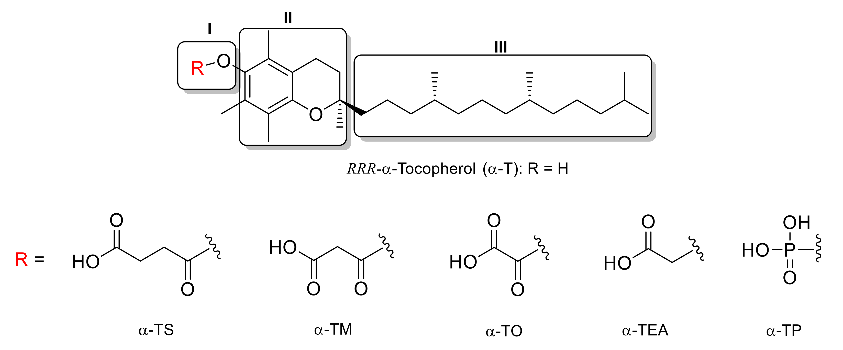
Three domains are distinguished in the structure of α-T and its derivatives (Figure 1). Domain I as the functional domain, is essential for the antioxidant or pro-apoptotic action of vitamin E. Acylation at the C-6 position with succinic anhydride makes α-tocopheryl succinate (α-TS) a dormant antioxidant with strong pro-apoptotic properties. Domain II consists of the chroman ring and is responsible for the signaling function affecting, among others, the activity of PP2A phosphatase and protein kinase C (PKC). Domain III includes the lipophilic side chain, which is responsible for docking tocopherol molecules in cell membranes and plasma lipoproteins [6,41,42].
The aim of the present study was to obtain some new α-tocopherol derivatives with higher anticancer activity than α-TS and α-TEA. For this purpose, we planned to modify the substituent at the C-6 position of tocopheroxyl moiety, which can modulate the original pro-apoptotic activity. We decided to introduce an additional group (carboxyl or alkyl) in the α-position relative to the terminal carboxyl group.
2. Results and Discussion
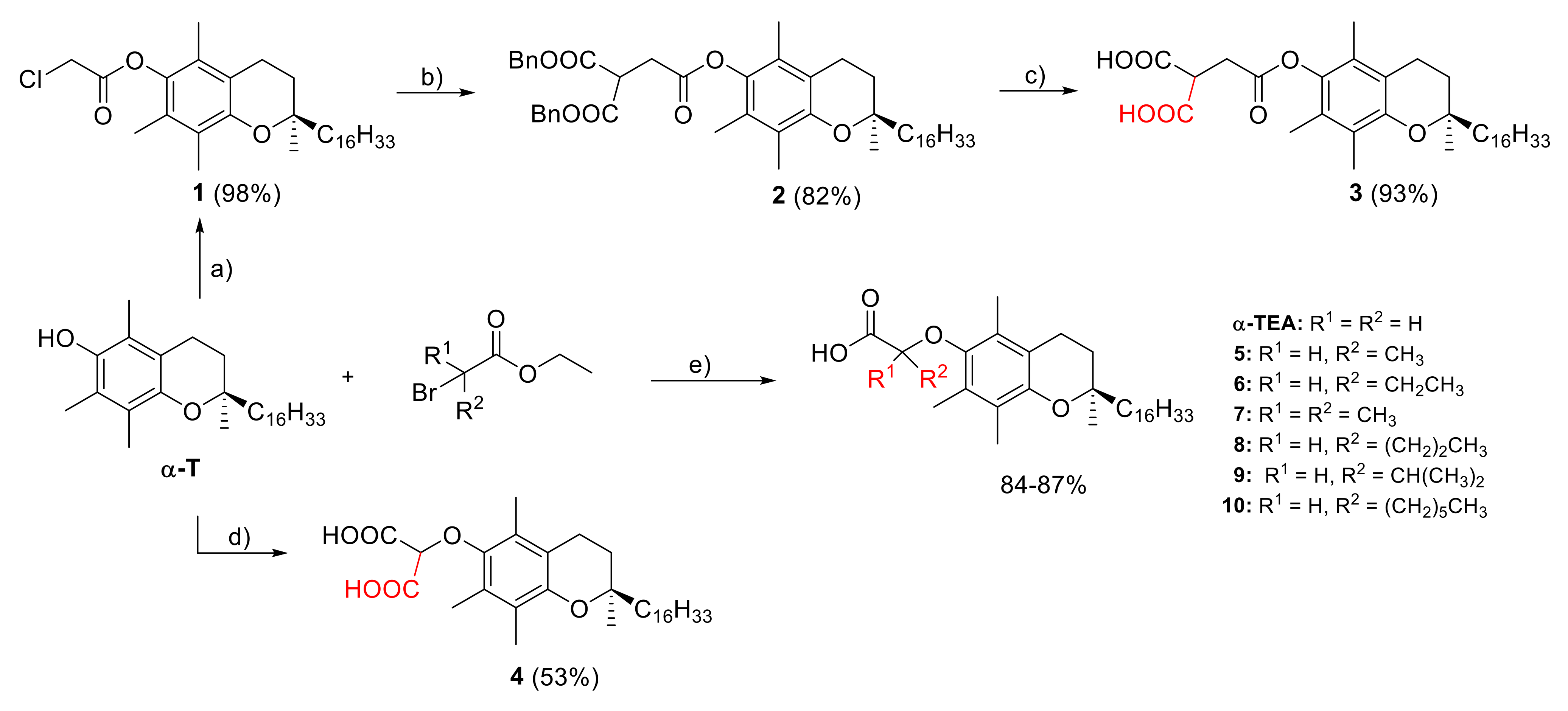
The synthesis of target vitamin E derivatives 3–10 was performed according to the synthetic route, as shown in Scheme 1. RRR-α-tocopherol (α-T) was the substrate in the described reactions. In the first variant, an additional carboxyl group was introduced into the α-position to the carboxyl group in the α-TS molecule. For this purpose, dibenzyl malonate was alkylated with α-tocopheryl chloroacetate (1), followed by hydrogenolysis of dibenzyl ester 2 to give compound 3. In another experiment, α-T was alkylated with diethyl bromomalonate followed by alkaline hydrolysis to give tocopheryloxymalonic acid (4). The analogs of α-TEA (5–10) with additional alkyl substituents in ether-linked acetic acid moiety were obtained by alkylation of α-T with corresponding α-bromoacid ethyl esters (i.e., 2-bromopropionate, 2-bromobutyrate, α-bromoisobutyrate, 2-bromovalerate, 2-bromo-3-methylbutyrate, 2-bromocaprylate, respectively). All reactions were carried out in mild conditions to give very good yields. The synthesized compounds were characterized by NMR, FT-IR, and HRMS analysis, and the obtained data were consistent with the desired structures.
To determine the cytotoxic activity, the synthesized compounds 3–10 were screened for their in vitro antiproliferative action against a panel of three different human cancer cell lines, namely human chronic lymphoblastic leukemia (CEM), human breast adenocarcinoma (MCF7), human cervical adenocarcinoma (HeLa), and normal human fibroblasts (BJ). For a better comparison, known compounds, namely α-TEA, α-TS, α-TM, α-TO, and α-TP, were also tested in our laboratory conditions. Furthermore, we used Doxorubicin (Dox), a known, anti-cancer drug, as a reference in cytotoxic assay. The obtained results are listed in Table 1.
As expected, Dox exhibited strong, non-selective cytotoxicity against all tested cell lines. α-T derivatives α-TEA, α-TS, α-MT, and α-TO showed moderate cytotoxicity against cancer cells and weak cytotoxicity toward normal human fibroblasts (BJ). This fact appears to be in accordance with the literature data, which reported a very limited or lack of toxicity toward non-malignant cells [13,42,43].
The selective toxicity of α-TS and α-TEA against malignant versus normal cells is probably related to the negative charge of the succinic or acetic part of the molecules at neutral pH [7,44]. This assumption was supported by an observation that pro-apoptotic action was increased in the acidic milieu [45]. The presence of a charged group appears to be essential for apoptosis induction. Methylation of the terminal carboxyl group deprives a compound of proapoptotic activity [46]. It was reported that pH of the interstitium of most tumors is 6.2–6.5, and for most normal tissues, it is in the range of 7.0–7.4 [47]. Thus, increasing the acidity in carboxylic moiety may result in higher apoptogenic activity. Such a tendency was observed for the α-tocopheryl dicarboxylic monoesters, increasing in the order: succinate < malonate < oxalate [12,13,48]. Following these observations, we decided to modify the acidic part in α-TS and α-TEA by introducing an additional carboxyl group into the α-position of the carboxyl group. Consequently, transformation into the malonic ending might result in stronger anticancer action due to increased acidity. Unexpectedly, the analog 3 (carboxylated α-TS) appeared inactive in the tests performed in three cancer cell lines. Similarly, α-tocopheryl phosphate (α-TP) was inactive against all investigated cancer cell lines. In turn, the compound 4 (carboxylated α-TEA) showed higher activity selectively against human cervical carcinoma cells (HeLa) compared to α-TEA. Particularly interesting anticancer profiles were obtained for the α-TEA analogs (5–10), in which alkyl substituents were introduced into the ether-linked acetic acid moiety. Only compound 10 containing the n-hexyl substituent in the α-position to the terminal carboxyl group was inactive against all tested cancer cell lines. 2-Tocopheryloxypropionic acid (5) showed higher cytotoxicity toward human chronic lymphoblastic leukemia CEM cells (24.5 ± 2.1) and much stronger action toward HeLa cells (17.2 ± 3.4) in comparison to the reference α-TEA. The replacing of methyl with ethyl group resulted in higher activity against HeLa (13.1 ± 0.1). Both compounds were also much less cytotoxic for normal fibroblasts (BJ). The most interesting results were obtained for compound 7 containing two geminal methyl groups (clofibric acid fragment), which revealed a considerable increase in activity against CEM cell line (14.1 ± 0.8). It also showed a relatively high therapeutic window because its cytotoxicity toward normal BJ cells was zero. Unexpectedly, it appeared inactive in assays in MCF7 and HeLa cell lines. Like most of the synthesized compounds, derivatives 8 and 9 showed moderate activity toward all cancer cell lines.
Based on the literature data, we have decided to determine the interaction of newly synthesized compounds with Complex II (CII), also known as succinate dehydrogenase (SDH), in silico via molecular docking simulation. Mitocans from the group of vitamin E derivatives interact with mitochondrial CII by interfering with ubiquinone (UbQ) functions, causing leakage of electrons and generation of reactive oxygen species (ROS), which induce selective apoptosis in cancer cells [3,49,50,51]. In the present work, the calculations were carried out in the Complex II active site for α-T derivatives 3–10 and, to compare, other species considered here, as detailed in the Section 3.3. The docking scores (affinities) of species tested here against their cytotoxicity are listed in Table 1. Additionally, for the sake of comparison, simulations were performed for α-T and ubiquinone-2 (UQ2) with affinities of −7.1 and −8.0 kcal/mol, respectively. Scores of newly synthesized species, changing in the range of −7.0 kcal/mol for 4 to −7.5 kcal/mol for 7, are not particularly different from each other, making it difficult to decide explicitly which is the most active. Additionally, affinities for compounds already reported in the literature (α-TS, α-TO, α-TM, α-TEA, and α-TP) are within a similar scope. Interestingly, derivatives with higher anticancer activity (5–8) simultaneously possess affinities slightly higher than that for α-TEA. Although not large, the differences may, however, contribute to the observed increased cytotoxicity toward cancer cells. While the bonding score for the native ligand UQ2 of −8.0 kcal/mol is more favorable, the one for the α-T (−7.1 kcal/mol) is noticeably worse. This indicates that substitution has a beneficial effect on the molecule’s ability to penetrate Complex II and thus confirms the consideration of α-T derivatives as new drug candidates. As shown below (Figure 2), ubiquinone-2 is bound to the active center of the protein via two hydrogen bonds to tryptophan (Trp164) from the subunit B of NEK1 and tyrosine (Tyr83, subunit C).
The key moieties involved in these interactions are N atoms from the pyrrole ring and the hydroxyl group from Tyr83 as hydrogen bond donors and ketone groups as acceptors, which further confirms the localization of ubiquinone-2 between three subunits of Complex 2 (B–D). As it is clearly seen in Figure 2, the arrangement of hydrogen bonds changes for α-T and its derivatives, replacing ubiquinone-2 in the protein. The peptide group from Gln78 is a H bond donor for α-T. In contrast, α-T derivatives interact with the N atoms from the aromatic rings of amino acids, as is the case for ubiquinone.
Figure 3 visualizes the exact spatial location of the best docking poses for the ligands in Figure 2. It is interesting to observe that the position of α-T differs significantly from that of the other three ligands. In fact, α-T seems to penetrate deeper into the structure of Complex II; in particular, it burrows more deeply into the D subunit. This could have significant consequences. Since it is reasonable to assume that the action of a ligand is most effective when it is located precisely in the active center of the enzyme itself, it can be inferred that the biological activity of α-T may be significantly less than that of its derivatives, which locate almost exactly at the site of ubiquinone-2 (UQ2), which is the native ligand. Indeed, similar conclusions also emerge from the experiments described in the previous section. As such, the in silico experiments also confirm the possible utility of the newly obtained compounds as drug candidates.
3. Materials and Methods
3.1. Chemistry
3.1.1. General
All reagents were purchased from commercial sources and used without further purification. Anhydrous solvents (i.e., THF, DCM, DMF, toluene, acetone, pyridine) were dried by distillation over appropriate drying agents under an argon atmosphere. D-α-tocopherol (RRR-α-tocopherol, α-T), D-α-tocopheryl succinate (α-TS), and α-bromoacid ethyl esters (i.e., 2-bromopropionate, 2-bromobutyrate, α-bromoisobutyrate, 2-bromovalerate, 2-bromo-3-methylbutyrate, 2-bromocaprylate) were purchased from Merck Life Sciences Sigma-Aldrich (Darmstadt, Germany). α-Tocopheryl malonate (α-TM) and α-tocopheryl oxalate (α-TO) were synthesized according to procedures in the literature [48]. α-Tocopheryl phosphate (α-TP) was prepared using phase transfer catalyzed phosphorylation method of phenols in a two-phase system (see Supplementary Materials file) [52].
Thin layer chromatography (TLC) was performed using Merck silica gel plates (0.25 mm, 60F-254) and visualized with CAM stain (ceric-ammonium-molybdate solution containing H2SO4). Standard flash chromatography (FC) was accomplished using silica gel (230–400 mesh size, J. T. Baker). Solvents for FC were freshly distilled before use.
NMR spectra were recorded at room temperature on a Bruker Avance DPX 200 or Avance II 400 spectrometers operating at 200 and 400 MHz, respectively. The chemical shifts (δ) are given in parts per million (ppm) relative to tetramethylsilane (TMS) using a residual solvent signal as an internal standard: CHCl3 = 7.26 ppm (1H NMR) and CDCl3 = 77.0 ppm (13C NMR). The following abbreviations are used to describe peak patterns: s = singlet, br = broad signal, d = doublet, t = triplet, q = quartet, m = multiplet. Only selected signals in the 1H NMR spectra are reported. The mean values of chemical shifts were given for signals for which the dynamic effects in 13C NMR spectra were observed. The original 1H and 13C NMR spectra are contained in the Supplementary Materials. FT-IR spectra were recorded in the range between 4000 and 500 cm−1 on a Nicolet 6700 spectrometer. HRMS data were acquired on an Agilent Technologies 6530 Accurate-Mass Q-TOF LC/MS.
3.1.2. Steps for the Preparation of Compound 3
Synthesis of RRR-α-tocopheryl chloroacetate (1)
A solution of chloroacetyl chloride (0.24 mL, 3 mmol) in dry toluene (4 mL) was added to a stirred solution of RRR-α-T (861 mg, 2 mmol) in dry toluene (10 mL) and dry pyridine (0.5 mL, 6 mmol) at room temperature. After 30 min, the resulting precipitate was filtrated through a pad of Celite, and the filtrate was washed with brine (15 mL), dried over anhydrous Na2SO4, and concentrated under reduced pressure. The crude product was purified using FC (hexane/ethyl acetate, 98:2, v/v) to yield compound 1 as a light-yellow oil (995 mg, 98% yield). 1H and 13C NMR data were consistent with those available in the literature [53].
Synthesis of dibenzyl RRR-α-tocopheryloxy-2-oxoethylmalonate (2)
To a stirred solution of dibenzyl malonate (341 mg, 1.2 mmol) in dry THF (10 mL), t-BuOK (146 mg, 1.3 mmol) was added at room temperature. After 30 min, solution of chloroacetate 1 (510 mg, 1 mmol) in dry THF (2 mL) was added. The reaction mixture was stirred overnight (16 h), poured into saturated solution of NH4Cl (20 mL), and extracted with diethyl ether (3 × 20 mL). The combined organic layers were washed with brine (20 mL), dried over anhydrous Na2SO4, and concentrated under vacuum. The crude product was purified using FC (hexane/ethyl acetate, 95:5, v/v) to yield compound 2 as a colorless oil (622 mg, 82% yield).
Compound 2: 1H NMR (200 MHz, CDCl3) δ 7.38–7.31 (m, 10H, H-Ar), 5.23–5.16 (m, 4H, CH2Ph), 4.11 (t, J = 7.5 Hz, 1H, CHCOOBn), 3.30 (d, J = 7.5 Hz, 2H, CH2CHCOOBn), 2.59 (t, J = 6.6 Hz, 2H, H-4), 2.09, 1.99, 1.95 (3s, 9H, H-5a, H-7a, H-8b), 1.89–1.72 (m, 2H, H-3), 0.90–0.86 (m, 12H, H-4′a, H-8′a, H-12′a, H-13′); 13C{1H} NMR (100 MHz, CDCl3) δ 169.5 (C), 168.0 (2 × C), 149.5 (C), 140.4 (C), 135.1 (2 x C), 128.9–128.1 (10 x CH), 126.6 (C), 124.9 (C), 123.0 (C), 117.4 (C), 75.05 (C), 67.6 (2 x CH2), 47.9 (CH), 40.2 (CH2), 39.4 (CH2), 37.6, 37.5, 37.4, 37.3 (4 × CH2), 32.8 (CH2), 32.8 (CH), 32.7 (CH), 31.0 (CH2), 28.0 (CH), 24.8 (CH2), 24.4 (CH2), 23.6 (CH3), 22.7 (CH3), 22.6 (CH3), 21.0 (CH2), 20.6 (CH2), 19.73 (CH3), 19.67 (CH3), 12.9 (CH3), 12.0 (CH3), 11.8 (CH3); IR (ATR) νmax 2928, 1748, 1456, 1239, 1154 cm−1.
Synthesis of RRR-α-tocopheryloxy-2-oxoethylmalonic acid (3)
The mixture of compound 2 (500 mg, 0.66 mmol) and 10% Pd/C (100 mg) in ethyl acetate (30 mL) was hydrogenated under atmospheric pressure at room temperature. After total conversion (4 h, TLC control), the reaction mixture was filtered through a pad of Celite, and the filtrate was evaporated under vacuum. The crude product was purified using flash chromatography (ethyl acetate) to yield compound 3 as a colorless oil (354 mg, 93% yield).
Compound 3: 1H NMR (400 MHz, CDCl3): δ 9.41 (bs, 1H, COOH), 4.04 (t, J = 7.0 Hz, 1H, CHCOOH), 3.31 (d, J = 7.0 Hz, 2H, CH2CHCOOH), 2.58 (t, J = 6.6 Hz, 2H, H-4), 2.09, 2.01, 1.96 (3s, 9H, H-5a, H-7a, H-8b), 1.84–1.71 (m, 2H, H-3), 0.89–0.85 (m, 12H, H-4′a, H-8′a, H-12′a, H-13′); 13C{1H}NMR (100 MHz, CDCl3) δ 173.0 (2 × C), 169.5 (C), 149.6 (C), 140.4 (C), 126.6 (C), 124.9 (C), 123.2 (C), 117.5 (C), 75.1 (C), 47.2 (CH), 40.4 (CH2), 39.4 (CH2), 37.6, 37.5, 37.4, 37.3 (4 × CH2), 32.8 (CH), 32.7 (CH), 32.5 (CH2), 31.0 (CH2), 28.0 (CH), 24.8 (CH2), 24.5 (CH2), 23.8 (CH3), 22.7 (CH3), 22.6 (CH3), 21.0 (CH2), 20.6 (CH2), 19.7 (CH3), 19.6 (CH3), 12.8 (CH3), 12.0 (CH3), 11.8 (CH3); IR (ATR): 3448, 2927, 1752, 1735, 1655, 1459, 1261, 1156, 1107 cm−1; HRMS m/z 575.3938 (calcd for C34H55O7 ([M+H]+ 575.3942).
3.1.3. Steps for the Preparation of Compound 4
Synthesis of ethyl RRR-α-tocopheryloxymalonate (4-S)
To a stirred solution of RRR-α-T (470 mg, 1.1 mmol) in dry acetone (20 mL), K2CO3 (1.2 g, 8.8 mmol) was added at room temperature. After 10 min, diethyl bromomalonate (981 mg, 4.1 mmol) was added to the reaction mixture. The resulting yellow suspension was refluxed for 18 h (TLC control). After cooling to room temperature, the inorganic material was filtered off, and the filtrate was concentrated under reduced pressure. The residue was dissolved in diethyl ether (20 mL), washed with brine (2 × 20 mL) and water (1 × 20 mL), dried over Na2SO4, and concentrated under vacuum. The crude product was purified by FC using hexane/ethyl acetate (gradient 50:1 → 10:1, v/v) as an eluent to give pure product as a colorless oil (530 mg, 87% yield).
Compound 4-S: 1H NMR (400 MHz, CDCl3) δ 4.69 (s, 1H, OCH), 4.34–4.26 (m, 4H, CH2CH3), 2.55 (t, J = 6.7 Hz, 2H, H-4), 2.15, 2.11, 2.06 (3s, 9H, H-5a, H-7a, H-8b), 1.80–1.76 (m, 2H, H-3), 0.88–0.84 (m, 12H, H-4′a, H-8′a, H-12′a, H-13′); 13C{1H} NMR (100 MHz, CDCl3) δ 166.5 (2 × C), 148.3 (C), 148.0 (C), 127.3 (C), 125.6 (C), 123.1 (C), 117.6 (C), 81.4 (CH), 74.8 (C), 62.0 (2 × CH2), 39.9 (CH2), 39.3 (CH2), 37.39–37.22 (4 × CH2), 32.7 (CH), 32.6 (CH), 31.2 (CH2), 27.9 (CH), 24.8 (CH2), 24.4 (CH2), 23.8 (CH3), 22.7 (CH3), 22.6 (CH3), 21.0 (CH2), 20.6 (CH2), 19.7 (CH3), 19.6 (CH3), 14.0 (2 × CH3), 13.2 (CH3), 12.3 (CH3), 11.8 (CH3).
Synthesis of RRR-α-tocopheryloxymalonic acid (4)
A solution of KOH (190 mg, 3.39 mmol) in water (2.5 mL) was added to a solution of compound 4-S (400 mg, 0.68 mmol) in THF (10 mL,) and the resulting mixture was stirred vigorously at room temperature. After 2 h, 6M HCl was added to reach pH 2 and extracted with ethyl acetate (3 × 50 mL). The combined organic extracts were washed with water (2 × 30 mL) and brine (2 × 30 mL), and dried over Na2SO4. The crude product was purified by FC using ethyl acetate as an eluent to yield compound 4 as a light-yellow oil (220 mg, 61% yield) and dried in vacuo for 48 h.
Compound 4: 1H NMR (400 MHz, CDCl3): δ 7.27 (bs, 2H, COOH), 4.80 (s, 1H, OCH), 2.55 (t, J = 6.5 Hz, 2H, H-4), 2.15, 2.12, 2.07 (3s, 9H, H-5a, H-7a, H-8b), 1.83–1.72 (m, 2H, H-3), 0.88–0.84 (m, 12H, H-4′a, H-8′a, H-12′a, H-13′); 13C{1H} NMR (100 MHz, CDCl3) δ: 169.8 (2 × C), 148.7 (C), 147.6 (C), 127.2 (C), 125.5 (C), 123.5 (C), 118.0 (C), 80.7 (CH), 75.1 (C), 40.2 (CH2), 39.4 (CH2), 37.6, 37.5, 37.4, 37.3 (4 × CH2), 32.8 (CH), 32.7 (CH), 31.1 (CH2), 28.0 (CH), 24.8 (CH2), 24.4 (CH2), 23.7 (CH3), 22.7 (CH3), 22.6 (CH3), 21.0 (CH2), 20.8 (CH2), 19.7 (CH3), 19.6 (CH3), 13.3 (CH3), 12.4 (CH3), 11.8 (CH3); IR (ATR): 3415, 2924 (broad), 2867, 1763, 1737, 1572, 1462, 1411, 1374, 1246, 1094 cm−1; HRMS m/z 533.3863 (calcd for C32H53O6 [M+H]+ 533.3858).
3.1.4. General Procedure for the Synthesis of α-TEA and Compounds 5–10
A solution of respective α-bromoester (4.51 mmol, 8.5 eq) in dry DMF (2 mL) and powdered NaOH (635 mg, 15.9 mmol, 30 eq) were added to a solution of α-T (230 mg, 0.53 mmol, 1 eq) in dry DMF (8 mL). The resulting yellow suspension was stirred overnight (18–24 h) at room temperature. Then, the reaction mixture was poured into water, acidified with 6M HCl to pH 2 and extracted with diethyl ether (3 × 30 mL). The combined organic extracts were washed with water (2 × 30 mL) and brine (2 × 30 mL), dried over Na2SO4, and concentrated under reduced pressure. The crude product was purified using flash column chromatography (gradient 60–100% ethyl acetate in hexane). The pure product was dried in vacuo for 48 h.
α-TEA (RRR-α-tocopheryloxyacetic acid): yellowish waxy solid; yield: 87%; 1H NMR (400 MHz, CDCl3) δ 4.25 (brs, 2H, OCH2), 2.56 (t, J = 6.6 Hz, 2H, H-4), 2.14, 2.10, 2.08 (3s, 9H, H-5a, H-7a, H-8b), 1.81–1.75 (m, 2H, H-3), 0.88–0.84 (m, 12H, H-4′a, H-8′a, H-12′a, H-13′); 13C{1H} NMR (100 MHz, CDCl3) δ 173.9 (C), 148.5 (C), 146.7 (C), 127.3 (C), 125.4 (C), 123.2 (C), 117.7 (C), 75.0 (C), 69.4 (OCH2), 40.1 (CH2), 39.3 (CH2), 37.43–37.26 (4 × CH2), 32.8 (CH), 32.7 (CH), 31.1 (CH2), 28.0 (CH), 24.8 (CH2), 24.4 (CH2), 23.8 (2 × CH3), 22.7 (CH3), 22.6 (CH3), 21.0 (CH2) 20.6 (CH2), 19.7 (CH3), 19.6 (CH3), 12.7 (CH3), 11.8 (2 × CH3); IR (ATR) νmax 2927 (broad), 1724, 1446, 1410, 1248, 1093, 1060 cm−1; HRMS m/z 487.3795 (calcd for C31H51O4 [M–H]− 487.3793).
Compound 5 (2-(RRR-α-tocopheryloxy)propanoic acid): yellowish waxy solid; yield: 85%; 1H NMR (400 MHz, CDCl3) δ 4.44 (q, 1H, J = 6.8 Hz, OCH), 2.56 (t, J = 6.4 Hz, 2H, H-4), 2.16, 2.12, 2.10 (3s, 9H, H-5a, H-7a, H-8b), 1.83–1.79 (m, 2H, H-3), 0.88–0.84 (m, 12H, H-4′a, H-8′a, H-12′a, H-13′); 13C{1H} NMR (100 MHz, CDCl3) δ 148.4 (C), 145.4 (C), 127.7 (C), 125.9 (C), 123.4 (C), 117.9 (C), 77.0 (OCH), 75.0 (C), 40.0 (CH2), 39.4 (CH2), 37.54–37.23 (4 × CH2), 32.8 (CH), 32.7 (CH), 31.2 (CH2), 28.0 (CH2), 24.8 (CH2), 24.4 (CH2), 23.8 (CH3), 22.7 (CH3), 22.6 (CH3), 21.0 (CH2), 20.7 (CH2), 19.7 (CH3), 19.6 (CH3), 17.4 (CH3), 13.8 (CH3), 12.9 (CH3), 11.9 (CH3); IR (ATR) νmax 2923 (broad), 2867, 1715, 1460, 1409, 1375, 1249, 1094 cm−1; HRMS m/z 501.3953 (calcd for C32H53O4 [M–H]− 501.3949).
Compound 6 (2-(RRR-α-tocopheryloxy)butanoic acid): white waxy solid; yield: 84%; 1H NMR (400 MHz, CDCl3) δ 4.28 (t, J = 5.7 Hz, 1H, OCH), 2.57 (t, J = 6.6 Hz, 2H, H-4), 2.17, 2.14, 2.09 (3s, 9H, H-5a, H-7a, H-8b), 1.95–1.92 (m, 2H), 1.80–1.78 (m, 2H), 0.89–0.85 (m, 12H, H-4′a, H-8′a, H-12′a, H-13′); 13C{1H} NMR (100 MHz, CDCl3): δ 175.0 (C), 148.2 (C), 146.8 (C), 127.6 (C), 125.7 (C), 123.3 (C), 117.8 (C), 82.5 (OCH), 74.9 (C), 40.0 (CH2), 39.4 (CH2), 37.54–37.27 (4 × CH2), 32.8 (CH), 32.7 (CH), 31.2 (CH2), 28.0 (CH), 24.9 (CH2), 24.8 (CH2), 24.4 (CH2), 23.8 (CH3), 22.7 (CH3), 22.6 (CH3), 21.0 (CH2), 20.8 (CH2), 19.7 (CH3), 19.6 (CH3), 13.7 (CH3), 12.9 (CH3), 11.9 (CH3), 8.9 (CH3); IR (ATR) νmax 2924 (broad), 2867, 1722, 1459, 1407, 1374, 1248, 1085 cm−1; HRMS m/z 517.4248 (calcd for C33H57O4 [M+H]+ 517.4251).
Compound 7 (2-(RRR-α-tocopheryloxy)-2-methylpropanoic acid): white waxy solid; yield: 86%; 1H NMR (400 MHz, CDCl3) δ 6.58 (bs, 1H, COOH), 2.57 (t, J = 6.4 Hz, 2H, H-4), 2.14, 2.10, 2.09 (3s, 9H, H-5a, H-7a, H-8b), 1.85–1.76 (m, 2H), 0.89–0.85 (m, 12H, H-4′a, H-8′a, H-12′a, H-13′); 13C{1H} NMR (100 MHz, CDCl3) δ 177.5 (C), 148.2 (C), 143.9 (C), 129.6 (C), 127.7 (C), 122.9 (C), 117.5 (C), 81.8 (O-C), 74.9 (C), 39.9 (CH2), 39.3 (CH2), 37.54–37.27 (4 × CH2), 32.8 (CH), 32.6 (CH), 31.3 (CH2), 28.0 (CH), 24.9 (2 × CH3), 24.8 (CH2), 24.4 (CH2), 23.7 (CH3), 22.7 (CH3), 22.6 (CH3), 21.0 (CH2), 20.8 (CH2), 19.7 (CH3), 19.6 (CH3), 14.9 (CH3), 14.0 (CH3), 11.9 (CH3); IR (ATR) νmax 2924 (broad), 1708, 1569, 1461, 1405, 1249, 1148, 1083 cm−1; HRMS m/z 515.4109 (calcd for C33H55O4 [M–H] 515.4106).
Compound 8 (2-(RRR-α-tocopheryloxy)-pentanoic acid): yellowish waxy solid; yield: 85%; 1H NMR (400 MHz, CDCl3) δ 9.63 (bs, 1H, COOH), 4.32 (t, J = 5.8 Hz, 1H, CH-O), 2.57 (t, J = 6.7 Hz, 2H, H-4), 2.17, 2.16, 2.09 (3s, 9H, H-5a, H-7a, H-8b), 1.88–1.73 (m, 2H), 2.96 (t, J = 7.3 Hz, 3H, CH2CH3), 0.89–0.85 (m, 12H, H-4′a, H-8′a, H-12′a, H-13′); 13C{1H} NMR (100 MHz, CDCl3) δ 175.2 (C), 148.2 (C), 146.9 (C), 127.5 (C), 125.7 (C), 123.3 (C), 117.8 (C), 81.4 (OCH), 74.9 (C), 40.0 (CH2), 39.4 (CH2), 37.45–37.28 (4 x CH2), 34.0 (CH2), 32.8 (CH), 32.7 (CH), 31.2 (CH2), 28.0 (CH), 24.8 (CH2), 24.4 (CH2), 23.8 (CH3), 22.7 (CH3), 22.6 (CH3), 21.0 (CH2), 20.7 (CH2), 19.7 (CH3), 19.6 (CH3), 17.9 (CH2), 14.0 (CH3), 13.7 (CH3), 12.8 (CH3), 11.9 (CH3); IR (ATR) νmax 2927 (broad), 1708, 1572, 1466, 1409, 1086 cm−1; HRMS m/z 529.4267 (calcd for C34H57O4 [M–H]− 529.4262).
Compound 9 (2-(RRR-α-tocopheryloxy)-3-methylbutanoic acid): yellowish waxy solid; yield: 86%; 1H NMR (400 MHz, CDCl3) δ 8.90 (bs, 1H, COOH), 4.21 (d, J = 4.7 Hz, 1H, CH-O), 2.58 (t, J = 6.7 Hz, 2H, H-4), 2.19, 2.15, 2.10 (3s, 9H, H-5a, H-7a, H-8b), 1.84–1.75 (m, 2H), 0.90–0.86 (m, 12H, H-4′a, H-8′a, H-12′a, H-13′); 13C NMR{1H} (100 MHz, CDCl3) δ 175.3 (C), 147.9 (C), 147.2 (C), 127.5 (C), 125.6 (C), 123.2 (C), 117.7 (C), 85.9 (OCH), 74.8 (C), 40.0 (CH2), 39.4 (CH2), 37.44–37.27 (4 × CH2), 32.8 (CH), 32.7 (CH), 31.3 (CH2), 31.1 (CH), 28.0 (CH), 24.8 (CH2), 24.4 (CH2), 23.8 (CH3), 22.7 (CH3), 22.6 (CH3), 21.0 (CH2), 20.7 (CH2), 19.7 (CH3), 19.6 (CH3), 18.6 (CH3), 17.3 (CH3), 13.9 (CH3), 13.0 (CH3), 11.9 (CH3); IR (ATR) νmax 2927 (broad), 1709, 1573, 1467, 1409, 1085 cm−1; HRMS m/z 529.4269 (calcd for C34H57O4 [M–H]− 529.4262).
Compound 10 (2-(RRR-α-tocopheryloxy)-octanoic acid): yellowish waxy solid; yield: 84%; 1H NMR (400 MHz, CDCl3) δ 10.09 (bs, 1H, COOH), 4.31 (t, J = 5.8 Hz, 1H, CH-O), 2.57 (t, J = 6.4 Hz, 2H, H-4), 2.17, 2.13, 2.09 (3s, 9H, H-5a, H-7a, H-8b), 0.89–0.85 (m, 15H, H-4′a, H-8′a, H-12′a, H-13′, CH2CH3); 13C{1H} NMR (100 MHz, CDCl3) δ 175.3 (C), 148.1 (C), 147.0 (C), 127.5 (C), 125.7 (C), 123.2 (C), 117.8 (C), 81.5 (OCH), 74.9 (C), 40.0 (CH2), 39.4 (CH2), 37.44–37.27 (4 × CH2), 32.8 (CH), 32.7 (CH), 32.0 (CH2), 31.6 (CH2), 31.2 (CH2), 29.2 (CH2), 28.0 (CH), 24.8 (CH2), 24.4 (CH2), 23.8 (CH3), 22.7 (CH3), 22.6 (CH3), 22.5 (CH2), 21.0 (CH2), 20.7 (CH2), 19.7 (CH3), 19.6 (CH3), 14.0 (CH3), 13.7 (CH3), 12.8 (CH3), 11.9 (CH3) ppm; IR (ATR): 2929 (broad), 1710, 1572, 1467, 1411, 1087 cm−1; HRMS m/z 571.4738 (calcd for C37H59O4 [M–H]− 571.4732).
The NMR spectra of compounds 3-10, α-TEA, and α-TP are presented in the Supplementary Materials file (Figures S3–S24 from Supplementary Materials).
3.2. Cytotoxicity Test Assay
T-lymphoblastic leukemia CEM, cervix epithelioid carcinoma HeLa, and breast carcinoma MCF7 cell lines, all of which were obtained from the European Collection of Authenticated Cell Cultures (ECACC, London, UK) were used for screening. Human foreskin fibroblasts (BJ) were purchased from the American Type Culture Collection (Manassas, VA, USA). The cytotoxicity of tested compounds in the described cell lines and normal cells was determined using resazurin assay following the manufacturer’s protocol (Sigma Aldrich, St. Louis, MO, USA).
Stock solutions of compounds were prepared in DMSO at 7.5 mM and the highest tested concentration in cultivation medium in well was 50 µM. The concentration of DMSO in well never exceeded 0.6%. Briefly, 5.0 × 10⁴ cells·mL−1 cells were seeded into 96-well plates (TPP, Trasadingen, Switzerland) and incubated for 24 h. Then, compounds solved in DMSO were added into cultivation medium and incubated further for 72 h. Cell viability was determined using resazurin. The procedure has been reported [54].
3.3. Molecular Docking
In order to acquire a deeper understanding of the biological activity of α-tocopherol derivatives, a molecular docking study was conducted with the AutoDock Vina (version 1.1.3) [55] program, provided by Scripps Research Institute. The visualization was carried out in Pymol [56] and LigPlus [57] programs. The crystal structure of Complex II (succinate dehydrogenase, SDH) from Escherichia Coli was obtained from the Protein Data Bank (PDB:1NEK) [58]. The enzyme was prepared for docking by cleansing its structure of water molecules and other co-crystallized species. Additionally, polar hydrogens and Kollman charges [59] were added. The search box was set to cover the co-crystallized ubiquinone-2 molecule (PDb:UQ2), which is located on the edge of the subunit D of 1NEK. A cubical (20 × 0 × 20 Å) grid box was utilized during the simulations. For the sake of validation, a redocking procedure for the ubiquinone-2 (a native ligand) was conducted. To improve docking accuracy, the EXHAUSTIVENESS parameter had been set to 100 (default = 8), ensuring the best performance [60] in treating large ligands with long phytyl chains. Additionally, for the same purpose, the SPACING parameter was set to 0.15, which is far below its default value of 0.375. The best (in the sense of docking score) pose was compared to UQ2 from the crystal structure, obtained with the resolution of 2.6 Å. Both conformations were found to be in satisfactory agreement, with RMSD = 1.313Å. The aligned structures are pictured in Figure 4.
4. Conclusions
In this project, a series of α-TS and α-TEA analogs have been synthesized by modification of the substituent in the C-6 position of the α-T molecule. All compounds possess the structural requirements described for their proapoptotic activity of α-tocopherol esters, i.e., the combination of three structural domains: functional, signaling, and hydrophobic. All compounds were examined for their in vitro cytotoxic activity against three human cancer cell lines (CEM, MCF7, HeLa), and normal human fibroblasts (BJ). Our results showed a relationship between anticancer properties and the structure of the substituent located at the C-6 position in the α-T molecule. Surprisingly, the analogs of α-TEA and α-TS (3 and 4, respectively) in which the acidic parts (acetic or succinic, respectively) contained an additional carboxyl group proved to be inactive. On the other hand, the introduction of an alkyl substituent (e.g., methyl or ethyl) into the ether-linked acetic acid moiety in α-TEA increases cytotoxic activity. Promising drug candidates seem to be compounds 5–8. They all had moderate activity toward tested cancer cell lines and relatively high therapeutic window as their cytotoxicity toward normal fibroblasts was very low or zero. Molecular docking to Complex II has shown that the affinities of newly synthesized compounds as ligands are approximately on the same level, and there is no easy way to indicate which one is potentially most useful. However, the differences between α-T and its derivatives clearly indicate that, while behaving similarly in affinity to the native ligand (UQ2), they differ significantly from α-T in terms of the localization and interaction with the receptor. Combined with better affinities, this may explain their significantly higher anticancer activity and confirm their suitability as drug candidates. Our results demonstrate that it is possible to achieve good selectivity and higher proapoptotic activity with the introduction of larger substituents into the acidic ethereal part in the α-TEA molecule. However, it should be taken into account that the results of enzymatic assays may differ from cytotoxic ones and may shed new light on the mechanism of action of the tested compounds. Further investigations are in progress.
Supplementary Materials
The following supporting information can be downloaded at: https://www.mdpi.com/article/10.3390/pr11061860/s1. S2, Experimental procedures for the preparation of α-tocopheryl phosphate (α-TP); S3–S24, 1H and 13C NMR spectra of the synthesized compounds.
Author Contributions
Conceptualization, A.B. and S.W.; methodology, A.B., L.R. and S.W.; formal analysis, A.B., L.R. and A.R.; investigation, A.B., L.R. and A.R.; visualization, A.B. and A.R.; writing—original draft preparation, A.B.; writing—review and editing, A.B., L.R., A.R., M.S. and S.W.; supervision, S.W. and. M.S. All authors have read and agreed to the published version of the manuscript.
Funding
This work was carried out as part of the project “Synthesis of vitamin E derivatives with potential anticancer activity” (Research of Young Scientists, 2019–2020, Faculty of Chemistry, University of Bialystok) financed by the Ministry of Science and Higher Education, Poland (MNiSW).
Institutional Review Board Statement
Not applicable.
Informed Consent Statement
Not applicable.
Data Availability Statement
All relevant data that support the findings of this study are included in this article.
Acknowledgments
Authors are grateful to Leszek Siergiejczyk for recording the NMR spectra (Faculty of Chemistry, University of Bialystok) and Aneta M. Tomkiel (Faculty of Chemistry, University of Bialystok) for HRMS measurements. The authors would like to thank the Computational Center of the University of Bialystok (Grant GO-008) for providing access to the supercomputer resources and the AutoDock VINA program.
Conflicts of Interest
The authors declare no conflict of interest.
References
- Chorawala, M.R.; Oza, P.M.; Shah, G.B. Mechanism of Anticancer Drugs Resistance: An Overview. Int. J. Pharm. Sci. Drug Res. 2012, 4, 1–9. [Google Scholar]
- Panda, V.; Khambat, P.; Patil, S. Mitocans as Novel Agents for Anticancer Therapy: An Overview. Int. J. Clin. Med. 2011, 2, 515–529. [Google Scholar] [CrossRef] [Green Version]
- Dong, L.-F.; Jameson, V.J.A.; Tilly, D.; Cerny, J.; Mahdavian, E.; Marín-Hernández, A.; Hernández-Esquivel, L.; Rodríguez-Enríquez, S.; Stursa, J.; Witting, P.K.; et al. Mitochondrial Targeting of Vitamin E Succinate Enhances Its Pro-Apoptotic and Anti-Cancer Activity via Mitochondrial Complex II. J. Biol. Chem. 2011, 286, 3717–3728. [Google Scholar] [CrossRef] [Green Version]
- Neuzil, J.; Dyason, J.C.; Freeman, R.; Dong, L.-F.; Prochazka, L.; Wang, X.-F.; Scheffler, I.; Ralph, S.J. Mitocans as Anti-Cancer Agents Targeting Mitochondria: Lessons from Studies with Vitamin E Analogues, Inhibitors of Complex II. J. Bioenerg. Biomembr. 2007, 39, 65–72. [Google Scholar] [CrossRef] [PubMed]
- Dong, L.; Gopalan, V.; Holland, O.; Neuzil, J. Mitocans Revisited: Mitochondrial Targeting as Efficient Anti-Cancer Therapy. Int. J. Mol. Sci. 2020, 21, 7941. [Google Scholar] [CrossRef]
- Neuzil, J.; Tomasetti, M.; Mellick, A.; Alleva, R.; Salvatore, B.; Birringer, M.; Fariss, M. Vitamin E Analogues: A New Class of Inducers of Apoptosis with Selective Anti-Cancer Effects. CCDT 2004, 4, 355–372. [Google Scholar] [CrossRef] [PubMed]
- Neuzil, J.; Weber, T.; Schröder, A.; Lu, M.; Ostermann, G.; Gellert, N.; Mayne, G.C.; Olejnicka, B.; Nègre-Salvayre, A.; Stícha, M.; et al. Induction of Cancer Cell Apoptosis by α-Tocopheryl Succinate: Molecular Pathways and Structural Requirements. FASEB J. 2001, 15, 403–415. [Google Scholar] [CrossRef] [PubMed]
- Weber, T.; Lu, M.; Andera, L.; Lahm, H.; Gellert, N.; Fariss, M.W.; Korinek, V.; Sattler, W.; Ucker, D.S.; Terman, A.; et al. Vitamin E Succinate Is a Potent Novel Antineoplastic Agent with High Selectivity and Cooperativity with Tumor Necrosis Factor-Related Apoptosis-Inducing Ligand (Apo2 Ligand) In Vivo. Clin. Cancer Res. 2002, 8, 863. [Google Scholar]
- Yu, Z.-Q.; Wang, L.-M.; Yang, W.-X. How Vitamin E and Its Derivatives Regulate Tumour Cells via the MAPK Signalling Pathway? Gene 2022, 808, 145998. [Google Scholar] [CrossRef]
- Majima, D.; Mitsuhashi, R.; Fukuta, T.; Tanaka, T.; Kogure, K. Biological Functions of α-Tocopheryl Succinate. J. Nutr. Sci. Vitaminol. 2019, 65, S104–S108. [Google Scholar] [CrossRef]
- Wang, X.-F.; Dong, L.; Zhao, Y.; Tomasetti, M.; Wu, K.; Neuzil, J. Vitamin E Analogues as Anticancer Agents: Lessons from Studies with α-Tocopheryl Succinate. Mol. Nutr. Food Res. 2006, 50, 675–685. [Google Scholar] [CrossRef] [PubMed]
- Kogure, K.; Manabe, S.; Suzuki, I.; Tokumura, A.; Fukuzawa, K. Cytotoxicity of Alpha-Tocopheryl Succinate, Malonate and Oxalate in Normal and Cancer Cells in vitro and Their Anti-Cancer Effects on Mouse Melanoma in vivo. J. Nutr. Sci. Vitaminol. 2005, 51, 392–397. [Google Scholar] [CrossRef] [PubMed] [Green Version]
- Zhao, Y.; Neuzil, J.; Wu, K. Vitamin E Analogues as Mitochondria-Targeting Compounds: From the Bench to the Bedside? Mol. Nutr. Food Res. 2009, 53, 129–139. [Google Scholar] [CrossRef] [PubMed] [Green Version]
- Goh, S.H.; Hew, N.F.; Ong, A.S.H.; Choo, Y.M.; Brumby, S. Tocotrienols from Palm Oil: Electron Spin Resonance Spectra of Tocotrienoxyl Radicals. J. Am. Oil Chem. Soc. 1990, 67, 250–254. [Google Scholar] [CrossRef]
- Ghayour-Mobarhan, M.; Saghiri, Z.; Ferns, G.; Sahebkar, A. α-Tocopheryl Phosphate as a Bioactive Derivative of Vitamin E: A Review of the Literature. J. Diet. Suppl. 2015, 12, 359–372. [Google Scholar] [CrossRef] [PubMed]
- Ju, J.; Picinich, S.C.; Yang, Z.; Zhao, Y.; Suh, N.; Kong, A.-N.; Yang, C.S. Cancer-Preventive Activities of Tocopherols and Tocotrienols. Carcinogenesis 2010, 31, 533–542. [Google Scholar] [CrossRef] [Green Version]
- Rezk, B.M. Alpha-Tocopheryl Phosphate Is a Novel Apoptotic Agent. Front. Biosci. 2007, 12, 2013. [Google Scholar] [CrossRef] [Green Version]
- Bidossi, A.; Bortolin, M.; Toscano, M.; De Vecchi, E.; Romanò, C.L.; Mattina, R.; Drago, L. In Vitro Comparison between α-Tocopheryl Acetate and α-Tocopheryl Phosphate against Bacteria Responsible of Prosthetic and Joint Infections. PLoS ONE 2017, 12, e0182323. [Google Scholar] [CrossRef] [Green Version]
- Harper, R.A.; Saleh, M.M.; Carpenter, G.; Abbate, V.; Proctor, G.; Harvey, R.D.; Gambogi, R.J.; Geonnotti, A.; Hider, R.; Jones, S.A. Soft, Adhesive (+) Alpha Tocopherol Phosphate Planar Bilayers That Control Oral Biofilm Growth through a Substantive Antimicrobial Effect. Nanomed. Nanotechnol. Biol. Med. 2018, 14, 2307–2316. [Google Scholar] [CrossRef]
- Hama, S.; Kirimura, N.; Obara, A.; Takatsu, H.; Kogure, K. Tocopheryl Phosphate Inhibits Rheumatoid Arthritis-Related Gene Expression In Vitro and Ameliorates Arthritic Symptoms in Mice. Molecules 2022, 27, 1425. [Google Scholar] [CrossRef]
- Dong, L.-F.; Grant, G.; Massa, H.; Zobalova, R.; Akporiaye, E.; Neuzil, J. α-Tocopheryloxyacetic Acid Is Superior to α-Tocopheryl Succinate in Suppressing HER2-High Breast Carcinomas Due to Its Higher Stability. Int. J. Cancer 2012, 131, 1052–1058. [Google Scholar] [CrossRef] [PubMed] [Green Version]
- Lawson, K.A.; Anderson, K.; Menchaca, M.; Atkinson, J.; Sun, L.; Knight, V.; Gilbert, B.E.; Conti, C.; Sanders, B.G.; Kline, K. Novel Vitamin E Analogue Decreases Syngeneic Mouse Mammary Tumor Burden and Reduces Lung Metastasis. Mol. Cancer Ther. 2003, 2, 437. [Google Scholar] [PubMed]
- Lawson, K.A.; Anderson, K.; Simmons-Menchaca, M.; Atkinson, J.; Sun, L.; Sanders, B.G.; Kline, K. Comparison of Vitamin E Derivatives α-TEA and VES in Reduction of Mouse Mammary Tumor Burden and Metastasis. Exp. Biol. Med. 2004, 229, 954–963. [Google Scholar] [CrossRef] [PubMed]
- Hahn, T.; Akporiaye, E.T. α-TEA as a Stimulator of Tumor Autophagy and Enhancer of Antigen Cross-Presentation. Autophagy 2013, 9, 429–431. [Google Scholar] [CrossRef] [PubMed] [Green Version]
- Hahn, T.; Bradley-Dunlop, D.J.; Hurley, L.H.; Von-Hoff, D.; Gately, S.; Mary, D.L.; Lu, H.; Penichet, M.L.; Besselsen, D.G.; Cole, B.B.; et al. The Vitamin E Analog, Alpha-Tocopheryloxyacetic Acid Enhances the Anti-Tumor Activity of Trastuzumab against HER2/Neu-Expressing Breast Cancer. BMC Cancer 2011, 11, 471. [Google Scholar] [CrossRef] [Green Version]
- Kawamura, K.; Kume, A.; Umemiya-Shirafuji, R.; Kasai, S.; Suzuki, H. Effect of α-Tocopheryloxy Acetic Acid, a Vitamin E Derivative Mitocan, on the Experimental Infection of Mice with Plasmodium Yoelii. Malar. J. 2021, 20, 280. [Google Scholar] [CrossRef]
- Hahn, T.; Szabo, L.; Gold, M.; Ramanathapuram, L.; Hurley, L.H.; Akporiaye, E.T. Dietary Administration of the Proapoptotic Vitamin E Analogue Alpha-Tocopheryloxyacetic Acid Inhibits Metastatic Murine Breast Cancer. Cancer Res. 2006, 66, 9374–9378. [Google Scholar] [CrossRef] [Green Version]
- Kline, K.; Yu, W.; Sanders, B.G. Vitamin E and Breast Cancer. J. Nutr. 2004, 134, 3458S–3462S. [Google Scholar] [CrossRef] [Green Version]
- Tiwary, R.; Yu, W.; Sanders, B.G.; Kline, K. α-TEA Cooperates with Chemotherapeutic Agents to Induce Apoptosis of P53 Mutant, Triple-Negative Human Breast Cancer Cells via Activating P73. Breast Cancer Res. 2011, 13, R1. [Google Scholar] [CrossRef] [Green Version]
- Li, Y.; Hahn, T.; Garrison, K.; Cui, Z.-H.; Thorburn, A.; Thorburn, J.; Hu, H.-M.; Akporiaye, E.T. The Vitamin E Analogue α-TEA Stimulates Tumor Autophagy and Enhances Antigen Cross-Presentation. Cancer Res. 2012, 72, 3535–3545. [Google Scholar] [CrossRef] [Green Version]
- Neuzil, J.; Kågedal, K.; Andera, L.; Weber, C.; Brunk, U.T. Vitamin E Analogs: A New Class of Multiple Action Agents with Anti-Neoplastic and Anti-Atherogenic Activity. Apoptosis 2002, 7, 179–187. [Google Scholar] [CrossRef]
- Jia, L.; Yu, W.; Wang, P.; Sanders, B.G.; Kline, K. In Vivo and in vitro Studies of Anticancer Actions of α-TEA for Human Prostate Cancer Cells. Prostate 2008, 68, 849–860. [Google Scholar] [CrossRef]
- Yao, J.; Gao, P.; Xu, Y.; Li, Z. α-TEA Inhibits the Growth and Motility of Human Colon Cancer Cells via Targeting RhoA/ROCK Signaling. Mol. Med. Rep. 2016, 14, 2534–2540. [Google Scholar] [CrossRef] [Green Version]
- Wu, Y.; Zu, K.; Ni, J.; Yeh, S.; Kasi, D.; James, N.S.; Chemler, S.; Ip, C. Cellular and Molecular Effects of Alpha-Tocopheryloxybutyrate: Lessons for the Design of Vitamin E Analog for Cancer Prevention. Anticancer Res. 2004, 24, 3795–3802. [Google Scholar]
- Ni, J.; Mai, T.; Pang, S.-T.; Haque, I.; Huang, K.; DiMaggio, M.A.; Xie, S.; James, N.S.; Kasi, D.; Chemler, S.R.; et al. In Vitro and In Vivo Anticancer Effects of the Novel Vitamin E Ether Analogue RRR-Tocopheryloxybutyl Sulfonic Acid in Prostate Cancer. Clin. Cancer Res. 2009, 15, 898–906. [Google Scholar] [CrossRef] [PubMed] [Green Version]
- Khallouki, F.; Hajji, L.; Saber, S.; Bouddine, T.; Edderkaoui, M.; Bourhia, M.; Mir, N.; Lim, A.; El Midaoui, A.; Giesy, J.P.; et al. An Update on Tamoxifen and the Chemo-Preventive Potential of Vitamin E in Breast Cancer Management. JPM 2023, 13, 754. [Google Scholar] [CrossRef]
- Zhang, S.; Lawson, K.A.; Simmons-Menchaca, M.; Sun, L.; Sanders, B.G.; Kline, K. Vitamin E Analog α-TEA and Celecoxib Alone and Together Reduce Human MDA-MB-435-FL-GFP Breast Cancer Burden and Metastasis in Nude Mice. Breast Cancer Res. Treat. 2004, 87, 111–121. [Google Scholar] [CrossRef]
- Anderson, K.; Lawson, K.A.; Simmons-Menchaca, M.; Sun, L.; Sanders, B.G.; Kline, K. Alpha-TEA plus Cisplatin Reduces Human Cisplatin-Resistant Ovarian Cancer Cell Tumor Burden and Metastasis. Exp. Biol. Med. 2004, 229, 1169–1176. [Google Scholar] [CrossRef]
- Yu, W.; Shun, M.; Anderson, K.; Chen, H.; Sanders, B.G.; Kline, K. α-TEA Inhibits Survival and Enhances Death Pathways in Cisplatin Sensitive and Resistant Human Ovarian Cancer Cells. Apoptosis 2006, 11, 1813–1823. [Google Scholar] [CrossRef] [PubMed]
- Suntharalingam, K.; Song, Y.; Lippard, S.J. Conjugation of Vitamin E Analog α-TOS to Pt(Iv) Complexes for Dual-Targeting Anticancer Therapy. Chem. Commun. 2014, 50, 2465. [Google Scholar] [CrossRef] [PubMed] [Green Version]
- Neuzil, J. α-Tocopheryl Succinate Epitomizes a Compound with a Shift in Biological Activity Due to pro-Vitamin-to-Vitamin Conversion. Biochem. Biophys. Res. Commun. 2002, 293, 1309–1313. [Google Scholar] [CrossRef] [PubMed]
- Birringer, M.; EyTina, J.H.; Salvatore, B.A.; Neuzil, J. Vitamin E Analogues as Inducers of Apoptosis: Structure–Function Relation. Br. J. Cancer 2003, 88, 1948–1955. [Google Scholar] [CrossRef] [PubMed] [Green Version]
- Neuzil, J.; Tomasetti, M.; Zhao, Y.; Dong, L.-F.; Birringer, M.; Wang, X.-F.; Low, P.; Wu, K.; Salvatore, B.A.; Ralph, S.J. Vitamin E Analogs, a Novel Group of “Mitocans”, as Anticancer Agents: The Importance of Being Redox-Silent. Mol. Pharmacol. 2007, 71, 1185–1199. [Google Scholar] [CrossRef] [PubMed] [Green Version]
- Neuzil, J.; Weber, T.; Gellert, N.; Weber, C. Selective Cancer Cell Killing by Alpha-Tocopheryl Succinate. Br. J. Cancer 2001, 84, 87–89. [Google Scholar] [CrossRef] [Green Version]
- Neuzil, J.; Zhao, M.; Ostermann, G.; Sticha, M.; Gellert, N.; Weber, C.; Eaton, J.W.; Brunk, U.T. α-Tocopheryl Succinate, an Agent with in vivo Anti-Tumour Activity, Induces Apoptosis by Causing Lysosomal Instability. Biochem. J. 2002, 362, 709–715. [Google Scholar] [CrossRef] [PubMed]
- Mazzini, F.; Betti, M.; Canonico, B.; Netscher, T.; Luchetti, F.; Papa, S.; Galli, F. Anticancer Activity of Vitamin E-Derived Compounds in Murine C6 Glioma Cells. ChemMedChem 2010, 5, 540–543. [Google Scholar] [CrossRef]
- Gerweck, L.E.; Seetharaman, K. Cellular PH Gradient in Tumor versus Normal Tissue: Potential Exploitation for the Treatment of Cancer. Cancer Res. 1996, 56, 1194–1198. [Google Scholar]
- Kogure, K.; Hama, S.; Kisaki, M.; Takemasa, H.; Tokumura, A.; Suzuki, I.; Fukuzawa, K. Structural Characteristic of Terminal Dicarboxylic Moiety Required for Apoptogenic Activity of α-Tocopheryl Esters. Biochim. Biophys. Acta (BBA) Gen. Subj. 2004, 1672, 93–99. [Google Scholar] [CrossRef]
- Yan, B.; Stantic, M.; Zobalova, R.; Bezawork-Geleta, A.; Stapelberg, M.; Stursa, J.; Prokopova, K.; Dong, L.; Neuzil, J. Mitochondrially Targeted Vitamin E Succinate Efficiently Kills Breast Tumour-Initiating Cells in a Complex II-Dependent Manner. BMC Cancer 2015, 15, 401. [Google Scholar] [CrossRef] [Green Version]
- Dong, L.-F.; Low, P.; Dyason, J.C.; Wang, X.-F.; Prochazka, L.; Witting, P.K.; Freeman, R.; Swettenham, E.; Valis, K.; Liu, J.; et al. α-Tocopheryl Succinate Induces Apoptosis by Targeting Ubiquinone-Binding Sites in Mitochondrial Respiratory Complex II. Oncogene 2008, 27, 4324–4335. [Google Scholar] [CrossRef] [Green Version]
- Neuzil, J.; Cerny, J.; Dyason, J.C.; Dong, L.-F.; Ralph, S.J. Affinity of Vitamin E Analogues for the Ubiquinone Complex II Site Correlates with Their Toxicity to Cancer Cells. Mol. Nutr. Food Res. 2011, 55, 1543–1551. [Google Scholar] [CrossRef]
- Zwierzak, A. Phase-Transfer-Catalysed Phosphorylation of Alcohols in a Two-Phase System. Synthesis 1976, 1976, 305–306. [Google Scholar] [CrossRef]
- López, G.V.; Batthyány, C.; Blanco, F.; Botti, H.; Trostchansky, A.; Migliaro, E.; Radi, R.; González, M.; Cerecetto, H.; Rubbo, H. Design, Synthesis, and Biological Characterization of Potential Antiatherogenic Nitric Oxide Releasing Tocopherol Analogs. Bioorg. Med. Chem. 2005, 13, 5787–5796. [Google Scholar] [CrossRef]
- Rárová, L.; Steigerová, J.; Kvasnica, M.; Bartůněk, P.; Křížová, K.; Chodounská, H.; Kolář, Z.; Sedlák, D.; Oklestkova, J.; Strnad, M. Structure Activity Relationship Studies on Cytotoxicity and the Effects on Steroid Receptors of AB-Functionalized Cholestanes. J. Steroid Biochem. Mol. Biol. 2016, 159, 154–169. [Google Scholar] [CrossRef]
- Trott, O.; Olson, A.J. AutoDock Vina: Improving the Speed and Accuracy of Docking with a New Scoring Function, Efficient Optimization, and Multithreading. J. Comput. Chem. 2009, 31, 455–461. [Google Scholar] [CrossRef] [PubMed] [Green Version]
- Schrödinger, L.; DeLano, W. The PyMOL Molecular Graphics System, Version 1.3r1. PyMOL. Available online: http://www.pymol.org/pymol (accessed on 26 May 2023).
- Wallace, A.C.; Laskowski, R.A.; Thornton, J.M. LIGPLOT: A Program to Generate Schematic Diagrams of Protein-Ligand Interactions. Protein Eng. Des. Sel. 1995, 8, 127–134. [Google Scholar] [CrossRef] [PubMed]
- Yankovskaya, V.; Horsefield, R.; Törnroth, S.; Luna-Chavez, C.; Miyoshi, H.; Léger, C.; Byrne, B.; Cecchini, G.; Iwata, S. Architecture of Succinate Dehydrogenase and Reactive Oxygen Species Generation. Science 2003, 299, 700–704. [Google Scholar] [CrossRef] [PubMed] [Green Version]
- Singh, U.C.; Kollman, P.A. An Approach to Computing Electrostatic Charges for Molecules. J. Comput. Chem. 1984, 5, 129–145. [Google Scholar] [CrossRef]
- Agarwal, R.; Smith, J.C. Speed vs Accuracy: Effect on Ligand Pose Accuracy of Varying Box Size and Exhaustiveness in AutoDock Vina. Mol. Inform. 2023, 42, 2200188. [Google Scholar] [CrossRef]
Figure 1.
Chemical structure of α-tocopherol (α-T), α-tocopheryl succinate (α-TS), α-tocopheryl malonate (α-TM), α-tocopheryl oxalate (α-TO), α-tocopheryloxyacetic acid (α-TEA), and α-tocopheryl phosphate (α-TP) (Domains in vitamin E molecule and its analogs: I—functional, II—signaling, III—hydrophobic).

Scheme 1.
Synthesis of vitamin E derivatives (2–10) from RRR-α-tocopherol (α-T). Reagents and conditions: (a) chloroacetyl chloride, pyridine, toluene, rt; (b) CH2(COOBn)2, t-BuOK, THF, rt; (c) H2 (1 atm), Pd/C, ethyl acetate; (d) 1. diethyl bromomalonate, K2CO3, acetone, reflux, 2. KOHaq, THF, rt; (e) NaOH, DMF, rt.

Figure 2.
The pattern of hydrogen bonds for the best scored docking modes of ubiquinone-2, α-tocopherol (α-T), compound 5 and compound 7 to Complex II. Green dashed lines symbolize hydrogen bonds, whereas red half circles with outlines mean nonpolar interactions. Bond lengths are given in angstroms.

Figure 3.
The best docking mods to the active center of Complex II for: ubiquinone-2 (UQ2, green color), α-tocopherol (blue color), compounds 5 (light brown color) and 7 (magenta). Subunits of Complex II are colored as follows: A—green, B—cyan, C—magenta, D—yellow.
Figure 3.
The best docking mods to the active center of Complex II for: ubiquinone-2 (UQ2, green color), α-tocopherol (blue color), compounds 5 (light brown color) and 7 (magenta). Subunits of Complex II are colored as follows: A—green, B—cyan, C—magenta, D—yellow.

Figure 4.
The alignment of the native UQ2 ligand (green scaffold) compared to the best pose from the redocking procedure (cyan scaffold).
Figure 4.
The alignment of the native UQ2 ligand (green scaffold) compared to the best pose from the redocking procedure (cyan scaffold).


Table 1.
IC50 (μM) on three cancer cell lines (CEM, MCF7, HeLa) and normal human fibroblasts (BJ) after 72 h of incubation with the tested compounds. The data are means ± standard deviation (SD) obtained from at least three independent experiments performed in triplicate.
Table 1.
IC50 (μM) on three cancer cell lines (CEM, MCF7, HeLa) and normal human fibroblasts (BJ) after 72 h of incubation with the tested compounds. The data are means ± standard deviation (SD) obtained from at least three independent experiments performed in triplicate.
 | ||||||
| Compound | R | CEM | MCF7 | HeLa | BJ | Complex II Affinity (kcal/mol) |
|---|---|---|---|---|---|---|
| α-TS |  | 33.8 ± 0.0 | 48.6 ± 2.1 | 45.8 ± 0.8 | 43.3 ± 2.3 | −7.4 |
| α-TO |  | 24.6 ± 3.6 | 31.0 ± 7.4 | 28.1 ± 0.3 | 45.1 ± 2.4 | −6.9 |
| α-TM |  | 17.8 ± 1.2 | 41.1 ± 6.3 | 26.9 ± 7.4 | 45.0 ± 0.8 | −7.4 |
| α-TEA |  | 32.0 ± 1.2 | 48.2 ± 1.1 | 40.9 ± 3.9 | 40.4 ± 6.6 | −7.2 |
| α-TP |  | >50 | >50 | >50 | >50 | −7.5 |
| 3 |  | >50 | >50 | >50 | >50 | −7.1 |
| 4 |  | >50 | >50 | 35.3 ± 0.8 | >50 | −7.0 |
| 5 |  | 24.5 ± 2.1 | 47.3 ± 0.1 | 17.2 ± 3.4 | 43.6 ± 2.2 | −7.4 |
| 6 |  | 24.5 ± 6.3 | 46.6 ± 0.4 | 13.1 ± 0.1 | 41.8 ± 1.7 | −7.3 |
| 7 |  | 14.1 ± 0.8 | >50 | >50 | >50 | −7.5 |
| 8 |  | 31.5 ± 3.7 | 26.9 ± 4.5 | 31.5 ± 3.6 | >50 | −7.3 |
| 9 |  | 28.7 ± 1.0 | 21.6 ± 1.0 | 29.9 ± 2.0 | 35.5 ± 3.7 | −7.4 |
| 10 |  | >50 | >50 | >50 | >50 | −7.4 |
| Dox | – | 0.255 ± 0.022 | 0.273 ± 0.019 | 0.868 ± 0.054 | 0.278 ± 0.036 | – |
Disclaimer/Publisher’s Note: The statements, opinions and data contained in all publications are solely those of the individual author(s) and contributor(s) and not of MDPI and/or the editor(s). MDPI and/or the editor(s) disclaim responsibility for any injury to people or property resulting from any ideas, methods, instructions or products referred to in the content. |
© 2023 by the authors. Licensee MDPI, Basel, Switzerland. This article is an open access article distributed under the terms and conditions of the Creative Commons Attribution (CC BY) license (https://creativecommons.org/licenses/by/4.0/).
Share and Cite
MDPI and ACS Style
Baj, A.; Rárová, L.; Ratkiewicz, A.; Strnad, M.; Witkowski, S. Synthesis and Biological Evaluation of α-Tocopherol Derivatives as Potential Anticancer Agents. Processes 2023, 11, 1860. https://doi.org/10.3390/pr11061860
AMA Style
Baj A, Rárová L, Ratkiewicz A, Strnad M, Witkowski S. Synthesis and Biological Evaluation of α-Tocopherol Derivatives as Potential Anticancer Agents. Processes. 2023; 11(6):1860. https://doi.org/10.3390/pr11061860
Chicago/Turabian StyleBaj, Aneta, Lucie Rárová, Artur Ratkiewicz, Miroslav Strnad, and Stanislaw Witkowski. 2023. "Synthesis and Biological Evaluation of α-Tocopherol Derivatives as Potential Anticancer Agents" Processes 11, no. 6: 1860. https://doi.org/10.3390/pr11061860
Note that from the first issue of 2016, this journal uses article numbers instead of page numbers. See further details here.