Article

Positive Effect of Acetylation on Proteomic Analysis Based on Liquid Chromatography with Atmospheric Pressure Chemical Ionization and Photoionization Mass Spectrometry

1
Institute of Organic Chemistry and Biochemistry of the Czech Academy of Sciences, Flemingovo Náměstí 542/2, 16000 Prague, Czech Republic
2
Department of Analytical Chemistry, Faculty of Science, Charles University, Hlavova 2030/8, 12800 Prague, Czech Republic
*
Author to whom correspondence should be addressed.
Molecules 2023, 28(9), 3711; https://doi.org/10.3390/molecules28093711
Submission received: 27 March 2023 / Revised: 14 April 2023 / Accepted: 21 April 2023 / Published: 25 April 2023
(This article belongs to the Special Issue Derivatization in Analytical Chemistry-II)

Abstract

:
A typical bottom-up proteomic workflow comprises sample digestion with trypsin, separation of the hydrolysate using reversed-phase HPLC, and detection of peptides via electrospray ionization (ESI) tandem mass spectrometry. Despite the advantages and wide usage of protein identification and quantification, the procedure has limitations. Some domains or parts of the proteins may remain inadequately described due to inefficient detection of certain peptides. This study presents an alternative approach based on sample acetylation and mass spectrometry with atmospheric pressure chemical ionization (APCI) and atmospheric pressure photoionization (APPI). These ionizations allowed for improved detection of acetylated peptides obtained via chymotrypsin or glutamyl peptidase I (Glu-C) digestion. APCI and APPI spectra of acetylated peptides often provided sequence information already at the full scan level, while fragmentation spectra of protonated molecules and sodium adducts were easy to interpret. As demonstrated for bovine serum albumin, acetylation improved proteomic analysis. Compared to ESI, gas-phase ionizations APCI and APPI made it possible to detect more peptides and provide better sequence coverages in most cases. Importantly, APCI and APPI detected many peptides which passed unnoticed in the ESI source. Therefore, analytical methods based on chymotrypsin or Glu-C digestion, acetylation, and APPI or APCI provide data complementary to classical bottom-up proteomics.

Graphical Abstract

1. Introduction

Bottom-up proteomics is a widely used mass spectrometry method for identifying and quantifying proteins in complex mixtures, such as cell lysates [1,2]. A typical workflow starts with isolating proteins from a biological sample. The proteins are then enzymatically cleaved and the resulting peptide products are analyzed via HPLC/electrospray (ESI)-MS/MS. Search engines are finally used to match mass spectra with data from primary sequence databases. Although bottom-up proteomics is highly efficient and well-established, it has limitations. Some peptides may remain undetected, which reduces sequence coverage and the number of identified and quantified proteins. The factors limiting data quality can be traced in all steps of the analytical workflow. In the sample preparation, insufficient solubilization of highly hydrophobic proteins can hinder complete digestion and contribute to protein precipitation or adhesion to plastic surfaces [3,4,5]. Trypsin is a gold standard in proteomics; however, tryptic digestion of membrane proteins tends to reveal only peptides from soluble loops and terminal tails [6]. The enormous variability of protein properties and concentrations creates troubles during chromatographic separation. Highly hydrophobic peptides are strongly retained on reversed-phase columns; thus, they may not elute. In contrast, short hydrophilic peptides do not interact with the stationary phase sufficiently and are often lost in the void volume [7]. The separation efficiency can be further reduced for peptides with basic residues [8]. Electrospray is prone to matrix effects resulting from impurities from the sample, sample preparation, or chromatography (e.g., surfactants, plasticizers, salts, buffers, or ion-pairing reagents), which change the peptide signal intensities [9]. Residues of detergents used during sample preparation cause significant suppression of peptide signals [10]. MS/MS provides hundreds of spectra even for simple protein digests. Although search engine algorithms constantly improve, they may provide false positive or negative results due to the limited quality of spectra or inappropriate proteolytic cleavage.
Considerable progress is being made to overcome the limitations, especially for membrane proteins that are difficult to analyze due to their limited solubility in aqueous media. For such proteins, solubilization in methanol [11] or alternative detergents, including amphipols, fluorinated surfactants, fluorinated glucose, maltose-based detergents, and disulfide-containing amphiphiles [12,13,14,15], has been suggested. Protein identification and post-translational modifications (PTMs) detection can be improved via utilization of alternative proteases, such as chymotrypsin [6], glutamyl peptidase I (Glu-C) [16], pepsin [17], proteinase K [18], elastase [6] or chemical cleavage with cyanogen bromide [11]. New approaches in chromatographic separations of peptides are based on two-dimensional (2D) chromatography [19]. Retention of polar substances can be increased through matching the mobile phase pH to the peptide isoelectric point (pI). The approach is efficient; nevertheless, it is troublesome when detecting many peptides with variable pI values. Alternatively, ion-pairing reagents, such as alkylated fluorinated carboxylic acids (e.g., trifluoroacetic acid, trifluoro heptanoic acid, heptaflurorobutyric acid, or tridecafluoroheptanoic acid) [20] or alkylated sodium sulfates (e.g., hexyl sodium sulfate, octyl sodium sulfate) [21], can be used. Such reagents improve retention and peak shapes through forming neutral ion pairs and shielding cationic species from interaction with negatively charged silanols. However, significant suppression of the peptide signal occurs in the positive mode of electrospray ionization [22].
One of the possibilities for overcoming some of the limitations of bottom-up proteomics is the use of an alternative ionization technique. In addition to electrospray ionization, atmospheric pressure chemical ionization (APCI) [23,24] and photoionization (APPI) [25] have proved successful in many LC-MS applications. Ionization occurs via completely different mechanisms than in the case of electrospray ionization. In APCI, the cascade of gas-phase ionization reactions is initiated using electrons in the corona discharge plasma, while in APPI vacuum ultraviolet photons are used. The final ionization step is proton transfer, leading to mostly singly charged protonated molecules. The alkali metal or ammonium adduct ions forming more easily in APPI than APCI are attributed to thermospray ionization [26,27,28]. Multiply charged ions are rare in gas-phase ionizations [29]. APCI and APPI are less prone to matrix effects than electrospray ionization [30,31,32]. Both APCI and APPI have demonstrated the ability to detect peptides. APCI was previously efficiently utilized for peptides up to 600 Da [26], cyclic peptides [33], and iminodipeptides [34]. APPI proved effective for detecting hydrophobic peptides with a higher molecular weight [35,36], as well as PTMs such as palmitoylation [37,38]. In our recent study, both gas-phase ionization techniques efficiently detected peptides from Glu-C and chymotrypsin digests [39].
Chemical derivatization is widely used in proteomics to improve chromatographic behavior and detectability of peptides. Various structural motifs are targeted, including amino, carboxy, hydroxy, carbonyl, and thiol groups [40,41,42]. Derivatization of amino groups is carried out frequently to enhance de novo sequencing [43,44] or quantification [45,46] or reveal PTMs [45]. Many reagents, including N-hydroxysuccinimide (NHS)-esters, sulfo-NHS esters, aryl halides, fluorophenyl esters, and chloroformates, carry a permanent charge, which is beneficial for electrospray ionization. Introducing a permanent charge is not useful for gas phase ionizations, e.g., APCI [47]. Moreover, permanently charged molecules are more difficult to separate via reversed-phase chromatography [48].
Acetylation of amino groups is a fast, efficient, and well-established derivatization method that increases the hydrophobicity of analytes. The primary targets for the acetylation of proteins and peptides are ɛ amino groups in lysine residues and amino groups at the N-termini of the peptide chains. Other acids bearing amino groups in their side chains, such as arginine, glutamine, and asparagine, remain mostly unmodified. Acetylation was previously used to improve ionization efficiency in older methods, such as fast atom bombardment (FAB) mass spectrometry [49,50,51]. The higher [M + H]+ signals were linked to increased proton affinity (PA) and reduced hydrogen bonding to the FAB matrix [50,51]. The FAB spectra of acetylated peptides provided low-mass amino acid fragments, allowing the estimation of peptides’ amino acid composition [52]. Acetylation also improved the detection sensitivity of sterols in thermospray ionization [53]. The positive effect of acetylation was also observed with electrospray ionization [54,55,56,57]. Acetylated peptides showed improved fragmentation patterns, allowing for better sequence information [56]. The intensities of b [54,55] and y ions increased [56], while less informative fragments were suppressed. Acetylated peptides were used for de novo sequencing [55,57]. Acetylation is also used to improve analyte detection in APPI-MS [58]. Acetylated substance P and substance P in dopant APPI-MS provided full scan spectra with c, b, y ions and less abundant a ions [29]. Investigation of c-type fragmentations led to the proposal of electron transfer/electron capture dissociation mechanisms in APPI of peptides. LCAPCI-MS was used for analyzing acetylated amino acids in urine [59].
This work presents a study aimed at developing an alternative bottom-up proteomic workflow based on detecting acetylated peptides via HPLC coupled with APCI and APPI mass spectrometry. Mass spectra of acetylated peptides and intensities of peptide signals in various solvents were investigated. Two ways of achieving sample acetylation were compared: (i) derivatization at protein level; and (ii) derivatization at peptides level. Bovine serum albumin (BSA) and Glu-C or chymotrypsin digestion were used to show that sample acetylation, followed by APCI or APPI MS, is an efficient way of analyzing a proteomic sample.

2. Results and Discussion

2.1. Mass Spectra of Acetylated Peptide Standards

Full scan APCI and APPI mass spectra of acetylated peptides LAF, VASLF, SLGF, SLGE, SLGK, AWSVAF, VLASSAF, AWSVAE, VLASSAE, QTALVELLE, and QTALVELLF showed singly charged ions (Figure 1, Figure 2 and Figure 3, Figures S1, S4, S6, S9, S11, S13, S15 and S17). While APCI provided only protonated molecules, APPI spectra displayed protonated molecules and sodium adducts. All peptides were fully acetylated; no signals of non-acetylated species were detected. Free carboxy groups in acetylated peptides were partially esterified with methanol, which was used during the derivatization. Methyl esters were abundantly present in most samples of the peptides terminated with glutamic acid (VLASSAE, SLGE, and AWSVAE; Figure 3, Figures S1 and S11); significantly lower signals of methyl esters were detected in other samples. In-source fragmentation products were formed in both ionization modes and the fragments were more abundant for shorter peptides (Figure 1). The fragments were mostly b and y ions, as well as their H2O and NH3 loss products.
Protonated molecules and sodium adducts of acetylated peptides and selected methyl esters of acetylated peptides were fragmented with CID in an ion trap. The fragmentation spectra of acetylated LAF, AWSVAF, and VLASSAE are shown in Figure 4, Figure 5 and Figure 6; mass spectra of other acetylated peptides are reported in the Supplementary Materials (Figures S2, S3, S5, S7, S8, S10, S12, S14, S16, and S18). Full scan and CID spectra were somewhat similar. Some fragments in the full scan spectra could be related to the thermal decomposition of the peptides.
The MS2 spectra of acetylated peptides differed depending on the type of precursor ions. Protonated and sodiated precursors provided abundant b, y, and a ions. In agreement with previous reports [60], MS2 spectra of acetylated peptides showed higher numbers and significantly more abundant b ions compared to non-acetylated peptides. Protonated peptides in low-energy CID produce b and y ion series. The b ions are less stable than y ions and may undergo cyclization in the gas phase; the subsequent ring opening leads to the formation of non-sequence fragments [61,62,63]. The non-sequence fragment ions complicate spectra interpretation and may cause erroneous conclusions. The extent to which the non-sequence peptide ions are detected depends on the peptides’ amino acid composition [63]. As the reaction is initiated via an amino group’s nucleophile attack of carbonyl, acetylation effectively prevents cyclization. Consequently, acetylated peptides provide simpler spectra with pronounced b-ion series and fewer non-sequence fragments [64]. Under low-energy conditions, unmodified peptides produce b1 ions only rarely [65,66,67], which can be improved via modifying their N-termini [68]. A recent study [60] confirmed that acetylation improves b1 detection for various peptides. The acetylated LAF provided abundant b1 ion m/z 156.1 in both full scan and MS2 APCI spectra (Figure 1 and Figure 2). Detecting b1 fragments in spectra of other peptides was impossible because of the ion trap’s low mass range cutoff. Besides common water loss products, b + H2O ions were observed frequently for sodiated peptide precursors (AWSVAF, VLASSAE, VASLF, QTALVELLF, AWSVAE, SLGF, SLGK, and VLASSAF). The b + H2O fragments can be formed from peptide ions with a charge at the N-terminus, allowing a rearrangement reaction that leads to the loss of the C-terminal residue [69,70]. This fragmentation pattern is often observed for alkali metal-cationized peptides [71]. The sodiated peptide precursors also yielded a ions, likely due to the loss of CO from b ions [72]. Regarding eliminating small neutral molecules from the peptide precursors, NH3 and H2O are typical losses for non-acetylated peptides [39]. Neutral loss of NH3 was rarely detected for acetylated peptides; instead, elimination of H2O, CO, and HCHO occurred. A neutral loss of CH2CO was observed for diacetylated SLGK (Figure S5).
The methyl esters of acetylated SLGE and VASLF showed a similar behavior as nonmethylated acetylated peptides, providing abundant a, b, and y ions in MS2 (Figures S3 and S8). Abundant molecular and fragment ions corresponding to neutral losses of water and HCHO were observed.
The effect of acetylation on peptide response was tested for VLASSAF and QTALVELLE. Flow injection analysis peaks reconstructed for [M + H]+ (APCI, ESI) or [M + Na]+ (APPI) were used to compare signals of acetylated and non-acetylated peptides. In APCI and APPI, acetylation improved the detection of the peptides. The ratio of peak heights for the acetylated and non-acetylated peptides in APCI was 3.0:1.0 (for both VLASSAF and QTALVELLE). The effect of acetylation was less pronounced in APPI; the acetylated and non-acetylated peptides ratio was 1.2:1.0 for VLASSAF and 2.2:1.0 for QTALVELLE. In ESI, acetylation did not affect the signal of VLASSAF; however, in the case of QTALVELLE, acetylation increased the intensity of singly charged molecular adduct almost two-fold (1.9:1.0).

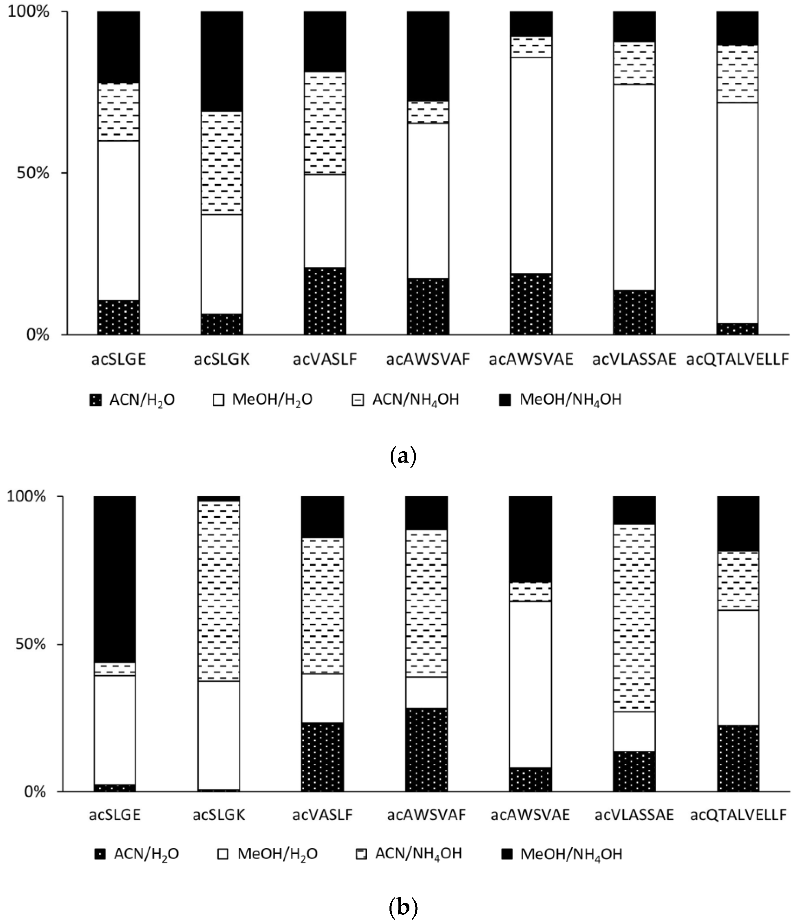
2.2. Effect of the Mobile Phase on the Ionization of Acetylated Peptides

The mobile phases for separating peptides usually contain acetonitrile or methanol and an aqueous buffer solution. In addition to providing separation, the mobile phase must also effectively support the ionization of the analytes. As regards gas-phase ionization methods, i.e., APCI and APPI, we have previously shown that ionization of peptides is most efficient when no buffer is present and the mobile phase is neutral or basic [39]. Here we tested the signals of acetylated SLGE, SLGK, VASLF, AWSVAF, AWSVAE, VLASSAE, and QTALVELLF in acetonitrile and methanol combined with water or aqueous ammonium hydroxide (pH 9). The organic and aqueous phase ratio was 4:1 (v/v). The relative intensities of peptide signals varied; however, all mobile phase combinations proved efficient for gas-phase ionization (Figure 7). In APCI, five out of seven tested peptides provided significantly higher signals in the aqueous mobile phases with no pH adjustment. In APPI, however, most peptides were efficiently ionized in the ammonia-treated mobile phase. The difference between methanol and acetonitrile can be related to the proton affinities of these solvents and the analyzed peptides. Methanol has lower PA than acetonitrile [73], facilitating efficient ionization of peptides with a relatively low PA.

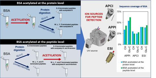
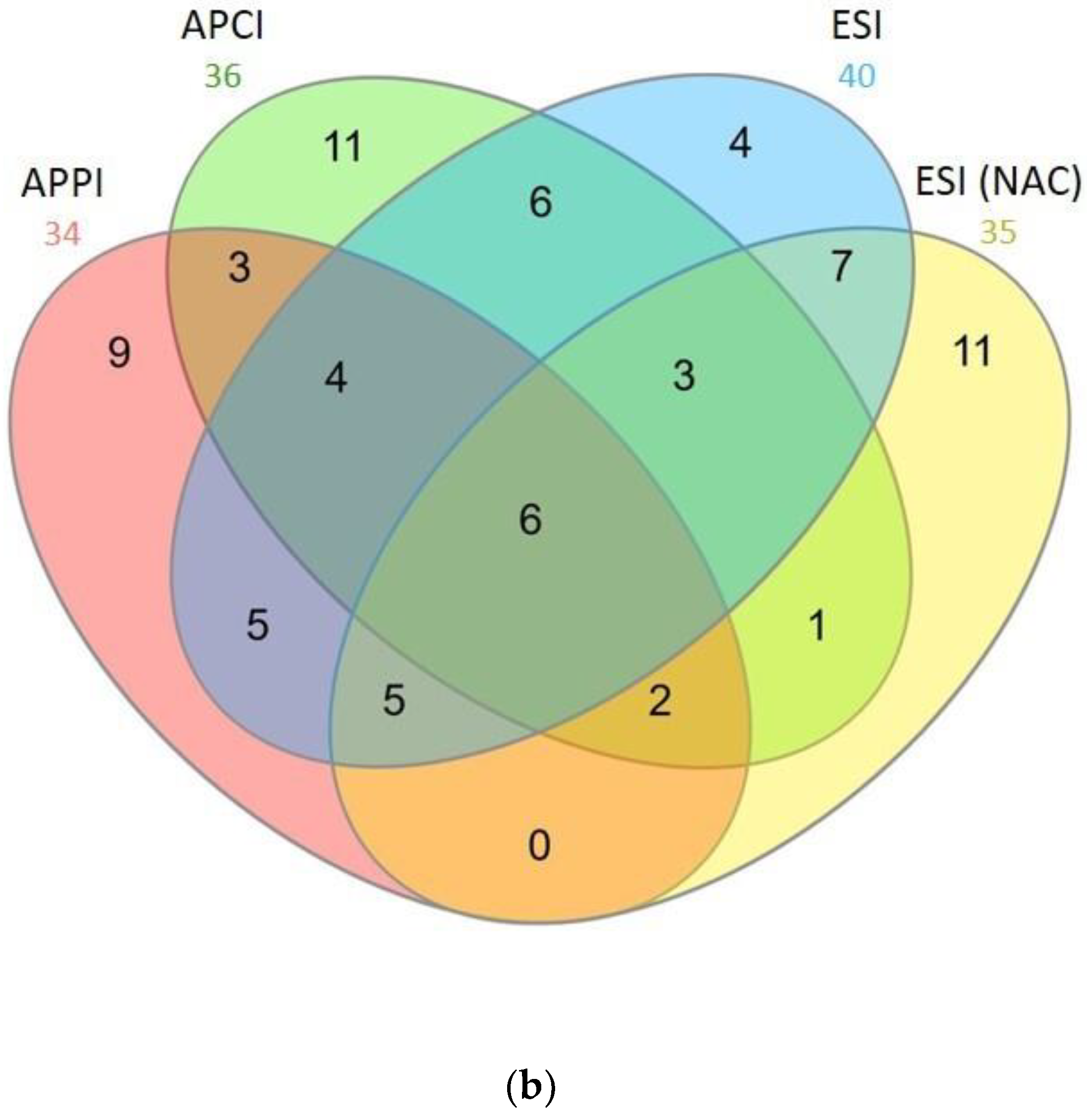
2.3. Bottom-Up Proteomics of BSA Digests

BSA digests were used to test the applicability of acetylation in bottom-up proteomic workflows based on APCI and APPI. The digests were prepared using chymotrypsin or Glu-C, which were shown to provide peptides with better properties than trypsin for gas-phase ionizations [39].
Two different ways of acetylation were compared. In the first case, the protein was acetylated before its enzymatic digestion. Acetylation occurred on ε amino groups of lysines and at the protein N-terminus. In the second approach, acetylation of digested peptides was performed, yielding peptides with modified lysines and N-termini. The third set of samples were enzymatic digests of unmodified BSA. The peptide samples were analyzed via HPLC/MS2 with ESI, APCI, and APPI sources.
The absence of unmodified peptides in the acetylated protein digests indicated a high derivatization efficiency. Acetylation occurred exclusively on lysine side chains and N-termini; no acetylation on other amino acids was detected. Due to the presence of methanol during the derivatization, a small amount of ester by-products was formed. As with the peptide standards, APCI and APPI provided singly charged protonated molecules and fragments, while sodiated molecules were readily formed in APPI. ESI showed singly and multiply charged peptide ions. The HPLC/MS2 data were processed using the Mascot search engine to identify peptides and evaluate sequence coverage.
After acetylation, sequence coverage increased, regardless of the method used for ionizing peptides (Table 1). In APCI, a moderate improvement in sequence coverage was observed for samples acetylated at protein and peptide levels. While unmodified peptides made it possible to describe 21–22% of the BSA sequence, acetylation improved the value by up to half. ESI provided high sequence coverage values, though the effect of acetylation was less important than in the case of APCI-MS. The highest values of sequence coverage were achieved in APPI for acetylated samples. Acetylation at the protein level increased the value from 22 to 34% for chymotrypsin and from 42 to 54% for Glu-C digestion. Acetylation of peptides resulted in an even more significant increase in sequence coverage to 43% and 62% for chymotrypsin and Glu-C digestion, respectively. The complete list of detected peptides is included in the Supplementary Materials (Tables S1–S6).
In APCI, acetylation improved the number of peptides detected in the chymotrypsin digest (Table 2). Although the number of acetylated peptides was relatively high, most of them were rather short; about 70–80% consisted of six or fewer amino acids. Half of the peptides detected via APCI in Glu-C digests were also short. Therefore, APCI demonstrated high sensitivity towards short peptides [26]. Data were also consistent with the nature of enzyme cleavages, i.e., the ability of chymotrypsin to form shorter peptides compared to Glu-C. In the case of BSA, chymotrypsin theoretically provides 55 short peptides (6 or fewer amino acids) from a total of 95 peptides, while Glu-C produces 39 short peptides from 86 peptides. Few methyl esters of acetylated peptides were detected; these were mostly detected in the sample derivatized at the peptides level, where the C-termini of the peptides were exposed to the reagent. In this sample, seven of 21 detected acetylated peptides were methylated at a significant level. Peptides with methylated carboxyl groups were also observed at comparable levels in APPI and ESI.
In APPI, acetylation did not increase the number of detected peptides in the chymotrypsin digest but made detecting more peptides with longer sequences possible (Table 3). As for the Glu-C digest, acetylation helped to increase the number of detected peptides significantly. This was attributed to a reduction in the polarity of the peptides which, thus, transferred more easily to the gas phase. APPI detected a higher number of longer peptides than APCI, particularly in the Glu-C digest. Sodium adducts were formed in APPI; approximately half of the acetylated peptides were detected as sodium adducts. The singly charged sodium adducts provided sequence-specific information [71] and improved the sensitivity compared to protonated molecules [74,75].
In ESI, similar sequence coverages and numbers of detected peptides were achieved for acetylated and non-acetylated digests. However, the structures of the individual peptides differed. The detection of different peptides can be explained using two factors influencing ionization. On one hand, increasing the hydrophobicity leads to easier migration of the peptides to the surface of the droplets, thus improving ionization efficiency [76,77,78]. On the other hand, decreasing the basicity of lysines leads to a lower ability to accept a proton. These two contending events had a different combined effect on various peptides (Table 4). The protein digests provided singly, doubly, triply, and quadruply charged ions. In the non-acetylated sample, doubly charged ions were mainly detected and singly and triply charged ions were formed less efficiently. As regards the acetylated samples, singly charged ions were formed the most, followed by doubly and, exceptionally, triply charged ions. Neutralization of the charge on lysines reduced the charge states of the peptides. For example, the non-derivatized form of DKLKHLVDEPQNL was detected as a 3+ ion, singly acetylated as 2+, and doubly acetylated as 1+. A charge state reduction resulting from acetylation was also observed for DEHVKL, GERAL, KDLGEEHF, QEAKDAF, SQKFPKAEF, FKADE, RMPCTE, CCHGDLLE, and LCKVASLRE. As shown in Table 4, multiply-charged peptides comprised about 70% of the peptides in the BSA digest but fewer than 50% in acetylated samples.
APPI, APCI, and ESI detected unique peptides in acetylated chymotrypsin and Glu-C digests. The most powerful ionization regarding the number of total and unique peptides in the chymotrypsin digest was APCI (Figure 8). However, the peptides were mostly short, meaning that the sequence coverage was unsatisfactory. On the contrary, the ionization that performed best for the Glu-C digest was APPI (Figure 9). APPI made it possible to detect more peptides, including many unique ones, than other ionizations. The detected peptides were mostly of high molecular weight (more than six amino acids). Workflows involving acetylation, Glu-C cleavage, and detection through APPI-MS also gave the best results in terms of sequence coverage (Table 1). The superior performance of APPI-MS for Glu-C digest can be related to the easier formation of sodium adducts of E- and D-terminated peptides. APPI-MS also easily detected longer acetylated peptides formed through Glu-C.
The detection of unique peptides can be utilized in novel proteomic workflows based on combining different ionization techniques. Such workflows describe primary protein sequences more comprehensively than the classical bottom-up approach. To test the hypothesis, we combined ESI, APCI, and APPI data to calculate sequence coverages (Table 5). The classical bottom-up proteomics based on ESI allowed us to describe 37% of the BSA sequence (Table 1). APCI made it possible to reveal an additional ~10% of the sequence. When ESI data were combined with APPI, sequence coverage increased by ~20% (chymotrypsin digest) and ~30% (Glu-C digest). Thus, APCI and APPI have yielded considerable complementary information to the classical ESI-based workflow. In addition to the ionization techniques, combining two or more proteases could further increase the number of detected peptides and sequence coverages [79].
The use of APCI and APPI in proteomics is limited due to the minimum flow rates that can be used with these ion sources. Commercial sources are designed for high mobile phase flow rates and operate optimally at around 1 mL·min−1. Nevertheless, some of the ion sources make it possible to reduce the flow rate down to 50 μL·min−1 and, thus, enable APCI and APPI MS detection in microflow HPLC. The use of commercial APCI and APPI sources with capillary or nanoflow HPLC is not possible; however, ion sources dedicated to capillary or nanoflow HPLC are being developed [80,81,82,83].

3. Materials and Methods

3.1. Chemicals and Reagent

Ammonium formate (reagent grade; 97%), ammonium bicarbonate (reagent grade), and bovine serum albumin (BSA; ≥98%) were purchased from Fluka Biochemica (Buchs, Switzerland). Trifluoroacetic acid (TFA) for HPLC (>99.9%), acetic anhydride (≥97%), ammonia solution (25%) for LC-MS, iodoacetamide (IAA; reagent grade), dithiothreitol (DTT; reagent grade), and triisopropylsilane (TIS; 98%) were obtained from Sigma Aldrich (St. Louise, WA, USA). Formic acid (FA; 98%) and acetic acid (99%) were purchased from Lach-Ner (Neratovice, Czech Republic). Methanol and acetonitrile (both OPTIMA LC-MS) were obtained from Fisher Chemical (Waltham, MA, USA). Sequencing grade chymotrypsin and Glu-C were purchased from Roche Diagnostics (Mannheim, Germany). Fmoc Arg(Pbf) WANG resin, Fmoc Lys(Boc) WANG resin, Fmoc Phe WANG, and Fmoc Glu(OtBu) WANG for peptide standards synthesis were purchased from Iris Biotech (Marktredwitz, Germany).

3.2. Peptide Standards

Peptide standards SLGK, SLGE, AWSVAE, VLASSAE, QTALVELLE, LAF, SLGF, VASLF, AWSVAF, VLASSAF, and QTALVELLF (all with free carboxyl on C-terminus) were synthesized in house. The peptides corresponded to BSA tryptic products and analogs with phenylalanine or glutamic acid at C-terminus, i.e., hypothetical peptides that could be formed using chymotrypsin or Glu-C cleavage. Peptide sequences were assembled on a Liberty Blue solid-phase synthesizer from CEM (Charlotte, NC, USA) through stepwise coupling of the corresponding Fmoc-amino acids to the growing chain on a Fmoc-Arg(Pbf)-WANG resin (100–200 mesh, 0.64 mmol·g−1); Fmoc-Lys (Boc)-WANG resin (100–200 mesh, 0.65 mmol·g−1); FmocGlu(OtBu)-WANG resin (200–400 mesh, 0.62 mmol·g−1), and Fmoc-Phe-WANG resin (200–400 mesh, 0.67 mmol·g−1). Fully protected peptides were synthesized according to a standard procedure involving cleavage of the Nα-Fmoc protecting group with 20% piperidine in dimethylformamide (DMF) (HPLC grade) and coupling, mediated with mixtures of coupling reagents (diisopropyl carbodiimide) DIC/(oxyma-ethylcyanohydroxyiminoacetate) Oxyma in DMF. On completion of synthesis, the deprotection and detachment of the linear peptides from the resins were carried out simultaneously using a TFA/H2O/TIS (95.0:2.5:2.5) cleavage mixture. Each resin was washed with dichloromethane and the combined TFA filtrates were evaporated at room temperature. The precipitated residues were triturated with tert-butyl methyl ether, collected via suction, and dried via lyophilization. The linear peptides were purified via HPLC using a Waters instrument with Delta 600 pump and a 2489 UV/VIS detector.

3.3. Protein Digestion

Proteins were digested using the enhanced filter-aided sample preparation (eFASP) method [84]. Protein dissolved in 80% acetonitrile 0.1% FA (125 μL; 1 mg·mL−1) was diluted with ammonium bicarbonate (50 mmol·L−1; pH 7.8) to a volume of 200 μL. The reduction was initiated by adding 15 μL of DTT (100 mmol·L−1 in water) and the sample was incubated for 30 min at 65 °C. Free thiol groups were alkylated with 45 μL of IAA (100 mmol·L−1 in water) and the sample was incubated in the dark for 30 min at 20 °C. The alkylation was terminated by adding 30 μL of DTT (100 mmol·L−1 in water). The sample was washed three times with 100 μL of 50 mmol·L−1 ammonium bicarbonate on Microcon centrifugal filters (Ultracel PL 10, Merck, Ireland) using Eppendorf Centrifuge 5417 Refrigerator (Boston, MA, USA). The digestion was performed on the same filter in 100 μL of 50 mmol·L−1 ammonium bicarbonate in the presence of protease (0.1 mg·mL−1) in a weight ratio of (chymotrypsin or Glu-C: protein) 1:50. Digestion of protein was performed with chymotrypsin at 30 °C and Glu-C for 16 h at 20 °C. Protein digest was washed twice with 75 μL of 50 mmol·L−1 ammonium bicarbonate via centrifugation and the filtrate was evaporated to dryness on a refrigerated centrifugal vacuum concentrator Labconco (Kansas City, MI, USA).

3.4. Acetylation

A synthetic peptide standard was dissolved in 80% ACN 0.1% FA (1 mg·mL−1). To 20 μL of the standard solution, 50 μL of acetylation reagent (acetic anhydride and methanol v/v 1:3) was added. Acetylation was carried out in an Eppendorf tube over 1 h at 25 °C in a thermoshaker. Acetic anhydride with methanol was evaporated on a refrigerated centrifugal vacuum concentrator.
Two approaches were used to acetylate BSA. Acetylation at the protein level caused the acetylation of lysine ɛ amines and protein N-termini. BSA was dissolved in 80% acetonitrile 0.1% FA (125 μL; 1 mg·mL−1), protein disulfide bonds were reduced via DTT, and thiol groups were alkylated via IAA according to the procedure described in Section 3.3. After the complete alkylation, 50 μL of the acetylating agent was added to 20 μg of BSA and the acetylation was achieved as described above for peptide standards. After the complete acetylation, the sample was washed three times with 100 μL of 50 mmol·L−1 ammonium bicarbonate on Microcon centrifugal filters using Eppendorf Centrifuge and the digestion proceeded as described in Section 3.3.
Acetylation at the peptide level generated peptides acetylated on lysine side chains and N-termini amino groups. BSA was processed according to the digestion protocol described in Section 3.3. Subsequently, the sample was re-dissolved in 80% acetonitrile 0.1% FA (125 μL; 1 mg·mL−1) and acetylated in the same way as peptide standards.

3.5. Mass Spectrometry

The experiments were performed on an LTQ Orbitrap XL (Thermo Scientific, Waltham, MA, USA) that combines a linear ion trap MS and an Orbitrap mass analyzer. The mass spectrometer was equipped with an IonMax source operating in ESI, APCI, or APPI mode. Full scan mass spectra were collected using the Orbitrap operated at a resolution of 30,000. Collision-induced dissociation (CID) and fragments detection were performed in the ion trap. The optimum normalized collision energy (NCE) values varied depending on the peptide structure. For acetylated peptides, NCEs of 20–30% were used. The isolation width for fragmentations was set to 1 Da.
The mass spectrometer was operated in the positive ion mode. In ESI, the ion source parameters were as follows: sheath and auxiliary gases were 35 and 5 arbitrary units, respectively; the source voltage was 4.80 kV; the capillary voltage was 7.0 V; the tube lens offset was 100.0 V; and the capillary temperature was 250 °C. In APCI, sheath and auxiliary gases were set at 50 and 10 arbitrary units, respectively; probe temperature was 480 °C; the corona discharge current was 10.00 µA; the, tube lens offset was 110.0 V; and the capillary temperature was 275 °C. In APPI, a krypton lamp from Syagen Technology, Inc. (Tustin, CA, USA) that emits photons at 10.0 eV and 10.6 eV (123.6 nm and 118.0 nm) was used. The sheath and auxiliary gases were at 90 and 10 arbitrary units, respectively; the probe temperature was 480 °C; the tube lens offset was 160 V; and the capillary temperature was 275 °C.

3.6. Flow Injection Analysis

The relative responses of acetylated and non-acetylated VLASSAF (41 μmol·L−1) and QTALVELLE (28 μmol·L−1) were examined via flow injection analysis. The peptide standards (2.5 μL) were injected into the mobile phase consisting of methanol and water (4:1, v/v; flow rate of 200 μL·min−1). Peak heights were obtained from data reconstructed for [M + H]+ or [M + Na]+ with a 0.2 Da extraction window.

3.7. Liquid Chromatography-Tandem Mass Spectrometry

The mass spectrometer (Section 3.5) was coupled to a liquid chromatograph consisting of a Rheos 2200 LC-MS Pump (Thermo Scientific/Flux Instruments), HTS PAL Autosampler (CTC Analytics, Zwingen, Switzerland), and DeltaChrom CTC 100 column thermostat (Watrex, Prague, Czech Republic). The analyses of protein digest (650 μmol·L−1, 10 μL injected) were performed on an Acquity UPLC BEH C18 reversed-phase column (1.7 μm; 2.1 × 50.0 mm, Waters) maintained at 50 °C. The mobile phases were prepared from solvent A (water or water adjusted to pH 9 with ammonia), solvent B (methanol), and solvent C (acetonitrile) as follows: sample loading 0–1.0 min (4.0% of B; 1.0% of C) continued via linear gradients 1.0–5.0 min (4.0–24.0% of B; 1.0–6.0% C), 5.0–10.0 min (24.0–25.5% of B; 6.0–6.5% of C), 10.0–12.5 min (25.5–76.5% of B; 6.5–18.5% of C), 12.5–16.5 min (76.5%–4.0 of B, 18.5–1.0% of C), and 17.5–20.0 min (4.0 of B; 1.0% of C). The mobile phase flow rate was 200 μL·min−1.
The data-dependent scanning with four scan events was set as follows: the NCE was 35%, the dynamic exclusion was 60 s with the exclusion list size limited to 500 precursor ions, the signal threshold was 75 counts, the isolation width was 3 Da, and the activation time was 30.0 ms.

3.8. Data Interpretation

MS data were processed in Xcalibur (version 4.1, Thermo Fisher Scientific, Waltham, MA, USA) using the Mascot search engine (Version 2.2, [85]). The search parameters were set as follows: In MS1, the mass tolerance was defined at 10 ppm, while in MS2 it was defined at 0.6 Da. Carbamidomethylation of C, deamidation of N and Q, and oxidation of M for non-derivatized peptides were selected as dynamic modifications. In the case of modified peptides, acetylation was either only on lysine residues or also on the N-termini of the peptides, depending on the method of the experiment. Furthermore, methylations of C termini were detected in some cases and, especially with APPI, sodium adducts at C-termini and D or E were detected.

4. Conclusions

This work shows the advantages of acetylation for proteomic analysis based on APCI and APPI mass spectrometry. Acetylated peptides provided many sequence-specific a, y, and b fragments, allowing for straightforward data interpretation. The applicability of acetylation in bottom-up proteomics was investigated in workflows encompassing derivatization of intact BSA or BSA digests. The enzymatic cleavage was accomplished with chymotrypsin or Glu-C. The acetylation targeted amino groups of lysines and N-termini; other functional groups remained unmodified. The only exception was the occasional formation of methyl esters on C-termini via a reaction with methanol in the reaction mixture.
In the experiments with non-derivatized and acetylated samples, sequence coverage and numbers of peptides detected using APCI, APPI, and ESI were compared. The most pronounced effect of acetylation was observed in APPI, particularly for a sample cleaved with Glu-C and acetylated at the peptides level. In APCI, acetylation led to a moderate increase in sequence coverage and significant improvement in the detection of short peptides. Peptides detected via ESI, APCI, and APPI overlapped only partially; all ionization methods generated unique peptides. The workflows based on acetylation and gas-phase ionizations represent complementary approaches to ESI-based methods. APCI and APPI offer access to missing pieces of information on proteomes, which are difficult to analyze via the conventional bottom-up approach.
Due to its robustness, reproducibility, and good sensitivity, micro-flow LC–MS2 is increasingly used in many proteomic applications [86]. As some APCI and APPI sources are compatible with microflow systems, including these ionizations into HPLC/MS2, proteomic protocols are suggested. Using gas-phase ionizations will make it possible to detect peptides that are difficult to analyze using electrospray and advance the possibilities of MS-based proteomics. The possibility of using APCI and APPI even in capillary HPLC can be expected in the future; the development of ion sources dedicated to low mobile phase flow rates is the subject of research [80,81,82,83].

Supplementary Materials

The following supporting information can be downloaded at: https://www.mdpi.com/article/10.3390/molecules28093711/s1, Figure S1. FTMS spectra of acetylated SLGE. Figure S2. MS2 spectra of acetylated SLGE. Figure S3. MS2 spectra of acetylated SLGE. Figure S4. FTMS spectra of acetylated SLGK. Figure S5. MS2 spectra of acetylated SLGK. Figure S6. FTMS spectra of acetylated VASLF. Figure S7. MS2 spectra of acetylated VASLF. Figure S8. MS2 spectra of acetylated VASLF. Figure S9. FTMS spectra of acetylated QTALVELLF. Figure S10. MS2 spectra of acetylated QTALVELLF. Figure S11. FTMS spectra of acetylated AWSVAE. Figure S12. MS2 spectra of acetylated AWSVAE. Figure S13. FTMS spectra of acetylated SLGF. Figure S14. MS2 spectra of acetylated SLGF. Figure S15. FTMS spectra of acetylated VLASSAF. Figure S16. MS2 spectra of acetylated VLASSAF. Figure S17. FTMS spectra of acetylated QTALVELLE. Figure S18. MS2 spectra of acetylated QTALVELLE. Table S1. Peptides detected via APPI in BSA digested using chymotrypsin. Table S2. Peptides detected via APCI in BSA digested using chymotrypsin. Table S3. Peptides detected via ESI in BSA digested using chymotrypsin. Table S4. Peptides detected via APPI in BSA digested using Glu-C. Table S5. Peptides detected via APCI in BSA digested using Glu-C. Table S6. Peptides detected via ESI in BSA digested using Glu-C.

Author Contributions

Conceptualization, J.C.; methodology, M.H., V.V.; resources, M.B.; investigation, formal analysis, and writing-original draft preparation S.S.; writing—review and editing, J.C., V.V., P.K., M.H.; supervision and funding acquisition, J.C. All authors have read and agreed to the published version of the manuscript.

Funding

This work was funded by the Czech Science Foundation (Project No. 20-09126S) and Charles University in Prague (Project SVV).

Institutional Review Board Statement

Not applicable.

Informed Consent Statement

Not applicable.

Data Availability Statement

The data presented in this study are available on request from the corresponding author.

Conflicts of Interest

The authors declare no conflict of interest.

Sample Availability

Samples of the compounds are not available from the authors.

References

  1. Link, A.J.; Eng, J.; Schieltz, D.M.; Carmack, E.; Mize, G.J.; Morris, D.R.; Garvik, B.M.; Yates, J.R. Direct Analysis of Protein Complexes Using Mass Spectrometry. Nat. Biotechnol. 1999, 17, 676–682. [Google Scholar] [CrossRef]
  2. Gygi, S.P.; Rist, B.; Gerber, S.A.; Turecek, F.; Gelb, M.H.; Aebersold, R. Quantitative Analysis of Complex Protein Mixtures Using Isotope-Coded Affinity Tags. Nat. Biotechnol. 1999, 17, 994–999. [Google Scholar] [CrossRef]
  3. Vit, O.; Petrak, J. Integral Membrane Proteins in Proteomics. How to Break Open the Black Box? J. Proteom. 2017, 153, 8–20. [Google Scholar] [CrossRef]
  4. Dupree, E.J.; Jayathirtha, M.; Yorkey, H.; Mihasan, M.; Petre, B.A.; Darie, C.C. A Critical Review of Bottom-Up Proteomics: The Good, the Bad, and the Future of This Field. Proteomes 2020, 8, 14. [Google Scholar] [CrossRef]
  5. Feist, P.; Hummon, A.B. Proteomic Challenges: Sample Preparation Techniques for Microgram-Quantity Protein Analysis from Biological Samples. Int. J. Mol. Sci. 2015, 16, 3537–3563. [Google Scholar] [CrossRef]
  6. Fischer, F.; Poetsch, A. Protein Cleavage Strategies for an Improved Analysis of the Membrane Proteome. Proteome Sci. 2006, 4, 2. [Google Scholar] [CrossRef] [PubMed]
  7. Lei, J.; Chen, D.A.; Regnier, F.E. Rapid Verification of Disulfide Linkages in Recombinant Human Growth Hormone by Tandem Column Tryptic Mapping. J. Chromatogr. A 1998, 808, 121–131. [Google Scholar] [CrossRef]
  8. Julka, S.; Regnier, F.E. Benzoyl Derivatization as a Method to Improve Retention of Hydrophilic Peptides in Tryptic Peptide Mapping. Anal. Chem. 2004, 76, 5799–5806. [Google Scholar] [CrossRef] [PubMed]
  9. Trufelli, H.; Palma, P.; Famiglini, G.; Cappiello, A. An Overview of Matrix Effects in Liquid Chrmatography-Mass Spectrometry. Mass Spectrom. 2010, 30, 491–509. [Google Scholar] [CrossRef]
  10. Loo, R.R.O.; Dales, N.; Andrews, P.C. Surfactant Effects on Protein Structure Examined by Electrospray Ionization Mass Spectrometry. Protein Sci. 1994, 3, 1975–1983. [Google Scholar] [CrossRef] [PubMed]
  11. Conrads, T.P.; Yu, L.; Terunuma, A.; Janini, G.M.; Issaq, H.J.; Vogel, J.C.; Veenstra, T.D. A Detergent- and Cyanogen Bromide-Free Method for Integral Membrane Proteomics: Application to Halobacterium Purple Membranes and The Human Epidermal Membrane Proteome. Proteomics 2004, 4, 31–45. [Google Scholar] [CrossRef]
  12. Xu, T.; Wang, H.; Wu, M.; Wang, W.; Tan, Q.; Zhao, F.; Zhou, F.; Hu, T.; Jiang, Z.; Liu, Z.; et al. Disulfide-Containing Detergents (DCDs) for the Structural Biology of Membrane Proteins Dongxiang. Chemistry 2019, 25, 11635–11640. [Google Scholar] [CrossRef]
  13. Donoghue, P.M.; Hughes, C.; Vissers, J.P.C.; Langridge, J.I.; Dunn, M.J. Nonionic Detergent Phase Extraction for the Proteomic Analysis of Heart Membrane Proteins Using Label-Free LC-MS. Proteomics 2008, 8, 3895–3905. [Google Scholar] [CrossRef] [PubMed]
  14. Breyton, C.; Pucci, B.; Popot, J. Amphiols and Fluorinated Surfactants: Two Alternatives to Detergents for Studying Membrane Proteins In Vitro. In Heterologous Expression of Membrane Proteins: Methods in Molecular Biology; Mus-Vetau, I., Ed.; Humana Press: Totowa, NJ, USA, 2010; Volume 601, pp. 219–245. ISBN 9781607613442. [Google Scholar]
  15. Wehbie, M.; Onyia, K.K.; Mahler, F.; Le Roy, A.; Deletraz, A.; Bouchemal, I.; Vargas, C.; Babalola, J.O.; Breyton, C.; Ebel, C.; et al. Maltose-Based Fluorinated Surfactants for Membrane-Protein Extraction and Stabilization. Langmuir 2021, 37, 2111–2122. [Google Scholar] [CrossRef]
  16. Gilmore, J.M.; Kettenbach, A.N.; Gerber, S.A. Increasing Phosphoproteomic Coverage through Sequential Digestion by Complementary Proteases. Anal. Bioanal. Chem. 2012, 402, 711–720. [Google Scholar] [CrossRef]
  17. Rietschel, B.; Bornemann, S.; Arrey, T.N.; Baeumlisberger, D.; Karas, M.; Meyer, B. Membrane Protein Analysis Using an Improved Peptic In-Solution Digestion Protocol. Proteomics 2009, 9, 5553–5557. [Google Scholar] [CrossRef]
  18. Wu, C.C.; MacCoss, M.J.; Howell, K.E.; Yates, J.R. A Method for the Comprehensive Proteomic Analysis of Membrane Proteins. Nat. Biotechnol. 2003, 21, 532–538. [Google Scholar] [CrossRef] [PubMed]
  19. Yu, D.; Wang, Z.; Cupp-Sutton, K.A.; Liu, X.; Wu, S. Deep Intact Proteoform Characterization in Human Cell Lysate Using High-PH and Low-PH Reversed-Phase Liquid Chromatography. J. Am. Soc. Mass Spectrom. 2019, 30, 2502–2513. [Google Scholar] [CrossRef]
  20. Petritis, K.; Brussaux, S.; Guenu, S.; Elfakir, C.; Dreux, M. Ion-Pair Reversed-Phase Liquid Chromatography-Electrospray Mass Spectrometry for the Analysis of Underivatized Small Peptides. J. Chromatogr. A 2002, 957, 173–185. [Google Scholar] [CrossRef]
  21. Kalghatgi, K.; Horvath, C. Rapid Peptide Mapping by High-Performance Liquid Chromatography. J. Chromatogr. 1988, 443, 343–354. [Google Scholar] [CrossRef]
  22. Gustavsson, S.Å.; Samskog, J.; Markides, K.E.; Långström, B. Studies of Signal Suppression in Liquid Chromatography-Electrospray Ionization Mass Spectrometry Using Volatile Ion-Pairing Reagents. J. Chromatogr. A 2001, 937, 41–47. [Google Scholar] [CrossRef] [PubMed]
  23. Bonmatin, J.M.; Moineau, I.; Charvet, R.; Fleche, C.; Colin, M.E.; Bengsch, E.R. A LC/APCI-MS/MS Method for Analysis of Imidacloprid in Soils, in Plants, and in Pollens. Anal. Chem. 2003, 75, 2027–2033. [Google Scholar] [CrossRef] [PubMed]
  24. Rosenberg, E. The Potential of Organic (Electrospray- and Atmospheric Pressure Chemical Ionisation) Mass Spectrometric Techniques Coupled to Liquid-Phase Separation for Speciation Analysis. J. Chromatogr. A 2003, 1000, 841–889. [Google Scholar] [CrossRef]
  25. Raffaelli, A.; Saba, A. Atmospheric Pressure Photoionization Mass Spectrometry. Mass Spectrom. Rev. 2003, 22, 318–331. [Google Scholar] [CrossRef]
  26. Cristoni, S.; Bernardi, L.R.; Biunno, I.; Guidugli, F. Analysis of Peptides Using Partial (No Discharge) Atmospheric Pressure Chemical Ionization Conditions with Ion Trap Mass Spectrometry. Rapid Commun. Mass Spectrom. 2002, 16, 1686–1691. [Google Scholar] [CrossRef] [PubMed]
  27. Robb, D.B.; Blades, M.W. State-of-the-Art in Atmospheric Pressure Photoionization for LC/MS. Anal. Chim. Acta 2008, 627, 34–49. [Google Scholar] [CrossRef]
  28. Delobel, A.; Halgand, F.; Laffranchise-Gosse, B.; Snijders, H.; Laprévote, O. Characterization of Hydrophobic Peptides by Atmospheric Pressure Photoionization-Mass Spectrometry and Tandem Mass Spectrometry. Anal. Chem. 2003, 75, 5961–5968. [Google Scholar] [CrossRef] [PubMed]
  29. Debois, D.; Giuliani, A.; Laprévote, O. Fragmentation Induced in Atmospheric Pressure Photoionization of Peptides. J. Mass Spectrom. 2006, 41, 1554–1560. [Google Scholar] [CrossRef]
  30. Matuszewski, B.K.; Constanzer, M.L.; Chavez-Eng, C.M. Matrix Effect in Quantitative LC/MS/MS Analyses of Biological Fluids: A Method for Determination of Finasteride in Human Plasma at Picogram Per Milliliter Concentrations. Anal. Chem. 1998, 70, 882–889. [Google Scholar] [CrossRef]
  31. Hsieh, Y.; Chintala, M.; Mei, H.; Agans, J.; Brisson, J.M.; Ng, K.; Korfmacher, W.A. Quantitative Screening and Matrix Effect Studies of Drug Discovery Compounds in Monkey Plasma Using Fastgradient Liquid Chromatography/Tandem Mass Spectrometry. Rapid Commun. Mass Spectrom. 2001, 15, 2481–2487. [Google Scholar] [CrossRef]
  32. McCulloch, R.D.; Robb, D.B. Field-Free Atmospheric Pressure Photoionization-Liquid Chromatography-Mass Spectrometry for the Analysis of Steroids within Complex Biological Matrices. Anal. Chem. 2017, 89, 4169–4176. [Google Scholar] [CrossRef]
  33. Shen, Y.; Han, C.; Chen, J.; Wang, X. Analysis of Cyclic Peptides in Pseudostellaria Heterophylla (Miq.) Pax by HPLC-APCI-MS. Chromatographia 2007, 66, 319–323. [Google Scholar] [CrossRef]
  34. Bose, U.; Hodson, M.; Shaw, P.; Fuerst, J.; Hewavitharana, A. Two Peptides, Cycloaspeptide A and Nazumamide A from a Sponge Associated Marine Actinobacterium Salinispora Sp. Nat. Prod. Commun. 2014, 9, 545–546. [Google Scholar] [CrossRef]
  35. Bagag, A.; Jault, J.M.; Sidahmed-Adrar, N.; Réfrégiers, M.; Giuliani, A.; Le Naour, F. Characterization of Hydrophobic Peptides in the Presence of Detergent by Photoionization Mass Spectrometry. PLoS ONE 2013, 8, e79033. [Google Scholar] [CrossRef] [PubMed]
  36. Bagag, A.; Giuliani, A.; Laprévote, O. Atmospheric Pressure Photoionization of Peptides. Int. J. Mass Spectrom. 2011, 299, 1–4. [Google Scholar] [CrossRef]
  37. Bagag, A.; Giuliani, A.; Réfrégiers, M.; Le Naour, F. Atmospheric Pressure Photoionization Study of Post-Translational Modifications: The Case of Palmitoylation. Int. J. Mass Spectrom. 2012, 328–329, 23–27. [Google Scholar] [CrossRef]
  38. Giuliani, A.; Giorgetta, J.L.; Ricaud, J.P.; Jamme, F.; Rouam, V.; Wien, F.; Laprévote, O.; Réfrégiers, M. Atmospheric Pressure Photoionization Using Tunable VUV Synchrotron Radiation. Nucl. Instrum. Methods Phys. Res. Sect. B Beam Interact. Mater. Atoms 2012, 279, 114–117. [Google Scholar] [CrossRef]
  39. Sedláčková, S.; Hubálek, M.; Vrkoslav, V.; Blechová, M.; Cvačka, J. Utility of Atmospheric-Pressure Chemical Ionization and Photoionization Mass Spectrometry in Bottom-Up Proteomics. Separations 2022, 9, 42. [Google Scholar] [CrossRef]
  40. Qiao, X.; Qin, X.; She, D.; Wang, R.; Zhang, X.; Zhang, L.; Zhang, Y. Mass Spectrometry-Based Tag and Its Application to High Efficient Peptide Analysis—A Review. Talanta 2014, 126, 91–102. [Google Scholar] [CrossRef] [PubMed]
  41. Leitner, A.; Lindner, W. Chemistry Meets Proteomics: The Use of Chemical Tagging Reactions for MS-Based Proteomics. Proteomics 2006, 6, 5418–5434. [Google Scholar] [CrossRef]
  42. Leitner, A.; Lindner, W. Current Chemical Tagging Strategies for Proteome Analysis by Mass Spectrometry. J. Chromatogr. B Anal. Technol. Biomed. Life Sci. 2004, 813, 1–26. [Google Scholar] [CrossRef]
  43. Miyagi, M.; Nakao, M.; Nakazawa, T.; Kato, I.; Tsunasawa, S. A Novel Derivatization Method with 5-Bromo-Nicotinic Acid N-Hydroxysuccinimide for Determination of the Amino Acid Sequences of Peptides. Rapid Commun. Mass Spectrom. 1998, 12, 603–608. [Google Scholar] [CrossRef]
  44. An, M.; Zou, X.; Wang, Q.; Zhao, X.; Wu, J.; Xu, M.; Shen, H.; Xiao, X.; He, D.; Ji, J.; et al. High-Confidence De Novo Peptide Sequencing Using Positive Charge Derivatization and Tandem MS Spectra Merging. Anal. Chem. 2013, 85, 4530–4537. [Google Scholar] [CrossRef] [PubMed]
  45. Yu, Q.; Shi, X.; Feng, Y.; Kent, K.C.; Li, L. Improving Data Quality and Preserving HCD-Generated Reporter Ions with EThcD for Isobaric Tag-Based Quantitative Proteomics and Proteome-Wide PTM Studies. Anal. Chim. Acta 2017, 968, 40–49. [Google Scholar] [CrossRef] [PubMed]
  46. Wu, Y.; Wang, F.; Liu, Z.; Qin, H.; Song, C.; Huang, J.; Bian, Y.; Wei, X.; Donga, J.; Zou, H. Five-Plex Isotope Dimethyl Labeling for Quantitative Proteomics. Chem. Commun. 2014, 50, 1708–1710. [Google Scholar] [CrossRef]
  47. Oldekop, M.L.; Herodes, K.; Rebane, R. Comparison of Amino Acid Derivatization Reagents for Liquid Chromatography Atmospheric Pressure Chemical Ionization Mass Spectrometric Analysis of Seven Amino Acids in Tea Extract. Int. J. Mass Spectrom. 2017, 421, 189–195. [Google Scholar] [CrossRef]
  48. Rebane, R.; Rodima, T.; Kütt, A.; Herodes, K. Development of Amino Acid Derivatization Reagents for Liquid Chromatography Electrospray Ionization Mass Spectrometric Analysis and Ionization Efficiency Measurements. J. Chromatogr. A 2015, 1390, 62–70. [Google Scholar] [CrossRef]
  49. Pohlentz, G.; Schlemm, S.; Klima, B.; Egge, H. Fast Atom Bombardment Mass Spectrometry of N-Acetylated Neoglycolipids of the 1-Deoxy-1-Phosphatidylethanolaminolactitol-Type. Chem. Phys. Lipids 1994, 70, 83–94. [Google Scholar] [CrossRef]
  50. Tsai, P.K.; Dell, A.; Ballou, C.E. Characterization of Acetylated and Acetolyzed Glycoprotein High-Mannose Core Oligosaccharides by Fast-Atom-Bombardment Mass Spectroscopy. Proc. Natl. Acad. Sci. USA 1986, 83, 4119–4123. [Google Scholar] [CrossRef]
  51. Claeys, M.; Claereboudt, J.; Uia, A. Fast Atom Bombardment Ionization in Mass Spectrometry, 3rd ed.; Elsevier Ltd.: Amsterdam, The Netherlands, 2017; ISBN 9780128032244. [Google Scholar]
  52. Yang, H.-J.; Hu, X.-Y.; Chen, Y.-Z. Amino Acid Identification and Sequence Analysis of Peptides by Reaction Mass Spectrometry. Chin. J. Chem. 1993, 11, 540–549. [Google Scholar] [CrossRef]
  53. Paulson, J.; Lindberg, C. Increasing Thermospray Response for Cortisol by Derivatization. J. Chromatogr. A 1991, 554, 149–154. [Google Scholar] [CrossRef]
  54. Cho, K.; Kang, W.; Choi, Y.; Kim, W.; Pyo, K. Effects of Peptide Acetylation and Dimethylation on Electrospray Ionization Efficiency. J. Mass Spectrom. 2016, 51, 105–110. [Google Scholar] [CrossRef]
  55. Zappacosta, F.; Wagner, C.D.; Della Pietra, A.; Gerhart, S.V.; Keenan, K.; Korenchuck, S.; Quinn, C.J.; Barbash, O.; McCabe, M.T.; Annan, R.S. A Chemical Acetylation-Based Mass Spectrometry Platform for Histone Methylation Profiling. Mol. Cell. Proteomics 2021, 20, 100067. [Google Scholar] [CrossRef]
  56. Kuchibhotla, B.; Kola, S.R.; Medicherla, J.V.; Cherukuvada, S.V.; Dhople, V.M.; Nalam, M.R. Combinatorial Labeling Method for Improving Peptide Fragmentation in Mass Spectrometry. J. Am. Soc. Mass Spectrom. 2017, 28, 1216–1226. [Google Scholar] [CrossRef]
  57. Samgina, T.Y.; Kovalev, S.V.; Gorshkov, V.A.; Artemenko, K.A.; Poljakov, N.B.; Lebedev, A.T. N-Terminal Tagging Strategy for De Novo Sequencing of Short Peptides by ESI-MS/MS and MALDI-MS/MS. J. Am. Soc. Mass Spectrom. 2010, 21, 104–111. [Google Scholar] [CrossRef]
  58. Straube, E.; Dekant, W.; Völkel, W. Enhanced Sensitivity for the Determination of Ambiphilic Polyaromatic Amines by LC-MS/MS after Acetylation. J. Chromatogr. A 2005, 1067, 181–190. [Google Scholar] [CrossRef] [PubMed]
  59. Sugahara, K.; Jianying, Z.; Kodama, H. Liquid Chromatographic-Mass Spectrometric Analysis of N-Acetylamino Acids in Human Urine. J. Chromatogr. B 1994, 657, 15–21. [Google Scholar] [CrossRef]
  60. Chandra, D.; Gayathri, P.; Vats, M.; Nagaraj, R.; Ray, M.K. Mass Spectral Analysis of Acetylated Peptides: Implications in Proteomics. Eur. J. Mass Spectrom. 2019, 26, 36–45. [Google Scholar] [CrossRef] [PubMed]
  61. Harrison, A.G.; Young, A.B.; Bleiholder, C. Scrambling of Sequence Information in Collision-Induced Dissociation of Peptides. J. Am. Chem. Soc. 2006, 128, 10364–10365. [Google Scholar] [CrossRef] [PubMed]
  62. Harrison, A.G. To b or Not to b: The Ongoing Saga of Peptide b Ions. Mass Spectrom. Rev. 2009, 28, 640–654. [Google Scholar] [CrossRef]
  63. Yaqüe, J.; Paradela, A.; Ramos, M.; Ogueta, S.; Marina, A.; Barahona, F.; López de Castro, J.A.; Vázquez, J. Peptide Rearrangement during Quadrupole Ion Trap Fragmentation: Added Complexity to MS/MS Spectra. Anal. Chem. 2003, 75, 1524–1535. [Google Scholar] [CrossRef]
  64. Harrison, A.G. Peptide Sequence Scrambling Through Cyclization of B5 Ions. J. Am. Soc. Mass Spectrom. 2008, 19, 1776–1780. [Google Scholar] [CrossRef] [PubMed]
  65. O’Hair, R.A.J.; Reid, G.E. Derivatization of Protonated Peptides via Gas Phase Ion-Molecule Reactions with Acetone. J. Am. Soc. Mass Spectrom. 2000, 11, 244–256. [Google Scholar] [CrossRef]
  66. Medzihradszky, K.F.; Chalkley, R.J. Lessons in De Novo Peptide Sequencing by Tandem Mass Spectrometry. Mass Spectrom. Rev. 2015, 34, 43–63. [Google Scholar] [CrossRef] [PubMed]
  67. Hiserodt, R.D.; Brown, S.M.; Swijter, D.F.H.; Hawkins, N.; Mussinan, C.J. A Study of B1+H2O and B1-Ions in the Product Ion Spectra of Dipeptides Containing N-Terminal Basic Amino Acid Residues. J. Am. Soc. Mass Spectrom. 2007, 18, 1414–1422. [Google Scholar] [CrossRef] [PubMed]
  68. Li, S.; Dabir, A.; Misal, S.A.; Tang, H.; Radivojac, P.; Reilly, J.P. The Impact of Amidination on Peptide Fragmentation and Identification in Shotgun Proteomics. J. Proteome Res. 2016, 15, 3656–3665. [Google Scholar] [CrossRef] [PubMed]
  69. Thorne, G.C.; Gaskell, S.J.; Gross, M.L. Elucidation of Some Fragmentations of Small Peptides Using Sequential Mass Spectrometry on a Hybrid Instrument. Rapid Commun. Mass Spectrom. 1989, 3, 217–221. [Google Scholar] [CrossRef]
  70. Thorne, G.C.; Ballard, K.D.; Gaskell, S.J. Metastable Decomposition of Peptide [M + H]+ Ions via Rearrangement Involving Loss of the C-Terminal Amino Acid Residue. J. Am. Soc. Mass Spectrom. 1990, 1, 249–257. [Google Scholar] [CrossRef]
  71. Newton, K.A.; McLuckey, S.A. Generation and Manipulation of Sodium Cationized Peptides in the Gas Phase. J. Am. Soc. Mass Spectrom. 2004, 15, 607–615. [Google Scholar] [CrossRef]
  72. Biemann, K.; Martin, S.A. Mass Spectrometric Determination of the Amino Acid Sequence of Peptides and Proteins. Mass Spectrom. Rev. 1987, 6, 1–75. [Google Scholar] [CrossRef]
  73. Hunter, E.P.L.; Lias, S.G. Evaluate Gas Phase Basicities and Proton Affinity of Molecules: An Update. J. Phys. Chem. Ref. Data 1998, 27, 413–656. [Google Scholar] [CrossRef]
  74. Schneider, R.P.; Lynch, M.J.; Ericson, J.F.; Fouda, H.G. Electrospray Ionization Mass Spectrometry of Semduramicin and Other Polyether Ionophores. Anal. Chem. 1991, 63, 1789–1794. [Google Scholar] [CrossRef] [PubMed]
  75. Gao, S.; Zhang, Z.P.; Karnes, H.T. Sensitivity Enhancement in Liquid Chromatography/Atmospheric Pressure Ionization Mass Spectrometry Using Derivatization and Mobile Phase Additives. J. Chromatogr. B 2005, 825, 98–110. [Google Scholar] [CrossRef]
  76. Fenn, J.B. Ion Formation from Charged Droplets: Roles of Geometry, Energy, and Time. J. Am. Soc. Mass Spectrom. 1993, 4, 524–535. [Google Scholar] [CrossRef] [PubMed]
  77. Iribarne, J.V.; Dziedzic, P.J.; Thomson, B.A. Atmospheric Pressure Ion Evaporation-Mass Spectrometry. Int. J. Mass Spectrom. Ion Phys. 1983, 50, 331–347. [Google Scholar] [CrossRef]
  78. Cech, N.B.; Enke, C.G. Practical Implications of Some Recent Studies in Electrospray Ionization Fundamentals. Mass Spectrom. Rev. 2001, 20, 362–387. [Google Scholar] [CrossRef]
  79. Sun, B.; Liu, Z.; Liu, J.; Zhao, S.; Wang, L.; Wang, F. The Utility of Proteases in Proteomics, from Sequence Profiling to Structure and Function Analysis. Proteomics 2022, 23, 2200132. [Google Scholar] [CrossRef]
  80. Kloudová, B.; Strmeň, T.; Vrkoslav, V.; Chára, Z.; Pačes, O.; Cvačka, J. Gas Dynamic Virtual Nozzle Sprayer for an Introduction of Liquid Samples in Atmospheric Pressure Ionization Mass Spectrometry. Anal. Chem. 2022, 95, 4196–4203. [Google Scholar] [CrossRef]
  81. Strmeň, T.; Vrkoslav, V.; Bosáková, Z.; Cvačka, J. Atmospheric Pressure Chemical Ionization Mass Spectrometry at Low Flow Rates: Importance of Ion Source Housing. Rapid Commun. Mass Spectrom. 2020, 34, e8722. [Google Scholar] [CrossRef]
  82. Vrkoslav, V.; Rumlová, B.; Strmeň, T.; Nekvasilová, P.; Šulc, M.; Cvačka, J. Applicability of Low-Flow Atmospheric Pressure Chemical Ionization and Photoionization Mass Spectrometry with a Microfabricated Nebulizer for Neutral Lipids. Rapid Commun. Mass Spectrom. 2018, 32, 639–648. [Google Scholar] [CrossRef]
  83. Strmeň, T.; Vrkoslav, V.; Pačes, O.; Cvačka, J. Evaluation of an Ion Source with a Tubular Nebulizer for Microflow Atmospheric Pressure Chemical Ionization. Mon. Für Chem. 2018, 149, 987–994. [Google Scholar] [CrossRef]
  84. Erde, J.; Loo, R.R.O.; Loo, J.A. Improving Proteome Coverage and Sample Recovery with Enhanced FASP (EFASP) for Quantitative Proteomic Experiments. Methods Mol. Biol. 2017, 1550, 11–18. [Google Scholar] [CrossRef] [PubMed]
  85. Gostian, A.O.; Hüttenbrink, K.B.; Luers, J.C.; Anagiotos, A.; Beutner, D. Probability-Based Protein Identification by Searching Sequence Databases Using Mass Spectrometry Data. Electrophoresis 1999, 20, 3551–3567. [Google Scholar] [CrossRef]
  86. Bian, Y.; Gao, C.; Kuster, B. On the Potential of Micro-Flow LC-MS/MS in Proteomics. Expert Rev. Proteom. 2022, 19, 153–164. [Google Scholar] [CrossRef] [PubMed]
Figure 1. APCI (left) and APPI (right) full scan mass spectra of acetylated LAF. The peak marked with an asterisk is m/z 120.1, y1 − H2O − CO.
Molecules 28 03711 g001
Figure 2. APCI (left) and APPI (right) full scan mass spectra of acetylated AWSVAF. The peak marked with an asterisk is m/z 736.3, [M (Me-ester) + H]+.
Molecules 28 03711 g002
Figure 3. APCI (left) and APPI (right) full scan mass spectra of acetylated VLASSAE.
Molecules 28 03711 g003
Figure 4. MS2 spectra of acetylated LAF: [M + H]+ m/z 392.2 in APCI (left) and [M + Na]+ m/z 414.2 in APPI (right).
Molecules 28 03711 g004
Figure 5. MS2 spectra of acetylated AWSVAF: [M + H]+ m/z 722.6 in APCI (left) and [M + Na]+ m/z 744.4 in APPI (right).
Molecules 28 03711 g005
Figure 6. MS2 spectra of acetylated VLASSAE: [M + H]+ m/z 718.3 in APCI (left) and [M + Na]+ m/z 740.4 in APPI (right). The peak marked with an asterisk is m/z 557.3, x6 – CO − H2O.
Molecules 28 03711 g006
Figure 7. Normalized intensities of [M + H]+ peaks of acetylated peptide standards in various mobile phases for (a) APCI and (b) APPI.
Molecules 28 03711 g007
Figure 8. Venn’s diagram with number of peptides detected in BSA chymotrypsin digests using APPI, APCI, and ESI-MS. Sample was acetylated at protein level (upper diagram, (a) and peptide level (lower diagram, (b). ESI data for non-acetylated sample (NAC) are shown for comparison. BSA peptides (10 μL injected; 0.65 mmol·L−1) were separated at 50 °C using a 20 min gradient and mobile phase flow rate was 200 μL·min−1. Data were evaluated using Mascot engine.
Molecules 28 03711 g008aMolecules 28 03711 g008b
Figure 9. Venn’s diagram with number of peptides detected in BSA Glu-C digests using APPI, APCI, and ESI-MS. Sample was acetylated at protein level (upper diagram, (a) and peptide level (lower diagram, (b). ESI data for non-acetylated sample (NAC) are shown for comparison. The BSA peptides (10 μL injected; 0.65 mmol·L−1) were separated at 50 °C by a 20 min gradient and mobile phase flow rate was 200 μL·min−1. Data were evaluated using Mascot engine.
Molecules 28 03711 g009aMolecules 28 03711 g009b
Table 1. Sequence coverages for BSA chymotrypsin and Glu-C digests analyzed via HPLC/MS2 with APCI, APPI, or ESI sources. Sample was acetylated at protein or peptide level. BSA digests (10 μL injected; 0.65 mmol·L−1) were separated at 50 °C using a 20 min gradient elution. Mobile phase consisted of water (APCI) or water adjusted to pH 9 with ammonia (ESI, APPI), methanol, and acetonitrile; flow rate was 200 μL·min−1. Data were evaluated using the Mascot engine.
Ion SourceProteaseSequence Coverage [%]
Acetylation at the Protein LevelAcetylation at the Peptides LevelNo Acetylation
APCIChymotrypsin302521
Glu-C252822
APPIChymotrypsin344322
Glu-C546242
ESIChymotrypsin404037
Glu-C394437
Table 2. Peptide types detected in BSA chymotrypsin and Glu-C digests via HPLC/MS2 with APCI source.
ProteaseType of PeptidesNumber of Peptides
Acetylation at the
Protein Level
Acetylation at the
Peptides Level
No Acetylation
ChymotrypsinAll413629
Acetylated methyl esters 110
Long peptides *787
Sodium adducts000
Glu-CAll 222321
Acetylated methyl esters 270
Long peptides *111613
Sodium adducts000
* Peptides with more than six amino acids.
Table 3. Peptide types detected in BSA chymotrypsin and Glu-C digests via HPLC/MS2 with APPI source.
ProteaseTypes of PeptideNumber of Peptides
Acetylation at the
Protein Level
Acetylation at the
Peptides Level
No Acetylation
ChymotrypsinAll273430
Acetylated methyl esters200
Long peptides *17196
Sodium adducts14160
Glu-CAll444736
Acetylated methyl esters2100
Long peptides *363928
Sodium adducts21264
* Peptides with more than six amino acids.
Table 4. Peptide types detected in BSA chymotrypsin and Glu-C digests via HPLC/MS2 with ESI source.
ProteaseType of Peptides Number of Peptides
Acetylation at the Protein LevelAcetylation at the
Peptides Level
No Acetylation
ChymotrypsinAll274035
Acetylated methyl esters340
Long peptides *202323
Sodium adducts312
Singly charged152811
Doubly charged101015
Triply charged228
Quadruply charged001
Glu-CAll253430
Acetylated methyl esters260
Long peptides *212423
Sodium adducts003
Singly charged15207
Doubly charged101418
Triply charged005
Quadruply charged000
* Peptides with more than six amino acids.
Table 5. Sequence coverages for BSA chymotrypsin and Glu-C digests analyzed via HPLC/MS2 and calculated via combining data from ESI, APCI, and APPI experiments. Sample for APCI and APPI was acetylated at protein or peptide level. BSA digests (10 μL injected; 0.65 mmol·L−1) were separated at 50 °C using a 20 min gradient elution. The mobile phase consisted of water (APCI) or water adjusted to pH 9 with ammonia (ESI, APPI), methanol, and acetonitrile; flow rate was 200 μL·min−1. Data were evaluated using Mascot engine.
ProteaseSequence Coverage [%]
Acetylated at the Protein Level *Acetylated at the Peptides Level *
ESI (NAC) + APCIESI (NAC) + APPIESI (NAC) + APCIESI (NAC) + APPI
Chymotrypsin49564754
Glu-C48704971
* The sample for ESI was not acetylated.
Disclaimer/Publisher’s Note: The statements, opinions and data contained in all publications are solely those of the individual author(s) and contributor(s) and not of MDPI and/or the editor(s). MDPI and/or the editor(s) disclaim responsibility for any injury to people or property resulting from any ideas, methods, instructions or products referred to in the content.

