
Etching and Doping of Pores in Polyethylene Terephthalate Analyzed by Ion Transmission Spectroscopy and Nuclear Depth Profiling
 ,
,  ,
,  ,
,  and
and 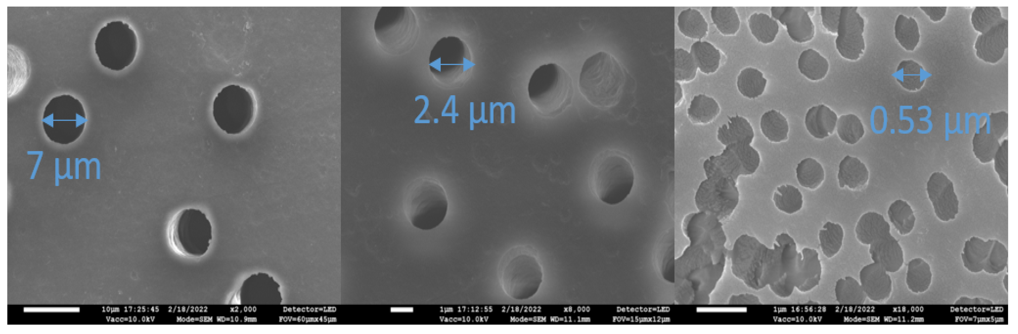
Abstract
:1. Introduction
2. Materials and Methods
2.1. Membrane Preparation
- (i)
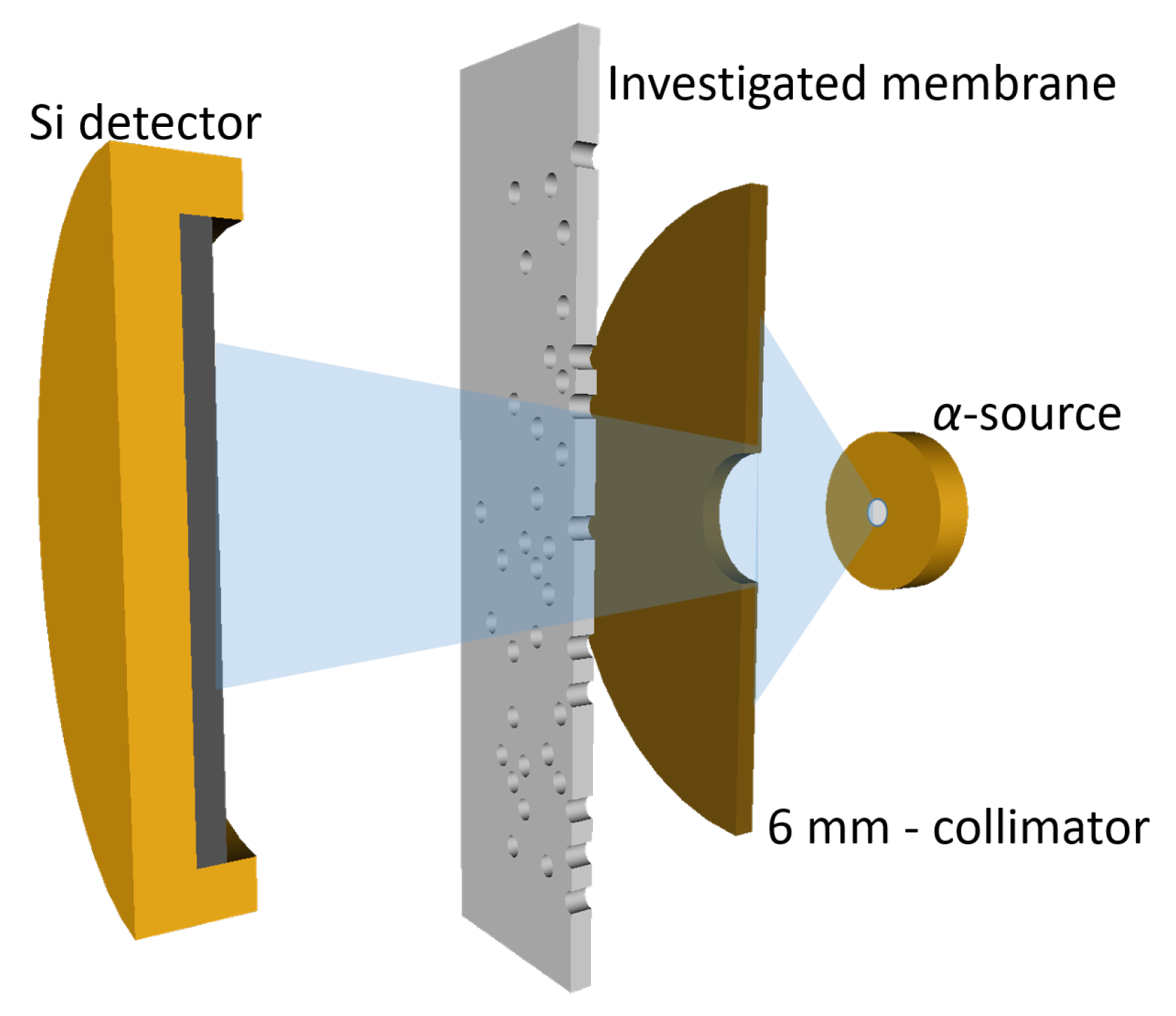
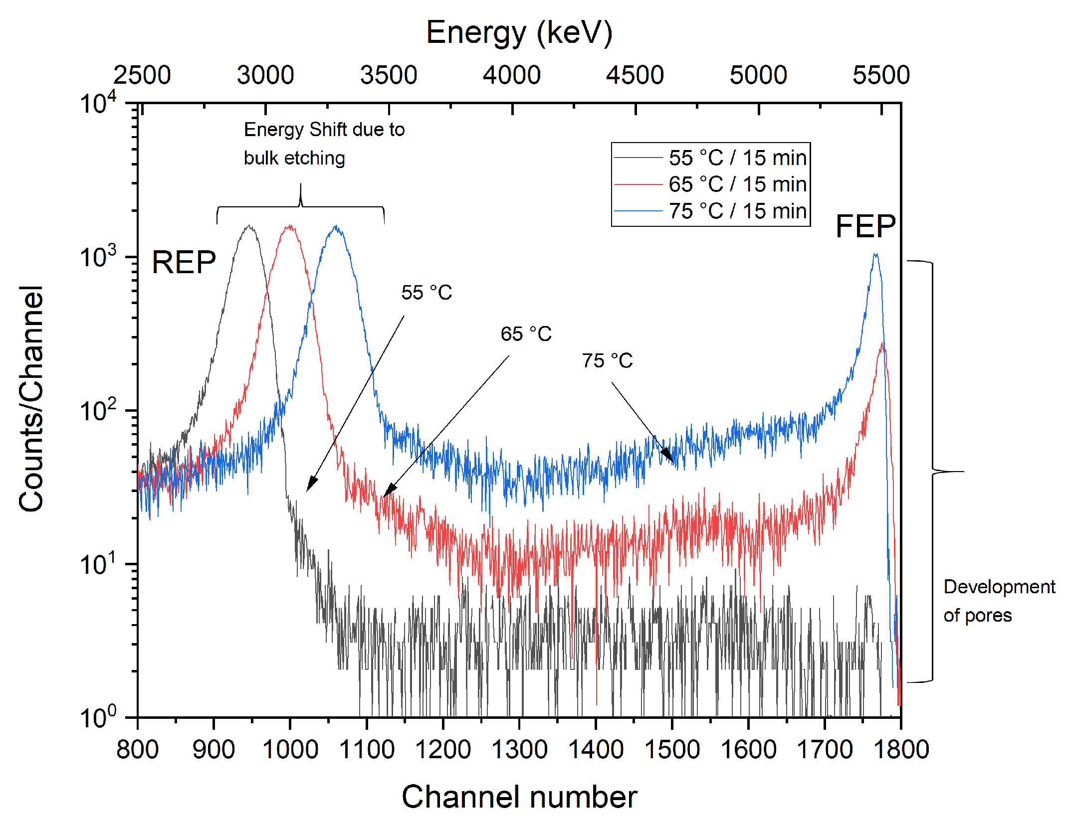
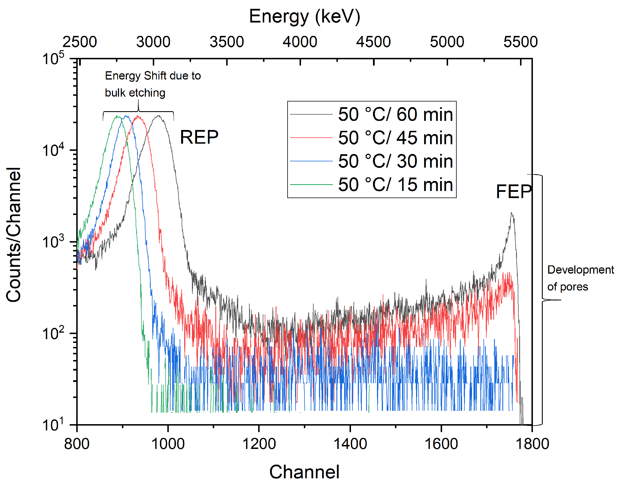
- Asymmetric etching (i.e., one-side etching protocol) of the irradiated foils, performed consecutively, was applied to study the gradual development of pores under both isochronal and isothermal conditions. Different pore shapes were obtained for different etching temperatures and exposure times. For the etching procedure, a 9M NaOH solution was used in a temperature range of 55–75 C and etching times 0–60 min. For subsequent doping, 5M LiCl solution was selected as a dopant, and doping was carried out for 24 h at RT only from the side of etching at different stages of the pore development. After removing the sample from the dopant vessel, the sample surface was gently dried and cleaned from the excess dopant solution by wiping a smooth cloth over the sample surface.
- (ii)
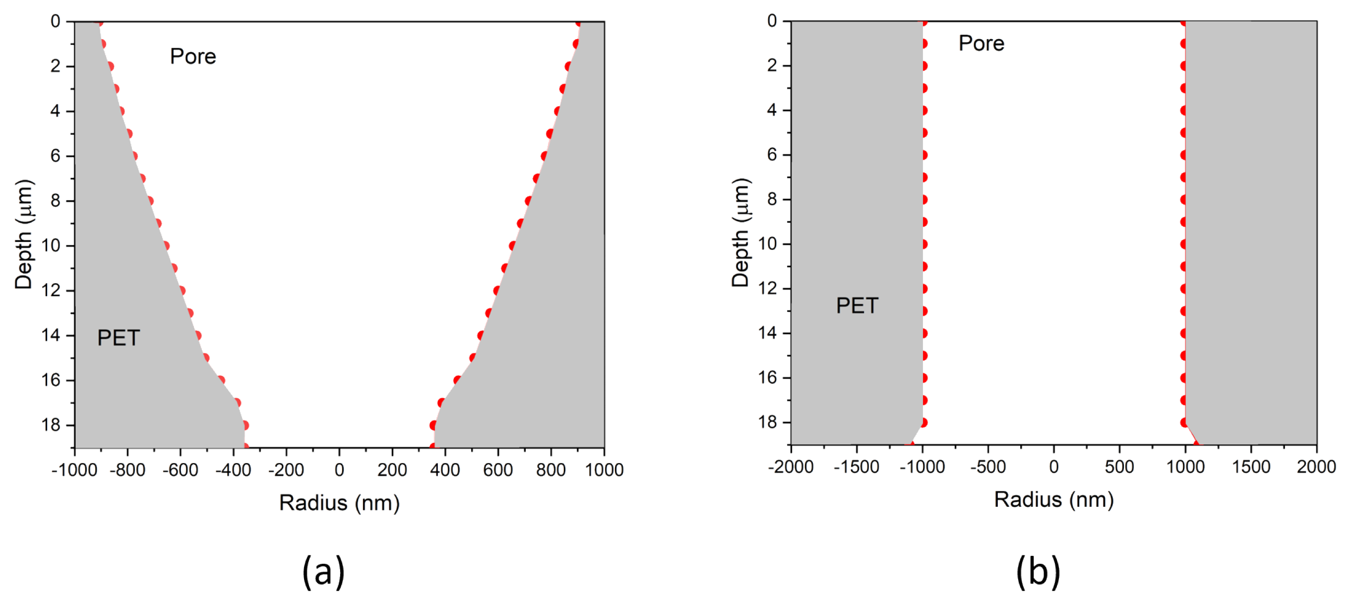
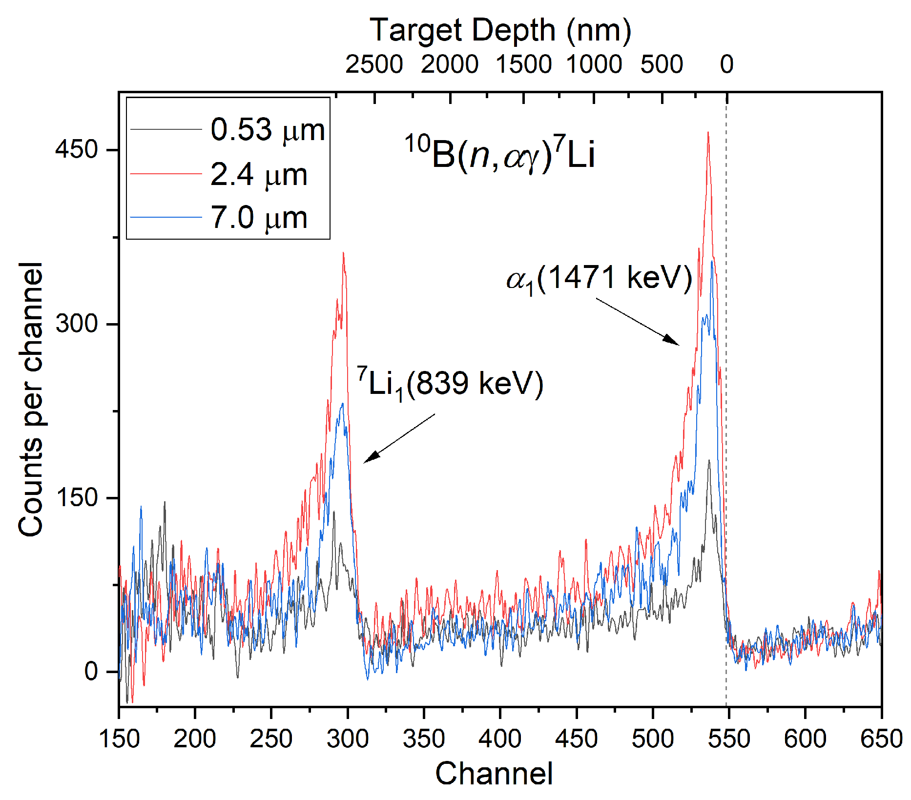
- In addition to one-side etching (performed in NPI Řež), a double-side etching protocol was also applied (JINR Dubna). Symmetric etching procedures made it possible to create membranes with cylindrical pores of several different diameters of 7 µm, 2.4 µm, and 0.53 µm (see Figure 1). Doping with boron was performed on only one side of the membrane (in addition to the PET films, a Si wafer was also used for the comparative analysis). Boron (99.9%, Kurt J. Lesker) was sputtered in Ar under a pressure of 5 Pa using a 3-inch planar magnetron powered by a radio frequency (13.56 MHz) power source. The power gradually increased (from 20 to 80 W) to avoid a thermal shock and cracking of the target. The total time of the deposition was 30 min, with a set thickness of 40 nm. Before deposition, the surface of the B target was pre-sputtered (cleaned) for 20 min.
2.2. Analytical Method
3. Results
4. Conclusions
Author Contributions
Funding
Institutional Review Board Statement
Data Availability Statement
Conflicts of Interest
References
- Apel, P. Ion-Track Membranes and Their Use in Biological and Medical Applications. AIP Conf. Proc. 2007, 912, 488–494. [Google Scholar]
- Kozlovskiy, A.; Borgekov, D.; Kenzhina, I.; Zdorovets, M.; Korolkov, I.; Kaniukov, E.; Kutuzau, M.; Shumskaya, A. PET ion-track membranes: Formation features and basic applications. In Proceedings of the International Conference on Nanotechnology and Nanomaterials, Kiev, Ukraine, 27–30 August 2018; Springer: Berlin/Heidelberg, Germany, 2018; pp. 461–479. [Google Scholar]
- Liu, F.; Wang, M.; Wang, X.; Wang, P.; Shen, W.; Ding, S.; Wang, Y. Fabrication and application of nanoporous polymer ion-track membranes. Nanotechnology 2018, 30, 052001. [Google Scholar] [CrossRef]
- Kaya, D.; Keçeci, K. Track-etched nanoporous polymer membranes as sensors: A review. J. Electrochem. Soc. 2020, 167, 037543. [Google Scholar] [CrossRef]
- Vasi, S.; Ceccio, G.; Cannavò, A.; Pleskunov, P.; Vacík, J. Study of Wettability of Polyethylene Membranes for Food Packaging. Sustainability 2022, 14, 5863. [Google Scholar] [CrossRef]
- Dutt, S.; Apel, P.; Lizunov, N.; Notthoff, C.; Wen, Q.; Trautmann, C.; Mota-Santiago, P.; Kirby, N.; Kluth, P. Shape of nanopores in track-etched polycarbonate membranes. J. Membr. Sci. 2021, 638, 119681. [Google Scholar] [CrossRef]
- Khulbe, K.; Feng, C.; Matsuura, T. The art of surface modification of synthetic polymeric membranes. J. Appl. Polym. Sci. 2010, 115, 855–895. [Google Scholar] [CrossRef]
- Korolkov, I.V.; Gorin, Y.G.; Yeszhanov, A.B.; Kozlovskiy, A.L.; Zdorovets, M.V. Preparation of PET track-etched membranes for membrane distillation by photo-induced graft polymerization. Mater. Chem. Phys. 2018, 205, 55–63. [Google Scholar] [CrossRef]
- Vacik, J.; Ceccio, G.; Cannavò, A.; Lavrentiev, V. Effects of UV irradiation and thermal annealing on LiCl derivatives encapsulation in porous PET membranes coated with a thin Au film. Radiat. Eff. Defects Solids 2022, 177, 112–123. [Google Scholar] [CrossRef]
- Mashentseva, A.A.; Korolkov, I.V.; Yeszhanov, A.B.; Zdorovets, M.V.; Russakova, A.V. The application of composite ion track membranes with embedded gold nanotubes in the reaction of aminomethylation of acetophenone. Mater. Res. Express 2019, 6, 115022. [Google Scholar] [CrossRef]
- Kotál, V.; Švorčík, V.; Slepička, P.; Sajdl, P.; Bláhová, O.; Šutta, P.; Hnatowicz, V. Gold coating of poly(ethylene terephthalate) modified by argon plasma. Plasma Process. Polym. 2007, 4, 69–76. [Google Scholar] [CrossRef]
- Agrawal, H.; Saraswat, V.K.; Awasthi, K. ZnO doping in PET matrix enhances conductivity of PET-ZnO nanocomposites. Adv. Electrochem. 2013, 1, 118–123. [Google Scholar] [CrossRef]
- Miao, J.; Zhao, K.; Guo, F.; Xu, L.; Xie, Y.; Deng, T. Novel LIS-doped mixed matrix membrane absorbent with high structural stability for sustainable lithium recovery from geothermal water. Desalination 2022, 527, 115570. [Google Scholar] [CrossRef]
- Liu, J.; Cao, D.; Yao, H.; Liu, D.; Zhang, X.; Zhang, Q.; Chen, L.; Wu, S.; Sun, Y.; He, D.; et al. Hexagonal Boron Nitride-Coated Polyimide Ion Track Etched Separator with Enhanced Thermal Conductivity and High-Temperature Stability for Lithium-Ion Batteries. ACS Appl. Energy Mater. 2022, 5, 8639–8649. [Google Scholar] [CrossRef]
- Lee, P.L.J.; Thangavel, V.; Guery, C.; Trautmann, C.; Toimil-Molares, M.E.; Morcrette, M. Etched ion-track membranes as tailored separators in Li–S batteries. Nanotechnology 2021, 32, 365401. [Google Scholar] [CrossRef]
- Siwy, Z.; Apel, P.; Dobrev, D.; Neumann, R.; Spohr, R.; Trautmann, C.; Voss, K. Ion transport through asymmetric nanopores prepared by ion track etching. Nucl. Instrum. Methods Phys. Res. Sect. B Beam Interact. Mater. Atoms 2003, 208, 143–148. [Google Scholar] [CrossRef]
- Kaniukov, E.; Shumskaya, A.; Yakimchuk, D.; Kozlovskiy, A.; Ibrayeva, A.; Zdorovets, M. Characterization of pet track membrane parameters. In Proceedings of the International Conference on Nanotechnology and Nanomaterials, Lviv, Ukraine, 24–27 August 2016; Springer: Berlin/Heidelberg, Germany, 2016; pp. 79–91. [Google Scholar]
- Apel, P.Y.; Ramirez, P.; Blonskaya, I.V.; Orelovitch, O.L.; Sartowska, B.A. Accurate characterization of single track-etched, conical nanopores. Phys. Chem. Chem. Phys. 2014, 16, 15214–15223. [Google Scholar] [CrossRef] [Green Version]
- Chander, M.; Kumar, S. Estimation of the nano-pores diameter by conductometric measurements. IOP Conf. Ser. Mater. Sci. Eng. 2022, 1221, 012050. [Google Scholar] [CrossRef]
- Álvarez-Arenas, T.G.; Apel, P.Y.; Orelovich, O. Characterization of ion-track membranes by non-contact ultrasonic magnitude and phase spectroscopy. J. Membr. Sci. 2007, 301, 210–220. [Google Scholar] [CrossRef]
- Apel, P. Track etching technique in membrane technology. Radiat. Meas. 2001, 34, 559–566. [Google Scholar] [CrossRef]
- George, J.; Irkens, M.; Neumann, S.; Scherer, U.; Srivastava, A.; Sinha, D.; Fink, D. Controlled ion track etching. Radiat. Eff. Defects Solids 2006, 161, 161–175. [Google Scholar] [CrossRef]
- UJF. ústav Jaderné Fyziky. Available online: http://www.ujf.cas.cz/cs/ (accessed on 22 September 2022).
- Vacik, J.; Hnatowicz, V.; Havranek, V.; Fink, D.; Apel, P.; Horak, P.; Ceccio, G.; Cannavo, A.; Torrisi, A. Ion track etching in polyethylene-terephthalate studied by charge particle transmission technique. Radiat. Eff. Defects Solids 2019, 174, 148–157. [Google Scholar] [CrossRef]
- Ceccio, G.; Vacik, J.; Trusso, S.; Cannavò, A.; Horak, P.; Hnatowicz, V.; Apel, P. Ion transmission spectroscopy of pores filled with Au nanoparticles. Nucl. Instrum. Methods Phys. Res. Sect. B Beam Interact. Mater. Atoms 2021, 491, 29–33. [Google Scholar] [CrossRef]
- Vacik, J.; Havranek, V.; Hnatowicz, V.; Horak, P.; Fink, D.; Apel, P. Study of ion tracks by micro-probe ion energy loss spectroscopy. Nucl. Instrum. Methods Phys. Res. Sect. B Beam Interact. Mater. Atoms 2014, 332, 308–311. [Google Scholar] [CrossRef]
- Vacík, J.; Červená, J.; Hnatowicz, V.; Pošta, S.; Fink, D.; Klett, R.; Strauss, P. Simple technique for characterization of ion-modified polymeric foils. Surf. Coat. Technol. 2000, 123, 97–100. [Google Scholar] [CrossRef]
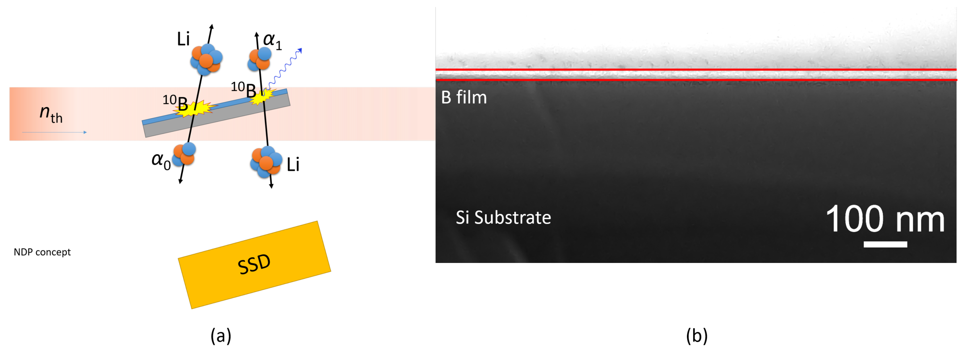
- Park, B.; Sun, G. Analysis of depth profiles of 10B and 6Li in Si wafers and lithium ion battery electrodes using the KAERI-NDP system. J. Radioanal. Nucl. Chem. 2016, 307, 1749–1756. [Google Scholar] [CrossRef]
- Chen-Mayer, H.H.; Lamaze, G.P. Depth distribution of boron determined by slow neutron induced lithium ion emission. Nucl. Instrum. Methods Phys. Res. Sect. B Beam Interact. Mater. Atoms 1998, 135, 407–412. [Google Scholar] [CrossRef]
- Hnatowicz, V.; Vacik, J.; Fink, D. Deconvolution of charged particle spectra from neutron depth profiling using Simplex method. Rev. Sci. Instrum. 2010, 81, 073906. [Google Scholar] [CrossRef]
- CVR. Centrum Výzkumu Řež. Available online: http://cvrez.cz/en/ (accessed on 22 September 2022).
- Froehlich, K.; Scheuerlein, M.C.; Ali, M.; Nasir, S.; Ensinger, W. Enhancement of heavy ion track-etching in polyimide membranes with organic solvents. Nanotechnology 2021, 33, 045301. [Google Scholar] [CrossRef]
- Yang, L.; Zhai, Q.; Li, G.; Jiang, H.; Han, L.; Wang, J.; Wang, E. A light transmission technique for pore size measurement in track-etched membranes. Chem. Commun. 2013, 49, 11415–11417. [Google Scholar] [CrossRef]


Publisher’s Note: MDPI stays neutral with regard to jurisdictional claims in published maps and institutional affiliations. |
© 2022 by the authors. Licensee MDPI, Basel, Switzerland. This article is an open access article distributed under the terms and conditions of the Creative Commons Attribution (CC BY) license (https://creativecommons.org/licenses/by/4.0/).
Share and Cite
Ceccio, G.; Vacik, J.; Siegel, J.; Cannavó, A.; Choukourov, A.; Pleskunov, P.; Tosca, M.; Fink, D. Etching and Doping of Pores in Polyethylene Terephthalate Analyzed by Ion Transmission Spectroscopy and Nuclear Depth Profiling. Membranes 2022, 12, 1061. https://doi.org/10.3390/membranes12111061
Ceccio G, Vacik J, Siegel J, Cannavó A, Choukourov A, Pleskunov P, Tosca M, Fink D. Etching and Doping of Pores in Polyethylene Terephthalate Analyzed by Ion Transmission Spectroscopy and Nuclear Depth Profiling. Membranes. 2022; 12(11):1061. https://doi.org/10.3390/membranes12111061
Chicago/Turabian StyleCeccio, Giovanni, Jiri Vacik, Jakub Siegel, Antonino Cannavó, Andrey Choukourov, Pavel Pleskunov, Marco Tosca, and Dietmar Fink. 2022. "Etching and Doping of Pores in Polyethylene Terephthalate Analyzed by Ion Transmission Spectroscopy and Nuclear Depth Profiling" Membranes 12, no. 11: 1061. https://doi.org/10.3390/membranes12111061