Ionic Environment Affects Biomolecular Interactions of Amyloid-β: SPR Biosensor Study


1
Institute of Photonics and Electronics of the Czech Academy of Sciences, Chaberská 1014/57, 182 51 Prague, Czech Republic
2
National Institute of Mental Health, Topolová 748, 250 67 Klecany, Czech Republic
*
Author to whom correspondence should be addressed.
Int. J. Mol. Sci. 2020, 21(24), 9727; https://doi.org/10.3390/ijms21249727
Submission received: 2 November 2020
/
Revised: 13 December 2020
/
Accepted: 17 December 2020
/
Published: 20 December 2020
(This article belongs to the Special Issue Amyloid-β: Structure, Function, and Pathophysiological Significance in Neurodegenerative Diseases 2.0)
Abstract
:In early stages of Alzheimer’s disease (AD), amyloid beta (Aβ) accumulates in the mitochondrial matrix and interacts with mitochondrial proteins, such as cyclophilin D (cypD) and 17β-hydroxysteroid dehydrogenase 10 (17β-HSD10). Multiple processes associated with AD such as increased production or oligomerization of Aβ affect these interactions and disbalance the equilibrium between the biomolecules, which contributes to mitochondrial dysfunction. Here, we investigate the effect of the ionic environment on the interactions of Aβ (Aβ1–40, Aβ1–42) with cypD and 17β-HSD10 using a surface plasmon resonance (SPR) biosensor. We show that changes in concentrations of K+ and Mg2+ significantly affect the interactions and may increase the binding efficiency between the biomolecules by up to 35% and 65% for the interactions with Aβ1–40 and Aβ1–42, respectively, in comparison with the physiological state. We also demonstrate that while the binding of Aβ1–40 to cypD and 17β-HSD10 takes place preferentially around the physiological concentrations of ions, decreased concentrations of K+ and increased concentrations of Mg2+ promote the interaction of both mitochondrial proteins with Aβ1–42. These results suggest that the ionic environment represents an important factor that should be considered in the investigation of biomolecular interactions taking place in the mitochondrial matrix under physiological as well as AD-associated conditions.
1. Introduction
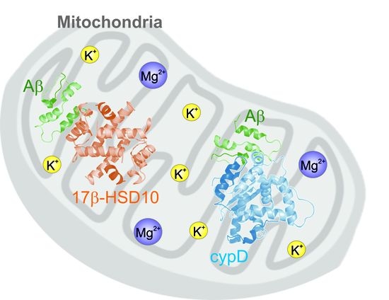
Alzheimer’s disease (AD) is currently the most common neurodegenerative disease of the elderly. It is characterized by extensive neuronal failure in which, according to the current understanding of AD, peptide amyloid beta (Aβ) plays a central role. Aβ is most commonly expressed as Aβ1–40 and Aβ1–42 fragments that are comprised of 40 and 42 amino acids, respectively [1]. During AD, the production of Aβ is increased and skewed towards Aβ1–42 [2]. Both fragments of Aβ are known to form oligomers (Aβ1–42 more readily than Aβ1–40 [3,4]) which have been shown to cause more substantial neuronal damage than monomers [5]. However, the exact molecular mechanisms of Aβ-induced pathological processes during AD remain poorly understood. Multiple hypotheses have been proposed [6], such as the mitochondrial cascade hypothesis [7], amyloid toxic oligomer hypothesis [8], calcium hypothesis [9] and lipid-chaperon hypothesis [10]. In this work, we follow the mitochondrial cascade hypothesis and investigate the Aβ-triggered processes taking place in mitochondria during AD.
In early stages of AD, Aβ accumulates in the mitochondrial matrix, where it interacts with multiple mitochondrial proteins, such as cyclophilin D (cypD) [11,12] and 17β-hydroxysteroid dehydrogenase type 10 (17β-HSD10) [13,14]. These interactions have been suggested to form a biomolecular link between Aβ and deteriorated mitochondrial functions [11,14], such as decreased energy metabolism [15,16], increased production of reactive oxygen species (ROS) [17] or formation of mitochondrial permeability transition pores (mPTPs) [12,18].
Interaction properties of Aβ have been the subject of multiple studies; in in vitro studies, synthetic Aβ oligomers generated in vitro have commonly been used [11,13,19,20]. Although there are differences in the biological characteristics of natural (obtained from biological samples) and synthetic oligomers, the differences are mainly in the efficiency of their action rather than in their function in biological processes [21,22]. In our recent work, we studied interactions of Aβ with cypD and 17β-HSD10, and with the use of a multi-interaction model, we demonstrated that the processes associated with early stages of AD such as the oligomerization of Aβ and increased/favored production of Aβ1–42 affect the equilibrium between these biomolecules in the mitochondrial matrix [23]. We also showed that 17β-HSD10 specifically binds cypD and thus regulates levels of free cypD in the mitochondrial matrix [24,25]. Our study also revealed that Aβ disrupts this regulation [24] which may result in the translocation of free cypD to the inner mitochondrial membrane and induce the formation of mPTPs [15]. In addition, our study showed that the ability of 17β-HSD10 to regulate cypD is significantly affected by the ionic environment in which the interaction takes place [24].
The ionic environment inside the mitochondrial matrix is established via multiple specialized channels through which ions are transferred between the cytosol and mitochondrial matrix. Physiological and pathological factors and processes (e.g., the temporal metabolic situation of the cell or the perturbations in the cytosolic environment) regulate this transfer and thus the concentrations of ions inside the mitochondrial matrix [26,27,28,29,30]. The most abundant ion present in the mitochondrial matrix is K+ which regulates/is regulated by the mitochondrial volume and the level of produced ROS [31,32]. It is assumed that K+ is present at a concentration of 140 mM both in the matrix and cytosol [31,33]. However, it has also been demonstrated that a decrease in the membrane potential (e.g., during the synthesis of ATP) may induce a decrease in K+ concentrations [34] and there are works that have reported considerably lower K+ concentrations (down to 15 mM) [32,35]. Mg2+ is the most abundant bivalent ion in the mitochondrial matrix and is typically present at concentrations of about 15–18 mM [36]. The majority of Mg2+ is bound by ATP and other Mg2+-binding biomolecules for which it serves as a cofactor [29] and only approximately 0.5–2.5 mM of the total Mg2+ is free [28,37]. In response to stimuli, such as the presence of particular hormones [26], increased energy demands of the cell [37] or perturbations in ionic strength inside the mitochondrial matrix [26], Mg2+ may dissociate from the complexes with biomolecules or leak from the matrix to the cytosol and thus increase or decrease the concentration of free Mg2+ in the mitochondrial matrix, respectively [38]. Moreover, it seems that the actions of different ions are interconnected and changes in the concentrations of one ion influence the distribution of the others [34]. In addition, the net ionic strength is also likely to fluctuate. Despite the number of studies on the ionic environment in the mitochondrial matrix, the knowledge of concentration ranges of ions and their relationship to particular biomolecular processes remains limited.
In this work, we investigate the effect of the ionic environment on the interactions of Aβ (Aβ1–40, Aβ1–42) with cypD and 17β-HSD10. The oligomeric forms of Aβ1–40 and Aβ1–42 pertinent to AD, i.e., monomeric Aβ1–40 and oligomeric Aβ1–42, are considered. In particular, we study the biomolecular interactions in the presence of K+ and Mg2+ using the surface plasmon resonance (SPR) biosensor method and show how changes in the concentrations of these ions affect the biomolecular interactions.
2. Results
2.1. Interactions of Aβ1–40 and Aβ1–42 with cypD at Different Concentrations of K+ and Mg2+
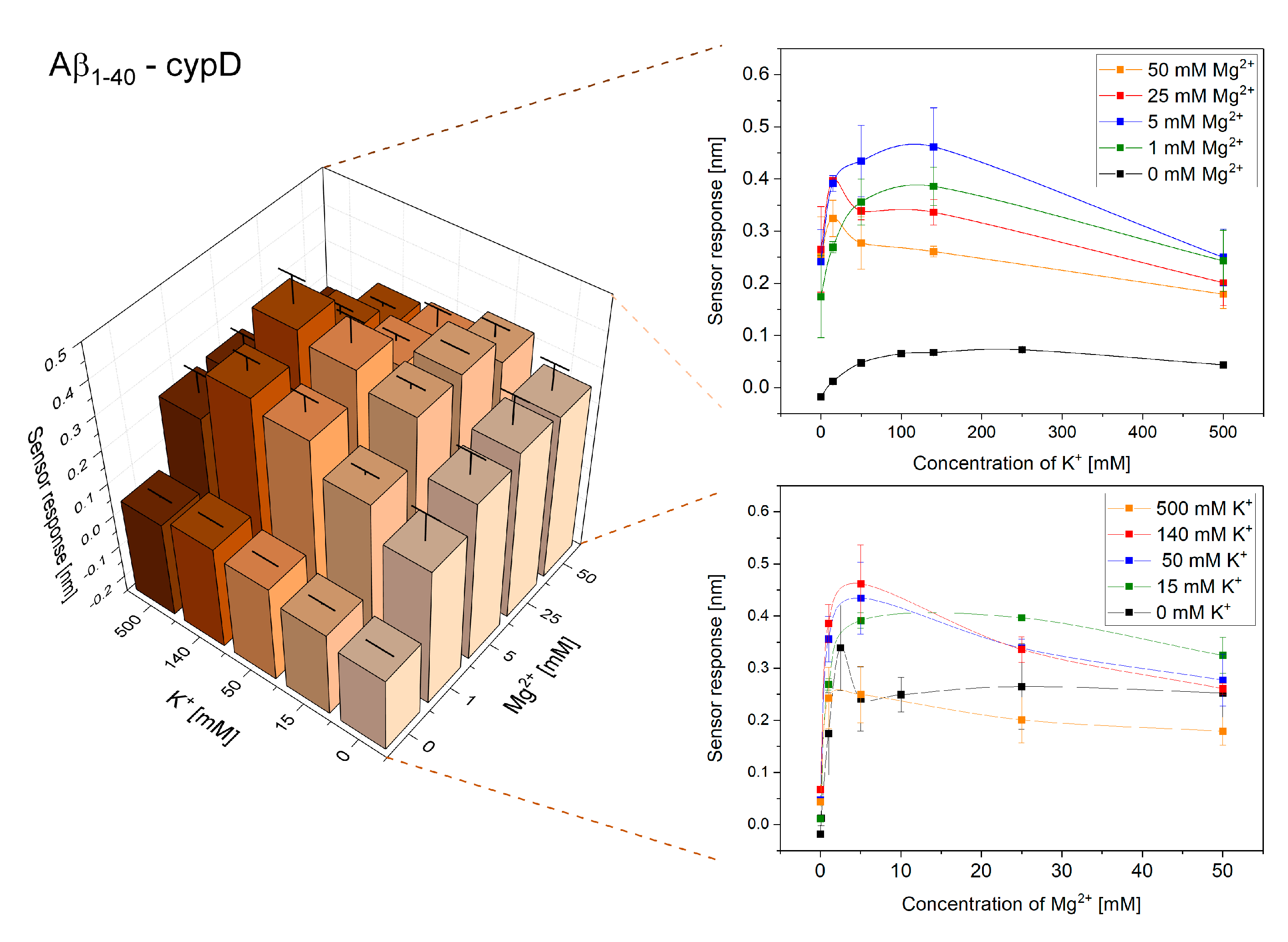
Initially, we investigated the effect of varying concentrations of K+ and Mg2+ on the interactions of cypD and Aβ. Figure 1 and Figure 2 show the obtained reference-compensated sensor responses corresponding to the binding of monomeric Aβ1–40 and oligomeric Aβ1–42 to the immobilized cypD in the presence of different concentrations of K+ and Mg2+, respectively, and demonstrate the significant effect of these ions on the interactions.
The binding efficiency of the interactions follows a complex trend. The binding efficiency is lowest in the absence of K+ and Mg2+, increases with increasing concentrations of ions until it reaches the maximum and then decreases. The maximum binding efficiency of the interactions between cypD and Aβ occurred at different ionic concentrations for Aβ1–40 and Aβ1–42. While the highest binding efficiency of the interaction between cypD and Aβ1–40 was observed at 140 mM K+ and 5 mM Mg2+, the highest binding efficiency of the interaction between Aβ1–42 and cypD occurred at 15 mM K+ and 25 mM Mg2+.
The different effects of ions on the binding efficiency between Aβ and cypD observed for Aβ1–40 and Aβ1–42 may originate from two main factors: (1) the different oligomerization state of the particular fragments of Aβ (Aβ1–40 in the monomeric form, Aβ1–42 in the oligomeric form), and (2) the structural differences between the two fragments (two additional hydrophobic amino acids in the structure of Aβ1–42). In order to evaluate the contribution of these factors, we investigate the effect of the oligomerization state of Aβ on the interactions (Section 2.2).
2.2. The Effect of the Oligomerization State of Aβ
In order to evaluate whether the different effects of ions on the binding between Aβ and cypD observed for Aβ1–40 and Aβ1–42 may originate from the different oligomerization states of the particular fragments of Aβ, we analyzed the binding of Aβ1–40 and Aβ1–42 in both monomeric and oligomeric forms (see Section 4.1.3 in Materials and Methods) to the immobilized cypD. The binding was observed in the presence of 5 mM Mg2+ and varying concentrations of K+, as under these conditions, the differences in trends obtained for the different fragments of Aβ were the most evident.
Figure 3 shows that oligomeric forms of Aβ exhibit higher sensor responses in comparison to the monomeric forms, which agrees well with the results of our previous study [14]. However, the maximum binding efficiency occurs for the same ionic environment for both oligomerization forms for both fragments of Aβ. It occurs in the presence of 140 mM K+ for the interaction between monomeric as well as oligomeric Aβ1–40 and cypD and in the presence of 50 mM K+ for the interaction between monomeric as well as oligomeric Aβ1–42 and cypD. Thus, we conclude that the observed trends are not affected by the different oligomerization states of the fragments but rather originate from the structural differences between Aβ1–40 and Aβ1–42.
2.3. Interactions of Aβ1–40 and Aβ1–42 with 17β-HSD10 at Different Concentrations of K+ and Mg2+
In this part of the study, we investigated the effect of the ionic environment on the interactions of 17β-HSD10 and Aβ. Figure 4 and Figure 5 show the obtained reference-compensated sensor responses to the binding of monomeric Aβ1–40 and oligomeric Aβ1–42 to the immobilized 17β-HSD10 in the presence of varying concentrations of K+ and Mg2+, respectively.
We observed a peak in the efficiency of the binding between 17β-HSD10 and Aβ for ionic compositions that were different for Aβ1–40 and Aβ1–42 (similarly as for the interaction between cypD and Aβ). The maximum binding efficiency for the interaction of 17β-HSD10 with Aβ1–40 and Aβ1–42 was observed for 50 mM K+ and 25 mM Mg2+ and for 15 mM K+ and 25 mM Mg2+, respectively.
3. Discussion
Our results demonstrate that perturbations of the concentrations of K+ and Mg2+ may considerably affect the interactions between biomolecules in the mitochondrial matrix (Figure 1, Figure 2, Figure 3, Figure 4 and Figure 5). For all the interactions investigated herein, we observed a peak in the maximum binding efficiency occurring at a particular ionic composition. The peak was sharper for interactions of Aβ1–42 with both cypD and 17β-HSD10 when compared to the interactions of Aβ1–40, suggesting that the interactions of Aβ1–42 are more sensitive to changes in concentrations of ions. Moreover, the position of the peak differed for different interactions. It occurred at 15 mM K+ and 25 mM Mg2+ for interactions of Aβ1–42 and at 50–140 mM K+ and 5–25 mM Mg2+ for interactions of Aβ1–40 with both cypD and 17β-HSD10. Assuming the physiological concentrations of ions in the mitochondrial matrix to be 140 mM K+ and 1 mM Mg2+, we conclude that the binding efficiency of the interactions of Aβ with cypD and 17β-HSD10 may increase by 20–35% for Aβ1–40 and by 40–65% for Aβ1–42 in comparison with the physiological conditions.
In our previous study, we showed that processes related to AD such as increased production of Aβ, unbalanced production of Aβ fragments favoring Aβ1–42 or oligomerization of Aβ1–42 significantly affect the interactions between Aβ and mitochondrial proteins and enhance the binding of Aβ1–42 to both cypD and 17β-HSD10 [23]. In this work, we demonstrate that the mitochondrial ionic environment also plays an important role in biomolecular processes taking place in the mitochondrial matrix, and we show that the conditions causing decreased concentrations of K+ and increased concentrations of Mg2+ may further enhance the binding of Aβ1–42 to both cypD and 17β-HSD10.
Despite the large number of studies on the ionic transport in mitochondria, it remains difficult to relate a specific ionic composition to a particular process taking place in the mitochondrial matrix. Due to the oxidative inactivation of ATP-related enzymes, decreased production of ATP has been observed in affected areas of the AD brain [39]. Bredshaw et al. associated the decreased levels of ATP with the increased concentration of free Mg2+ inside the mitochondrial matrix [26]. The excess of free Mg2+ may inhibit the function of mitoKATP, which may decrease the influx of K+ in mitochondria [40]. Therefore, we hypothesize that, under the circumstances of progressing AD, the deteriorated mitochondrial functions alter the ionic environment in mitochondria which promotes the interactions of Aβ1–42 with cypD and 17β-HSD10. These interactions lead to the inhibition of the enzymatic function of 17β-HSD10 [14] and contribute to an increased production of ROS [41] and thus further aggravate the pathology of AD.
4. Materials and Methods
4.1. Materials
4.1.1. Reagents
NaCl, NaOH, KCl, MgCl2, hexafluoroisopropanol (HFIP), NH4OH, bovine serum albumin (BSA) and all buffers were purchased from Sigma-Aldrich, Czech Republic. Thiols: 11-mercapto-hexa(ethyleneglycol)undecyloxy acetic acid (HS-C11-(EG)6-OCH2-COOH) and 11-mercapto-tetra(ethyleneglycol)undecanol (HS-C11-(EG)4-OH), were purchased from Prochimia, Poland. N-hydroxysuccinimide (NHS), 1-ethyl-3-(3-dimethylaminopropyl)-carbodiimide hydrochloride (EDC) and ethanolamine hydrochloride (EA) were purchased from GE Healthcare, Sweden. 17β-HSD10 (human, recombinant), cypD (human, recombinant) and an antibody against cypD (Ab(cypD)) were purchased from Fitzgerald, USA. An antibody against 17β-HSD10 (Ab(17β-HSD10)) was purchased from Biolegend, USA. Aβ1–40 and Aβ1–42 (human, synthetic) were obtained from AnaSpec, USA, dissolved in 1% NH4OH and diluted by PBS to obtain the stock concentration of 100 μM.
4.1.2. Buffers
The buffers used for the functionalization of SPR chips and the immobilization of cypD and 17β-HSD10 to the surface included the following: sodium acetate (SA10; 10 mM, pH 5.0), MES (10 mM, pH 5.0), phosphate-buffered saline (PBS; 10 mM phosphate, 2.7 mM KCl, 138 mM NaCl, pH 7.4), high-ionic strength PBS (PBSNa; 10 mM phosphate, 2.7 mM KCl, 750 mM NaCl, pH 7.4). All the buffers were prepared using deionized Milli-Q water (Merck, Czech Republic).
The running buffer (RB) with varying concentrations of K+ and Mg2+ was used for the binding of Aβ to the surface to simulate different ionic environments of the interactions of Aβ with cypD and 17β-HSD10. All the used RBs were prepared as 10 mM HEPES in Milli-Q with addition of BSA (200 μg/mL). The pH was adjusted by NaOH to 7.4 and NaCl was used to set the concentration of Na+ to 5 mM. Concentrations of K+ and Mg2+ in RBs were adjusted by the addition of aqueous solutions of KCl and MgCl2 at particular concentrations that ensured the same dilution (9:1) of RBs for all the concentrations of ions.
4.1.3. Preparation of Aβ Samples
In this work, Aβ1–40 and Aβ1–42 fragments with different oligomerization states were prepared using the procedures described in our previous study [23]. The oligomeric forms of Aβ1–40 and Aβ1–42 were prepared using Preparation A, whereas monomeric Aβ1–40 and monomeric Aβ1–42 were prepared using Preparation C and Preparation B, respectively. Briefly, in Preparation A, Aβ from the stock solution was diluted by RB to obtain the final concentration of Aβ of 1 μM. In Preparation B, Aβ from the stock solution was diluted by 12.5 mM NaOH in the volume ratio of 1:4, sonicated for 5 min and finally diluted by RB to obtain the final concentration of Aβ of 1 μM. In Preparation C, Aβ from the stock solution was mixed with HFIP in the volume ratio of 1:9, vortexed for 1 min and then the solvent was evaporated using a stream of nitrogen (solid Aβ was dissolved in RB to obtain the final concentration of Aβ of 1 μM).
4.2. Instrumentation
4.2.1. Surface Plasmon Resonance (SPR) Biosensor
We used a six-channel SPR biosensor platform based on wavelength spectroscopy of surface plasmons (Plasmon VI) developed at the Institute of Photonics and Electronics, Prague. In this SPR platform, the angle of incidence of the light beam is fixed and changes in the resonance wavelength of surface plasmons are measured by analyzing the spectrum of polychromatic light reflected from an SPR chip. The resonance wavelength is sensitive to changes in the refractive index caused by the binding of biomolecules to the surface of an SPR chip. A shift in the resonance wavelength of 1 nm represents a change in the protein surface coverage of 17 ng/cm2. The SPR chips used in this study were prepared by coating microscope glass slides obtained from Marienfeld, Germany, with thin layers of titanium (1–2 nm) and gold (48 nm) via e-beam evaporation in vacuum. The SPR platform was combined with a dispersionless microfluidic module [42]. The active temperature stabilization unit allowed maintaining the temperature in the system with a precision of 0.01 °C. All experiments reported in this study were performed at a temperature of 25 °C and a flow rate of 20 µL/min.
4.2.2. Functionalization of the SPR Chip
Prior to the experiments, the surface of an SPR chip was modified by a self-assembled monolayer of mixed thiols, on which specific antibodies (Ab(cypD) or Ab(17β-HSD10)) were immobilized using the amino-coupling method as described previously [43]. Briefly, a clean SPR chip was immersed in a 3:7 molar mixture of HS-C11-(EG)6-OCH2-COOH and HS-C11-(EG)4-OH (ethanol solution, total concentration of 0.2 mM), incubated for 10 min at 40 °C and then incubated for at least 12 h at room temperature in the dark. Prior to use, the chip was rinsed with ethanol and Milli-Q water, dried with a stream of nitrogen and mounted in the SPR platform. First, the mixture of 12.5 mM NHS and 62.5 mM EDC in Milli-Q water was injected (10 min) to activate the carboxylic groups. Then, Ab(cypD) or Ab(17β-HSD10) at a concentration of 10 μg/mL in SA10 was pumped through the flow cell until the response to the immobilized antibody leveled off (~15 min, surface coverage ~ 220 and 300 ng/cm2). Then, PBSNa was applied (5 min) to remove the antibody non-covalently attached to the surface. Finally, 500 mM EA in Milli-Q water was injected (5 min) to deactivate the unreacted carboxylic groups.
4.2.3. Immobilization of cypD to the Functionalized SPR Chip
The SPR chip functionalized with Ab(cypD) was washed with MES. Then, the detection channels were exposed to 100 nM cypD in MES until the response to the immobilized cypD leveled off (~15 min, surface coverage ~100 ng/cm2), while the reference channels were kept in MES. Then, all channels were washed with MES (> 20 min) and exposed to PBSNa (5 min) to remove the non-covalently attached cypD.
In order to compensate for the variations in the surface coverage of the immobilized cypD after the PBSNa washing, five different levels of cypD (surface coverage of 1.7–100 ng/cm2 before PBSNa washing) were immobilized on the surface of an SPR chip. Then, MES was exchanged for RB containing 5 mM Mg2+ and 140 mM K+ and monomeric Aβ1–40 or oligomeric Aβ1–42 at a concentration of 1 μM in RB were injected in both the reference and detection channels (5 min). Finally, RB was injected again. The sensor responses obtained in the reference channels 5 min after switching back to the RB were subtracted from those obtained in the detection channels. Such reference-compensated sensor responses were plotted as a function of cypD surface coverage and fitted by the Boltzmann function. The experiment was repeated three times and the parameters obtained from the fitting were averaged and used to normalize the measured sensor responses to the binding of Aβ to varying levels of cypD to those corresponding to the binding of Aβ to cypD with the surface coverage of 34 ng/cm2.
4.2.4. Immobilization of 17β-HSD10 to the Functionalized SPR Chip
The SPR chip functionalized with Ab(17β-HSD10) was washed with MES. Then, the detection channels were exposed to 100 nM 17β-HSD10 in MES until the response to the immobilized 17β-HSD10 leveled off (~15 min, surface coverage of 150 ng/cm2), while the reference channels were kept in MES. Then, all the channels were washed with MES (> 20 min). In contrast to the immobilization of cypD, the immobilization of 17β-HSD10 resulted in a stable surface that was resistant to changes in RB and thus no additional steps were necessary.
4.3. Interactions of Aβ1–40 and Aβ1–42 with cypD and 17β-HSD10 at Different Concentrations of K+ and Mg2+
In all the experiments performed in this study, RB was injected into both the detection and reference channels of the functionalized SPR chip and flowed along the surface until the stable baseline was reached. Then, the sample of Aβ (prepared according to Preparations A–C, see Section 4.1.3 in Materials and Methods) at a concentration of 1 μM in the particular RB was injected into both the detection (surface with immobilized cypD or 17β-HSD10) and the reference (surface without immobilized cypD or 17β-HSD10) channels (5 min). Then, RB was injected again. The final sensor response was determined as a difference between the responses of the detection and reference channels 5 min after switching back to RB. The reference channel accounts for interferences (e.g., due to the electrostatically induced effects) and therefore the final reference-compensated sensor response corresponds to the specific binding of Aβ to cypD or 17β-HSD10 in the particular ionic environment. Each experiment was repeated at least three times and at least three sensor response values were used to calculate the mean and standard deviation of the sensor responses for the particular concentration of ions.
The effect of the ionic environment on the interactions of monomeric Aβ1–40 and oligomeric Aβ1–42 with cypD and 17β-HSD10 was investigated using RBs containing K+ at concentrations of 0, 15, 50, 140 and 500 mM and Mg2+ at concentrations of 0, 1, 5, 25 and 50 mM. The effect of the oligomerization state of Aβ on the interactions of Aβ with cypD was investigated using RBs containing 5 mM Mg2+ and 0, 15, 50, 140 and 500 mM K+ and Aβ1–40 and Aβ1–42 prepared in both monomeric and oligomeric forms.
5. Conclusions
In this work, we demonstrate that the ionic environment significantly affects the interactions between proteins of the mitochondrial matrix (cypD, 17β-HSD10) and Aβ and show that the particular ionic environment may increase the binding efficiency of these interactions in comparison with the physiological state. As the ionic environment in the mitochondrial matrix fluctuates in response to different physiological as well as AD-associated processes, its effect on the biomolecular interactions should be considered in the studies of the processes taking place in the mitochondrial matrix.
Author Contributions
Conceptualization, E.H. and J.H.; methodology, E.H and T.Š.; investigation, data analysis and interpretation, E.H., T.Š., Z.K. and J.H.; writing, review and editing, E.H., T.Š., Z.K. and J.H.; project administration and funding acquisition, Z.K. and J.H. All authors have read and agreed to the published version of the manuscript.
Funding
This research was supported by the Czech Science Foundation (contracts 20-23787X and 19-02739S).
Acknowledgments
The authors would like to acknowledge the contribution of Lucie Peštová and Emma Etter who performed multiple SPR biosensing experiments.
Conflicts of Interest
The authors declare no conflict of interest.
References
- Murphy, M.P.; LeVine, H., 3rd. Alzheimer’s disease and the amyloid-beta peptide. J. Alzheimers Dis. Jad 2010, 19, 311–323. [Google Scholar] [CrossRef] [PubMed] [Green Version]
- Yan, Y.; Wang, C. Aβ42 is More Rigid than Aβ40 at the C Terminus: Implications for Aβ Aggregation and Toxicity. J. Mol. Biol. 2006, 364, 853–862. [Google Scholar] [CrossRef] [PubMed]
- Lührs, T. 3D structure of Alzheimer’s amyloid-β(1–42) fibrils. Proc. Natl. Acad. Sci. USA 2005, 102, 17342–17347. [Google Scholar] [CrossRef] [PubMed] [Green Version]
- Garai, K.; Frieden, C. Quantitative analysis of the time course of Aβ oligomerization and subsequent growth steps using tetramethylrhodamine-labeled Aβ. Proc. Natl. Acad. Sci. USA 2013, 110, 3321–3326. [Google Scholar] [CrossRef] [Green Version]
- Reddy, P.H. Amyloid beta, mitochondrial structural and functional dynamics in Alzheimer’s disease. Exp. Neurol. 2009, 218, 286–292. [Google Scholar] [CrossRef] [Green Version]
- Crouch, P.J.; Harding, S.-M.E.; White, A.R.; Camakaris, J.; Bush, A.I.; Masters, C.L. Mechanisms of Aβ mediated neurodegeneration in Alzheimer’s disease. Int. J. Biochem. Cell Biol. 2008, 40, 181–198. [Google Scholar] [CrossRef]
- Swerdlow, R.H.; Burns, J.M.; Khan, S.M. The Alzheimer‘s disease mitochondrial cascade hypothesis: Progress and perspectives. Biochim. Et Biophys. Acta (Bba)-Mol. Basis Dis. 2014, 1842, 1219–1231. [Google Scholar] [CrossRef] [Green Version]
- Cline, E.N.; Bicca, M.A.; Viola, K.L.; Klein, W.L. The Amyloid-β Oligomer Hypothesis: Beginning of the Third Decade. J. Alzheimers Dis. Jad 2018, 64, S567–S610. [Google Scholar] [CrossRef] [Green Version]
- Berridge, M.J. Calcium hypothesis of Alzheimer’s disease. Pflügers Arch. -Eur. J. Physiol. 2010, 459, 441–449. [Google Scholar] [CrossRef]
- Sciacca, M.F.; Lolicato, F.; Tempra, C.; Scollo, F.; Sahoo, B.R.; Watson, M.D.; García-Viñuales, S.; Milardi, D.; Raudino, A.; Lee, J.C. Lipid-Chaperone Hypothesis: A Common Molecular Mechanism of Membrane Disruption by Intrinsically Disordered Proteins. Acs Chem. Neurosci. 2020, 11, 4336–4350. [Google Scholar] [CrossRef]
- Du, H.; Guo, L.; Fang, F.; Chen, D.; Sosunov, A.A.; McKhann, G.M.; Yan, Y.; Wang, C.; Zhang, H.; Molkentin, J.D.; et al. Cyclophilin D deficiency attenuates mitochondrial and neuronal perturbation and ameliorates learning and memory in Alzheimer’s disease. Nat. Med. 2008, 14, 1097–1105. [Google Scholar] [CrossRef] [PubMed]
- Du, H.; Guo, L.; Zhang, W.; Rydzewska, M.; Yan, S. Cyclophilin D deficiency improves mitochondrial function and learning/memory in aging Alzheimer disease mouse model. Neurobiol. Aging 2011, 32, 398–406. [Google Scholar] [CrossRef] [PubMed] [Green Version]
- Yan, Y.; Liu, Y.; Sorci, M.; Belfort, G.; Lustbader, J.W.; Yan, S.S.; Wang, C. Surface Plasmon Resonance and Nuclear Magnetic Resonance Studies of ABAD−Aβ Interaction. Biochemistry 2007, 46, 1724–1731. [Google Scholar] [CrossRef]
- Lustbader, J.W.; Cirilli, M.; Lin, C.; Xu, H.W.; Takuma, K.; Wang, N.; Caspersen, C.; Chen, X.; Pollak, S.; Chaney, M.; et al. ABAD directly links Abeta to mitochondrial toxicity in Alzheimer’s disease. Science 2004, 304, 448–452. [Google Scholar] [CrossRef] [PubMed] [Green Version]
- Yan, S.D.; Stern, D.M. Mitochondrial dysfunction and Alzheimer‘s disease: Role of amyloid-β peptide alcohol dehydrogenase (ABAD). Int. J. Exp. Pathol. 2005, 86, 161–171. [Google Scholar] [CrossRef]
- Luo, Z.; Zhang, J.; Wang, Y.; Chen, J.; Li, Y.; Duan, Y. An aptamer based method for small molecules detection through monitoring salt-induced AuNPs aggregation and surface plasmon resonance (SPR) detection. Sens. Actuators B Chem. 2016, 236, 474–479. [Google Scholar] [CrossRef]
- Singh, P.; Suman, S.; Chandna, S.; Das, T.K. Possible role of amyloid-beta, adenine nucleotide translocase and cyclophilin-D interaction in mitochondrial dysfunction of Alzheimer‘s disease. Bioinformation 2009, 3, 440–445. [Google Scholar] [CrossRef] [Green Version]
- Rao, V.K.; Carlson, E.A.; Yan, S.S. Mitochondrial permeability transition pore is a potential drug target for neurodegeneration. Biochim. Et Biophys. Acta (Bba)-Mol. Basis Dis. 2014, 1842, 1267–1272. [Google Scholar] [CrossRef] [Green Version]
- Bartolini, M.; Naldi, M.; Fiori, J.; Valle, F.; Biscarini, F.; Nicolau, D.V.; Andrisano, V. Kinetic characterization of amyloid-beta 1–42 aggregation with a multimethodological approach. Anal. Biochem. 2011, 414, 215–225. [Google Scholar] [CrossRef]
- Hou, L.; Shao, H.; Zhang, Y.; Li, H.; Menon, N.K.; Neuhaus, E.B.; Brewer, J.M.; Byeon, I.-J.L.; Ray, D.G.; Vitek, M.P.; et al. Solution NMR Studies of the Aβ(1−40) and Aβ(1−42) Peptides Establish that the Met35 Oxidation State Affects the Mechanism of Amyloid Formation. J. Am. Chem. Soc. 2004, 126, 1992–2005. [Google Scholar] [CrossRef]
- Wang, Q.; Walsh, D.M.; Rowan, M.J.; Selkoe, D.J.; Anwyl, R. Block of Long-Term Potentiation by Naturally Secreted and Synthetic Amyloid β-Peptide in Hippocampal Slices Is Mediated via Activation of the Kinases c-Jun N-Terminal Kinase, Cyclin-Dependent Kinase 5, and p38 Mitogen-Activated Protein Kinase as well as Metabotropic Glutamate Receptor Type 5. J. Neurosci. 2004, 24, 3370–3378. [Google Scholar] [CrossRef] [PubMed] [Green Version]
- Kittelberger, K.A.; Piazza, F.; Tesco, G.; Reijmers, L.G. Natural Amyloid-Beta Oligomers Acutely Impair the Formation of a Contextual Fear Memory in Mice. PLoS ONE 2012, 7, e29940. [Google Scholar] [CrossRef] [PubMed]
- Hemmerová, E.; Špringer, T.; Krištofiková, Z.; Homola, J. Study of Biomolecular Interactions of Mitochondrial Proteins Related to Alzheimer’s Disease: Toward Multi-Interaction Biomolecular Processes. Biomolecules 2020, 10, 1214. [Google Scholar] [CrossRef] [PubMed]
- Hemmerová, E.; Špringer, T.; Krištofiková, Z.; Homola, J. In vitro study of interaction of 17β-hydroxysteroid dehydrogenase type 10 and cyclophilin D and its potential implications for Alzheimer’s disease. Sci. Rep. 2019, 9, 16700. [Google Scholar] [CrossRef] [Green Version]
- Krištofiková, Z.; Špringer, T.; Gedeonová, E.; Hofmannová, A.; Říčný, J.; Hromádková, L.; Vyhnálek, M.; Laczo, J.; Nikolai, T.; Hort, J.; et al. Interactions of 17β-Hydroxysteroid Dehydrogenase Type 10 and Cyclophilin D in Alzheimer’s Disease. Neurochem. Res. 2020, 45, 915–927. [Google Scholar]
- Bradshaw, P.C.; Pfeiffer, D.R. Release of Ca2+ and Mg2+ from yeast mitochondria is stimulated by increased ionic strength. Bmc Biochem. 2006, 7, 4. [Google Scholar] [CrossRef] [Green Version]
- Haumann, J.; Dash, R.K.; Stowe, D.F.; Boelens, A.D.; Beard, D.A.; Camara, A.K. Mitochondrial free [Ca2+] increases during ATP/ADP antiport and ADP phosphorylation: Exploration of mechanisms. Biophys. J. 2010, 99, 997–1006. [Google Scholar] [CrossRef] [Green Version]
- Jung, D.W.; Apel, L.; Brierley, G.P. Matrix free magnesium changes with metabolic state in isolated heart mitochondria. Biochemistry 1990, 29, 4121–4128. [Google Scholar] [CrossRef]
- Yamanaka, R.; Tabata, S.; Shindo, Y.; Hotta, K.; Suzuki, K.; Soga, T.; Oka, K. Mitochondrial Mg2+ homeostasis decides cellular energy metabolism and vulnerability to stress. Sci. Rep. 2016, 6, 30027. [Google Scholar] [CrossRef]
- O’Rourke, B.; Cortassa, S.; Aon, M.A. Mitochondrial Ion Channels: Gatekeepers of Life and Death. Physiology 2005, 20, 303–315. [Google Scholar] [CrossRef] [Green Version]
- Garlid, K.D.; Paucek, P. Mitochondrial potassium transport: The K+ cycle. Biochim. Et Biophys. Acta (Bba)-Bioenerg. 2003, 1606, 23–41. [Google Scholar] [CrossRef] [Green Version]
- Kaasik, A.; Safiulina, D.; Zharkovsky, A.; Veksler, V. Regulation of mitochondrial matrix volume. Am. J. Physiol. -Cell Physiol. 2007, 292, C157–C163. [Google Scholar] [CrossRef] [PubMed]
- Augustynek, B.; Wrzosek, A.; Koprowski, P.; Kielbasa, A.; Bednarczyk, P.; Lukasiak, A.; Dolowy, K.; Szewczyk, A. What we don’t know about mitochondrial potassium channels? Postepy Biochem. 2016, 62, 189–198. [Google Scholar] [PubMed]
- Szabò, I.; Leanza, L.; Gulbins, E.; Zoratti, M. Physiology of potassium channels in the inner membrane of mitochondria. Pflügers Arch. -Eur. J. Physiol. 2012, 463, 231–246. [Google Scholar] [CrossRef]
- Zoeteweij, J.P.; van de Water, B.; de Bont, H.J.; Nagelkerke, J.F. Mitochondrial K+ as modulator of Ca(2+)-dependent cytotoxicity in hepatocytes. Novel application of the K(+)-sensitive dye PBFI (K(+)-binding benzofuran isophthalate) to assess free mitochondrial K+ concentrations. Biochem. J. 1994, 299, 539–543. [Google Scholar] [CrossRef] [PubMed] [Green Version]
- Yamanaka, R.; Shindo, Y.; Oka, K. Magnesium Is a Key Player in Neuronal Maturation and Neuropathology. Int. J. Mol. Sci. 2019, 20, 3439. [Google Scholar] [CrossRef] [Green Version]
- Gout, E.; Rébeillé, F.; Douce, R.; Bligny, R. Interplay of Mg(2+), ADP, and ATP in the cytosol and mitochondria: Unravelling the role of Mg(2+) in cell respiration. Proc. Natl. Acad. Sci. USA 2014, 111, E4560–E4567. [Google Scholar] [CrossRef] [Green Version]
- Pilchova, I.; Klacanova, K.; Tatarkova, Z.; Kaplan, P.; Racay, P. The Involvement of Mg2+ in Regulation of Cellular and Mitochondrial Functions. Oxidative Med. Cell. Longev. 2017, 2017, 8. [Google Scholar] [CrossRef] [Green Version]
- Tramutola, A.; Lanzillotta, C.; Perluigi, M.; Butterfield, D.A. Oxidative stress, protein modification and Alzheimer disease. Brain Res. Bull. 2017, 133, 88–96. [Google Scholar] [CrossRef]
- Bednarczyk, P.; Dołowy, K.; Szewczyk, A. Matrix Mg2+ regulates mitochondrial ATP-dependent potassium channel from heart. Febs Lett. 2005, 579, 1625–1632. [Google Scholar] [CrossRef] [Green Version]
- Du, H.; Yan, S.S. Mitochondrial permeability transition pore in Alzheimer’s disease: Cyclophilin D and amyloid beta. Biochim. Et Biophys. Acta (Bba)-Mol. Basis Dis. 2010, 1802, 198–204. [Google Scholar] [CrossRef] [PubMed] [Green Version]
- Špringer, T.; Piliarik, M.; Homola, J. Surface plasmon resonance sensor with dispersionless microfluidics for direct detection of nucleic acids at the low femtomole level. Sens. Actuators B Chem. 2010, 145, 588–591. [Google Scholar]
- Špringer, T.; ChadtováSong, X.; Ermini, M.L.; Lamačová, J.; Homola, J. Functional gold nanoparticles for optical affinity biosensing. Anal. Bioanal. Chem. 2017, 409, 4087–4097. [Google Scholar]
Figure 1.
Reference-compensated sensor response to the binding of monomeric Aβ1–40 to the immobilized cypD as a function of concentration of K+ and Mg2+.
Figure 1.
Reference-compensated sensor response to the binding of monomeric Aβ1–40 to the immobilized cypD as a function of concentration of K+ and Mg2+.

Figure 2.
Reference-compensated sensor response to the binding of oligomeric Aβ1–42 to the immobilized cypD as a function of concentration of K+ and Mg2+.
Figure 2.
Reference-compensated sensor response to the binding of oligomeric Aβ1–42 to the immobilized cypD as a function of concentration of K+ and Mg2+.

Figure 3.
Reference-compensated sensor response to the binding of Aβ in different oligomerization forms to the immobilized cypD as a function of concentration of K+ in the presence of 5 mM Mg2+.
Figure 3.
Reference-compensated sensor response to the binding of Aβ in different oligomerization forms to the immobilized cypD as a function of concentration of K+ in the presence of 5 mM Mg2+.

Figure 4.
Reference-compensated sensor response to the binding of monomeric Aβ1–40 to the immobilized 17β-HSD10 as a function of concentration of K+ and Mg2+.
Figure 4.
Reference-compensated sensor response to the binding of monomeric Aβ1–40 to the immobilized 17β-HSD10 as a function of concentration of K+ and Mg2+.

Figure 5.
Reference-compensated sensor response to the binding of oligomeric Aβ1–42 to the immobilized 17β-HSD10 as a function of concentration of K+ and Mg2+.
Figure 5.
Reference-compensated sensor response to the binding of oligomeric Aβ1–42 to the immobilized 17β-HSD10 as a function of concentration of K+ and Mg2+.

Publisher’s Note: MDPI stays neutral with regard to jurisdictional claims in published maps and institutional affiliations. |
© 2020 by the authors. Licensee MDPI, Basel, Switzerland. This article is an open access article distributed under the terms and conditions of the Creative Commons Attribution (CC BY) license (http://creativecommons.org/licenses/by/4.0/).
Share and Cite
MDPI and ACS Style
Hemmerová, E.; Špringer, T.; Krištofiková, Z.; Homola, J. Ionic Environment Affects Biomolecular Interactions of Amyloid-β: SPR Biosensor Study. Int. J. Mol. Sci. 2020, 21, 9727. https://doi.org/10.3390/ijms21249727
AMA Style
Hemmerová E, Špringer T, Krištofiková Z, Homola J. Ionic Environment Affects Biomolecular Interactions of Amyloid-β: SPR Biosensor Study. International Journal of Molecular Sciences. 2020; 21(24):9727. https://doi.org/10.3390/ijms21249727
Chicago/Turabian StyleHemmerová, Erika, Tomáš Špringer, Zdeňka Krištofiková, and Jiří Homola. 2020. "Ionic Environment Affects Biomolecular Interactions of Amyloid-β: SPR Biosensor Study" International Journal of Molecular Sciences 21, no. 24: 9727. https://doi.org/10.3390/ijms21249727
Note that from the first issue of 2016, this journal uses article numbers instead of page numbers. See further details here.