Article

Molecular Characterization of Mitogenome of Cacopsylla picta and Cacopsylla melanoneura, Two Vector Species of ‘Candidatus Phytoplasma mali’

by
Dana Šafářová
1,*,
Erika Zrníková
1,
Kateřina Holušová
2,
Jana Ouředníčková
3,
Martin Starý
4 and
Milan Navrátil
1
1
Department of Cell Biology and Genetics, Faculty of Science, Palacký University in Olomouc, Šlechtitelů 27, 779 00 Olomouc, Czech Republic
2
Institute of Experimental Botany of the Czech Academy of Sciences, Centre of the Region Haná for Biotechnological and Agricultural Research, Šlechtitelů 31, 779 00 Olomouc, Czech Republic
3
Research and Breeding Institute of Pomology Holovousy Ltd., Holovousy 129, 508 01 Holovousy, Czech Republic
4
Department of Botany and Zoology, Faculty of Science, Masaryk University, Kotlářská 2, 602 00 Brno, Czech Republic
*
Author to whom correspondence should be addressed.
Agronomy 2023, 13(9), 2210; https://doi.org/10.3390/agronomy13092210
Submission received: 27 July 2023 / Revised: 21 August 2023 / Accepted: 22 August 2023 / Published: 24 August 2023
(This article belongs to the Section Horticultural and Floricultural Crops)

Abstract

:
The mitochondrial genomes of two vector psyllids of the ‘Candidatus Phytoplasma mali’, Cacopsylla picta and C. melanoneura, were sequenced using high-throughput sequencing on the Illumina platform. The main objective of the study was to describe their mitogenome and characterize their genetic variability and the potential changes in the context of the observed global warming. The four complete sequences for C. picta, 14,801 bp and 14,802 bp in length, two complete and one partial sequence for C. melanoneura, ranging from 14,879 bp to 14,881 bp in length, were obtained for the first time for these European apple psyllids. The detected intraspecies mtDNA identity was highly similar (99.85–99.98%), the identity’s similarity with other Cacopsylla species varied between 79.79 and 86.64%. The mitogenomes showed a typical mitochondrial DNA structure with 13 protein-coding genes, 2 rRNA genes and 22 tRNA genes; the presence of CGGA motif in the ND1-trnS2 junction was detected in both species. Phylogenetic analysis placed both species in close relationship with C. burckhardti within the Cacopsylla clade-I O group. The analysis of complete mitogenomes and of partial COI sequences of fifty-two Cacopsylla individuals showed a high homogeneity of genotypes over 15 years and among the different localities in the Czech Republic.

1. Introduction

Psyllids, or jumping plant lice, are phytophagous insects that comprise about 4000 species with a worldwide distribution, the majority of which are found in tropical and subtropical areas, but only 100 which are considered to be economically important pests in agriculture or forestry. They are hemimetabolous phloem feeders with stylets that penetrate intercellularly into phloem sieve elements [1,2,3]. Some species of this group are vectors of serious bacterial pathogens. Among them, one of the most important are phytoplasmas (‘Candidatus Phytoplasma sp.’), which are the causal agents of serious diseases of more than 1000, mainly dicotyledonous, herbaceous plants and trees [4,5].
The genus Cacopsylla includes the univoltine or polyvoltine psyllids that are vectors of fruit phytoplasmas [6,7]. Among them, Cacopsylla picta and Cacopsylla melanoneura are univoltine apple psyllids found in Europe. Both species are vectors of ‘Ca. Phytoplasma mali’ (apple proliferation phytoplasma) [8,9,10], the causal agent of apple proliferation disease with typical symptoms such as witches’ broom, enlarged stipules, tasteless, and dwarf fruits [4,7,9]. Both species complete their life cycle on different plants of the Rosaceae family. The overwintering adults (re-migrants) migrate in early spring to the host feeding plants where they complete their life cycle, and the new generation migrates in early summer to the sheltering conifers in the highlands [6,11,12,13].
C. picta (Föerster, 1848) is considered to be a monophagous species feeding on the Malus plants [2,14], but recent studies based on high-throughput sequencing (HTS) analysis have shown that it can also feed on herbaceous plants such as squash (Cucurbita) and dandelion (Taraxacum sp.), and other trees such as birch (Betula sp.), oak (Quercus sp.), ash (Fraxinus sp.) or even spruce (Picea sp.), pine (Pinus sp.) or cedar (Cedrus sp.) [15]. This psyllid is the main vector of ‘Ca. Phytoplasma mali’ in Europe, in terms of abundance and the relatively high infectivity of the psyllids [16,17,18]. C. picta is able to transmit the phytoplasma at all stages of its life cycle, including vertical and transovarial transmission [17,19]. The spring migration of C. picta begins in mid-March or early April, depending on the country and year, and peaks in the second half of April–early May with adults of the new generation gradually emigrating to shelter plants during July [6,12,20].
C. melanoneura (Förster, 1848) is an oligophagous species feeding on the hawthorn (Crataegus sp.), apple (Malus sp.), mosses (Mespilus sp.) and pear (Pyrus sp.) [2,9]. The life cycle is shifted about 3 weeks earlier compared to C. picta; the re-migrants begin to infest feeding plants in mid-February and the emigrants move to the overwintering area in the late May [11,20,21]. However, its efficacy in phytoplasma transmission and its importance in the spread of apple proliferation disease is still ambiguous. It seems that although it has been reported from orchards in almost all of Europe, its efficiency in spreading phytoplasma depends on the combination of the local biotic and abiotic conditions. As the main vector of apple proliferation phytoplasma has been detected only in the northwestern Italy [8,11,22], its importance in other European countries is probably low in terms of psyllid abundance and infectivity [6,12,16,21].
The determination of psyllids to date is mainly based on morphological observation, which is difficult in the case of females and almost impossible for the larval stages. For this reason, the methods of molecular determination and DNA barcoding based mainly on the analysis of the mitochondrial cytochrome oxidase subunit I gene have been repeatedly applied for their determination [22,23,24,25,26,27,28]. The evolution of psyllids has also been intensively studied in recent years, suggesting a polyphyletic origin of this taxonomic group [29]. Surprisingly, however, only a few complete mitochondrial genomes of these agronomically important psyllids are publicly available. To date, six mitogenomes have been characterized, mostly from pear or Asian psyllids—C. burckhardti, C. jukyungi, C. pyri, C. coccinae, C. citrisuga, and recently C. fuscicella [29,30,31,32,33,34].
The main objective of this work was to characterize the mitogenome of important apple psyllids, vectors of ‘Ca. Phytoplasma mali’, C. picta and C. melanoneura, in order to increase the knowledge of these agronomically important pests and their evolution, and to contribute to a better understanding of the epidemiology of the apple proliferation phytoplasma. The hypothesis that led to this study was that the currently observed global warming and the increasing average temperature as well as the changing character of the year seasons, i.e., mild winters and warm springs and summers generally reported in Central Europe [35,36], could lead to the expansion of different psyllid genotypes. These could then have a different/higher ability to spread the phytoplasma that causes severe apple proliferation disease in susceptible apple cultivars. The different genetic structure of C. melanoneura populations could be a reason for the ambiguous and still discussed vector status and the importance of this psyllid in the effective phytoplasma spread in Central Europe.

2. Materials and Methods

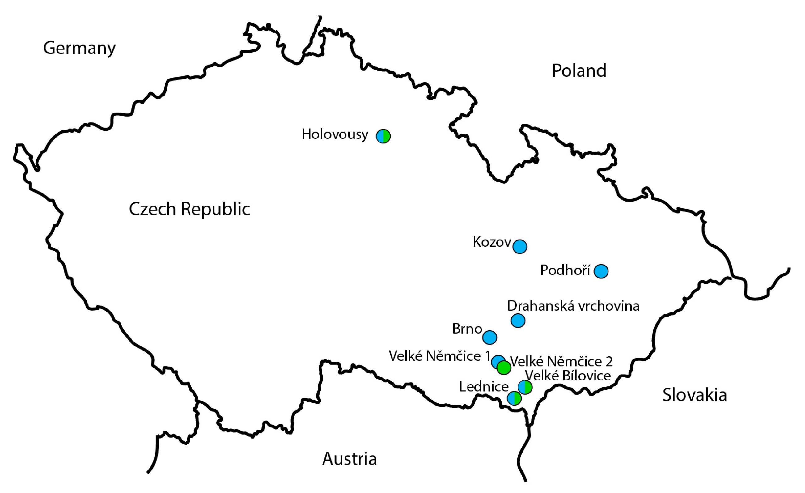
2.1. Biological Material

Adult psyllids were collected via sweep netting from localities in southern Moravia in 2005, 2006 and 2022, and in eastern Bohemia in 2022 (See the list in Table 1, Figure 1), Czech Republic. Collected samples were kept in 96% ethanol, at −20 °C, and they were determined based on their morphological characteristics.

2.2. DNA Isolation

Individual adult psyllids were homogenized in ATL buffer in 1.5 mL microtube using a micropestle homogenizer and total DNA was isolated using the QIAmp DNA Blood mini kit (Qiagen, Hilden, Germany, Cat. No. 51106) and eluted in 100 µL of deionized water. Purified DNA was stored at a temperature of −20 °C until used in HTS or PCR assays.

2.3. PCR Amplification and Sanger Sequencing

For the species confirmation and to study of the variability of the larger sample set, the partial COI gene was amplified using the universal primer pair CPF4 (5′-TAAGAACTAACCATAAGATTATCGG-3′) and CPR4 (5′-CACTTCAGGGTGTCCAAAGAATC-3′) according to the work of Kang et al. [24]. The PCR reaction mix consisted of MyTaq polymerase buffer (1×), MyTaq DNA polymerase (1U) (both Bioline, London, UK, Cat. No. BIO-21105) and 0.2 mM of each primer, and 2 µL of isolated DNA in a concentration of 50–200 ng, in total volume of 25 µL. The PCR reaction was conducted under the following conditions: 95 °C for 3 min, followed by 40 cycles of 94 °C for 30 s, 54 °C for 30 s, 72 °C for 1 min, and a final extension 72 °C for 5 min. Amplicons were separated in 1% agarose gel in TAE buffer and visualized using GelRed nucleic acid stain (Biotium, Fremont, CA, USA, Cat. No. 41002-0.5).
The amplicons were purified using NucleoSpin Gel and PCR Clean-up (Macherey-Nagel, Dueren, Germany, Cat. No. 740609.50) and Zymo-Spin III columns (Zymo Research, Irvine, CA, USA, Cat. No. C1005-250) and directly sequenced via bi-directional Sanger sequencing using BigDye Terminator v3.1 Cycle Sequencing Kit (Applied Biosystems-Thermo Fisher Scientific, Waltham, MA, USA, Cat. No. 4337455) and Genetic Analyzer ABI Prism 3730 at the Sequencing Centre of the Institute of Experimental Botany of the Czech Academy of Sciences, v. v. i. and Palacký University, Olomouc (CRH-UPOL), Czech Republic. The obtained Sanger sequences were analyzed and assembled into final contigs using Geneious Prime assembler 2023.1.2.

2.4. High-Throughput Sequencing

The 50 ng of DNA was fragmented in 50 µL of Sonication buffer [TE buffer (10 mM Tris, 1 mM EDTA), pH 7.5–8.0] using the Bioruptor Plus (Diagenode, Denville, NJ, USA) five times for 30 s at the HIGH setting. The sheared DNA was used to prepare a sequencing library using the NEBNext Ultra™ II DNA Library Prep Kit for Illumina (New England Biolabs, Ipswich, MA, USA, Cat. No. E7645L) with a mean fragment size of 500 bp. The library was sequenced on an Illumina NovaSeq 6000 to generate 2 × 160 or 2 × 250 bp paired-end reads to achieve at least 100-fold mitochondrial genome coverage. The samples were sequenced in multiple runs using the NovaSeq6000 at CATRIN-UPOL, Czech Republic.

2.5. Assembly of HTS Reads and the Genome Annotation

The high-throughput reads were paired by name, trimmed using BBDuk, merged and de novo assembled using Spades assembler, under standard conditions, all algorithms implemented in Geneious Prime version 2023.1.2. The resulting contigs were screened using the BLASTN algorithm [37] against the NCBI Cacopsylla mitochondrion sequences and circularized based on the identical 5′- and 3′- overhangs. The derived mtDNAs were subjected to mitogenome analysis using the MITOS Web server (http://mitos.bioinf.uni-leipzig.de, accessed on 22 June 2023) [38] under the invertebrate genetic code (code 5), and the annotation files and tRNA structure analyses were generated. The position of the identified genes was manually verified based on the additional comparisons with the available Cacopsylla mitogenomes using MEGA11 version 11.0.9 [39] and Geneious Prime 2023.1.2 software. Geneious Prime 2023.1.2 was also used for the visualization of the mitogenome map.

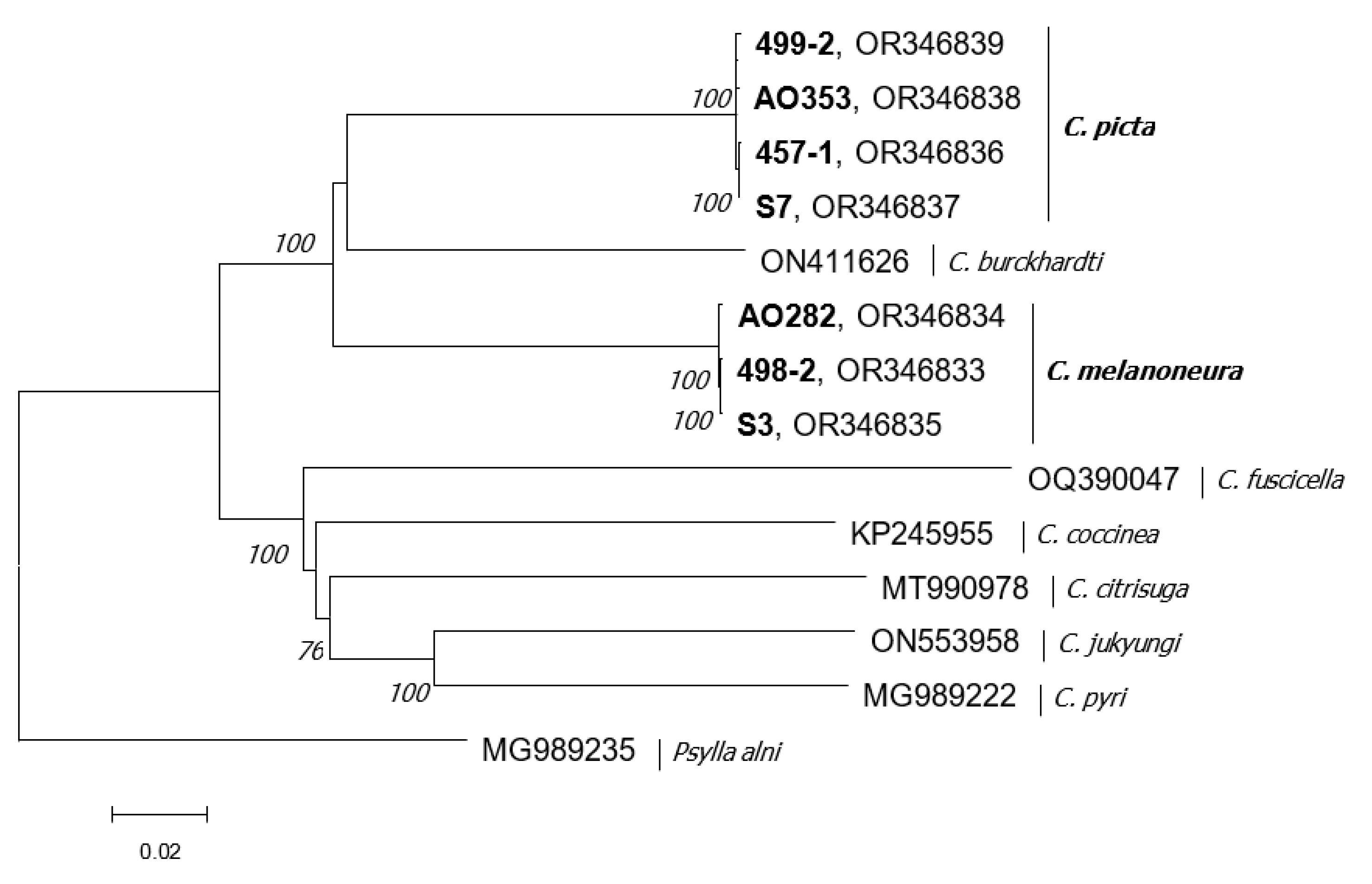
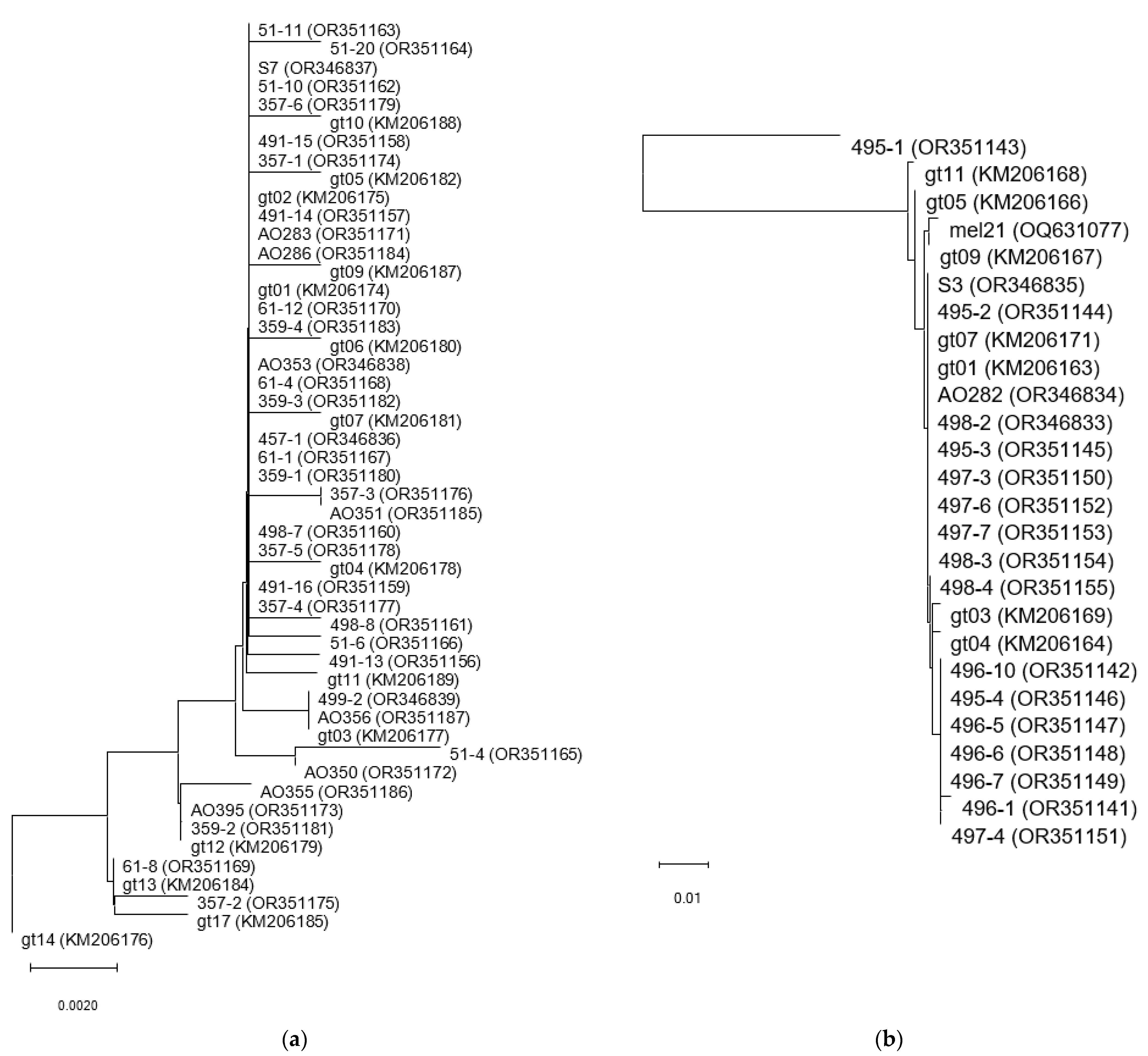
2.6. Phylogenetic Analysis

Multiple alignments of the obtained HTS mtDNA sequences and complete mtDNA sequences of Cacopsylla sp. available in the GenBank were performed using ClustalW algorithm. The obtained nt matrices were used for the estimation of genetic variability and phylogenetic relationships: (1) genetic variability was calculated using p-distance algorithm; (2) frequency of synonymous and nonsynonymous codons was calculated using distance analysis and the Nei–Gojobori model; (3) the phylogenetic trees were constructed on nucleotide sequences of complete mitogenome and discrete genes using the neighbor-joining method and the Tajima–Nei (G+I) model (selected using the model algorithm). The 1000 bootstrap replicates were used for all analyses, and trees were rooted using the appropriate Psylla alni sequences. The phylogenetic tree was visualized using Tree Explorer. All of the above analyses were performed using MEGA11 version 11.0.9 [39]. A similar strategy was used for the analysis of partial COI sequences except for the selection of the Tamura–Nei three-parametric model (G+I) used in the phylogenetic analysis.

3. Results

In order to gain a broader insight into the structure of the mitogenome of the ‘Candidatus Phytoplasma mali’ vector species, Cacopsylla picta and Cacopsylla melanoneura, four individuals of C. picta and three of C. melanoneura collected in 2005, 2006 and 2022 in the southern Moravia and the eastern Bohemia of the Czech Republic were subjected to high-throughput sequencing.

3.1. HTS Analysis

The four complete mtDNAs of C. picta, and two complete and one partial mtDNAs of C. melanoneura were obtained after de novo assembly of the HTS reads (Table 2); one overlapping contig was obtained for each sample. The sequences were deposited in the GenBank under Acc. Nos. OR346833-OR346839, OR351141-OR351187.
The length of the C. picta mtDNA varied between 14,801 and 14,802 bp, with an indel -/A present at position 1314. These mtDNA sequences showed 99.85–99.98% similarity with each other and 79.79–86.51% similarity with complete mitochondrial sequences of other Cacopsylla species available in GenBank, namely C. burckhardti, C. pyri, C. coccinea, C. yukiungi, C. citrinella, C. fuscicella. The correct morphological determination of the analyzed individuals was confirmed via BLASTN analysis, in which 99.32–100% similarity was found with the different partial COI sequences of C. picta available in the GenBank database.
Two complete mitochondrial sequences (gt S3 and 498-2) and one partial sequence (gt AO282), lacking the complete A+T rich control region, measuring 14,879–14,881 bp in length were obtained for C. melanoneura samples. The sequences differ according to the presence of specific indels in non-coding regions, with A/- at position 6034, AT/-- at position 14,400, T/- at position 14,608; and they showed 99.86–99.98% similarity with each other, 80.02–86.64% with the complete mitochondrial sequences of the other Cacopsylla sp. and 99.73–100% similarity with various C. melanoneura sequences available in GenBank, respectively.

3.2. Mitogenome Structure of C. picta and C. melanoneura

All molecules showed the standard organization of the mitogenome consisting of 36 typical genes, 13 protein-coding genes, 2 ribosomal (rRNA) RNA genes and 22 transfer RNA (tRNA) genes (Figure 2, Table 3 and Table S1). All mtDNAs obtained showed high frequency of A and T bases, with 39.1% of A, 35% of T, 16.3% of C, 9.6% of G and a GC content of 25.8% for the complete sequence and slightly higher, 26.9%, for protein-coding genes for the C. picta. A similar situation was found for the C. melanoneura with the frequencies 38.8% of A, 35.1 of T, 16.1% of C, 10.1 of G, and 26.2% of GC in the complete sequence and 27.5% of GC, in protein-coding genes.
The mtDNA of both psyllid studies is similar to those described to date, namely for Cacopsylla species, i.e., C. burckhardti, C. pyri, C. coccinea, C. yukiungi, C. citrinella, C. fuscicella psyllids shows a standard structure typical for the Cacopsylla mitogenome. The 13 protein-coding genes have been identified there, of which 9 (ND2, COI, COII, APT6, ATP8, COIII, ND3, ND6 and CYTB) are localized on the H strand and 4 (ND5, ND4, ND4L, ND1) on the L strand. The genes are flanked by AT- start codons, mainly ATG, followed by ATA and ATT (specific TTG as a start codon has been detected for the ND5 gene, at the 5′ end) and usually by TAA and less frequently TAG termination codons at the 3′ end. The incomplete T- termination, which is thought to be converted to TAA after specific adenylation is present in the COII, COIII, ND4 and ND5 genes (Table 4). The coding region consists of 3604 codons, and the most frequent codons were ATT (9.40 and 9.49%), TTA (8.70% and 8.46%), TTT (8.34% and 8.10%) and ATA (5.55% and 5.55%) for C. picta and C. melanoneura, respectively. The analysis of the overall codon usage shows a clear preference for codons with A or T in the third position compared to G or C (Figure S1). As expected, the shortest gene is ATP8 with a length of 153 bp for both species, and the longest genes are ND5 with length of 1618 bp, followed by the COI gene, which varies in length from 1530 bp in C. picta to 1533 bp in C. melanoneura. The length of the protein coding genes was identical for the both species, except for the COI gene mentioned above and the ND4L gene (1249 and 1246 bp). Minimal nucleotide variability was found among the genes within each Cacopsylla species. The interspecies comparisons reflected the species differences. The lowest diversity (12.3%) was found for the COI gene, while the most variable genes were ND4L (21.7–24.1% diversity), ATP8 (27.2%), and ND6 (28.9–28.7%). The analysis of the frequency of synonymous and nonsynonymous mutations showed the more frequent presence of synonymous codons at intra-species and inter-species levels, with intra-species dN-dS values close to zero and inter-species dN-dS values ranging from −0.225 to −1.175 (Table 4), implying the effect of negative selection in all genes, except for ND6 in C. melanoneura.
The analysis of the variability of the mtDNAs of both species did not show any significant differences between the psyllids collected in 2005, 2006 and 2022, and between the samples collected in the Czech Republic in the regions of Eastern Bohemia and Southern Moravia.
For the both Cacopsylla species, 22 tRNA genes were identified in the each mitogenome, 17 tRNAs carrying anticodon specific to unique amino acid, and 2 tRNAs carrying specific Leu anticodon (L1, L2) and 2 carrying Ser anticodon (S1, S2).
The mitogenome harbors two ribosomal RNA genes, both in the reverse orientation positioned between the ND1 and A+T rich Control region, the 16S rRNA gene of 1157 bp in length for C. picta and 1154 bp in length for C. melanoneura, and the 12S rRNA gene of 747 bp and 748 bp in length, respectively. Both species differ in the length of the A+T rich control regions, which are 689 bp and 757–758 bp in length, respectively, and contain a 20 bp and 23 bp long poly-T motif.
Phylogenetic analysis of the complete mitochondrial sequences using neighbor-joining analysis allowed the construction of a phylogenetic tree, whose topology placed C. picta and C. melanoneura sequences in relationship to C. burckhardti (Figure 3). Both species formed a homogeneous branch that differed significantly from the other sequences analyzed. Phylogenetic trees of similar topology with the strong support for the C. picta and C. melanoneura branches were obtained not only for the COI, 16S rRNA and CYTB genes used for taxonomic classification, but also for all other protein-coding genes.
With the aim of mapping the variability of the studied psyllids and the change in the Czech population structure in the context of global warming and the possible spread of different genotypes in the area over more than 15 years, the partial COI sequence analysis was performed on the set of C. picta and C. melanoneura psyllids collected in the 2005–2022 period from different localities in the Czech Republic. The species-specific sequences were almost identical, showing 99.7–100% similarity between each other and compared to Italian samples available in GenBank for C. picta, and 99.3–100% for C. melanoneura, respectively. The only exception was the sample adult genotype 495-1, which was determined to be C. melanoneura and showed only 91.5–92.0% similarity to other C. melanoneura sequences.
In the phylogenetic analysis using neighbor-joining analysis (Figure 4), samples of C. picta collected from different localities in Moravia and eastern Bohemia formed one major and four minor clusters, but without the statistical support of bootstrap values, among which the sequences were distributed together with the Italian sequences available in GenBank without correlating with their origin or the date of their collection. The C. melanoneura psyllids formed a homogeneous cluster covering all analyzed sequences without any correlation with the origin or date of collection. The specific situation of adult male 495-1 was also confirmed in this analysis, when this sequence formed a significant single branch (bootstrap value 97) that was distinct from the other C. melanoneura and C. picta psyllids.

4. Discussion

In our study, we have obtained the first complete mitochondrial genomes of two apple psyllids Cacopsylla picta and Cacopsylla melanoneura. The length of the mitogenomes, 14,801–14,802 bp and 14,879–14,881 bp is similar to the mitogenomes of Cacopsylla species described so far, with the detected length of around 14.8–14.9 kb [29,30,31,33,40]. The phylogenetic analysis of the complete mtDNA places the two species characterized here in a close relationship with C. burckhardti. Despite the lack of complex data publicly available for further comparisons, it could be deduced that both of them belong to the Cacopsylla clade I of the O-group of psyllids, psyllids of the Old and New World [29], which is in agreement with the expectation based on their distribution limited to the Europe and the variability of the partial cytochrome c oxidase subunit I gene used for the DNA barcoding and molecular identification of psyllid species [11,22,25,26,27,28,41].
The A/T content is the same for mitochondrial DNA from other members of the Cacopsylla species, typically ranging from 72.04 for C. citrisuga to 73.85% for C. pyri, and 73.69% for C. burckhardti, respectively. The codon bias, with a preference for A/T in the third position of the codon, also reflects this situation. TTA, ATT, TTT and ATA codons are the most frequently observed codons in their mitogenomes, and their frequency in C. picta (31.99%) and C. melanoneura (31.6%) belongs to the upper limit of their frequency, showing again a high similarity with the C. burckhardti (31.89–32% frequency of the mentioned codons) and the use of these codons in the range of 28.91–31.69% by the other Cacopsylla psyllid species [33].
The variability of all protein-coding genes and the observed identical topology of the phylogenetic tree constructed for each of them support the previous suggestion that all of them and not only COI could be used for the species discrimination and their molecular identification. Thus, the most variable genes, ND6 and ATP8, could be efficient in the study of population variability at least for the C. picta and C. melanoneura. On the other hand, the conservative character and low variability of the COI gene, also confirmed in our study, regardless of whether we discuss the complete or partial sequences, shows that this gene is not so suitable for intrapopulation variability studies, although it is still used for them. The combination of chromosomal and mitochondrial markers might be more useful [24,26,28,42]. In addition to this gene, the mitochondrial genes, such as ND5, COIII or ND4L, are recommended for the study of interspecies variability [33]; however, their different variability among the species or higher taxonomic groups shows that the further studies of the mitogenome structure of the other Cacopsylla species are necessary for a better understanding of their evolution and their adaptations to the environment and life strategies. Their study is still under progress.
The finding of genetic and phylogenetic distance of sample genotype 495-1, male determined as Cacopsylla melanoneura (Pavel Lauterer, personal communication), which shows almost 8.5% distance from any other C. melanoneura plant lice and even higher with the other representatives of the genus Cacopsylla, is surprising. The significant phylogenetic position between C. melanoneura and C. burckhardti and its distance from them raises questions about the identity of this insect. The two distinct lineages have been previously observed for the Cacopsylla chinensis in Japan, and China and Taiwan [27,42], and one of them has recently been described as a new species, Cacopsylla jukyungi [33]. Given our high respect for the morphological determination by Dr. P. Lauterer, the possible existence of the two cryptic C. melanoneura species could not be excluded, similar to the existence of two cryptic species of C. pruni and the existence of A and B genotypes [43,44]. However, further efforts to clarify the situation could be complicated by the age of the sample analyzed and by the fact that individuals could occur quite randomly in the area, in minimal abundance, due to their random transfer by air currents [13] during the re-emigration from their from their wintering grounds and that their natural population occurs in the other area. Their different ability to transmit phytoplasma ‘Ca. Phytoplasma mali’ could not be excluded.

5. Conclusions

The complete mitogenome of two apple plant lice, C. picta and C. melanoneura, is characterized for the first time for these species. It brings comprehensive information about the mitogenome of European members of the genus Cacopsylla, and its stability on the set of samples collected more than seventeen years apart, at least in the center of Europe, Czech Republic. Both species share common characteristics with other members of the genus described to date, such as length of mtDNA, its molecular and genetic structure, including codon bias, structure of the A+T rich control region, and carry the CGGTA motif in the non-coding region between ND1 and tRNA-S2 (- orientation). On the other hand, the observed variability in the length of this region between the individuals within the same species of C. picta implies that the length and conservation of this repetitive region might not be so strict. The obtained results show a high genetic homogeneity of the mtDNA of both psyllid species occurring in the studied Czech localities and clear differences over a 15-year period were not detected.
Increasing the knowledge about the structure of the mitogenome of other European psyllids of the genus Cacopsylla, not only C. picta and C. melanoneura described in our study, could be beneficial in the future.

Supplementary Materials

The following supporting information can be downloaded at: https://www.mdpi.com/article/10.3390/agronomy13092210/s1, Figure S1: Characterization of codon usage by C. picta and C. melanoneura. The frequencies calculated for each amino acid; Table S1: Characterization of the mitogenome of selected genotypes of C. picta (457-1, S7) and C. melanoneura (498-2, AO282).

Author Contributions

Conceptualization, D.Š.; methodology, D.Š.; formal analysis, D.Š., E.Z. and M.N.; investigation, D.Š., E.Z., J.O., K.H., M.N. and M.S.; resources, D.Š. and M.N.; data curation, D.Š.; writing—original draft preparation, D.Š.; writing—review and editing, D.Š., E.Z., J.O., K.H., M.N. and M.S.; visualization, D.Š.; supervision, M.N.; project administration, D.Š.; funding acquisition, D.Š. and M.N. All authors have read and agreed to the published version of the manuscript.

Funding

This research was funded by the National Agency for Agricultural Research (NAZV) project of Ministry of Agriculture of the Czech Republic, grant number QK21020395.

Data Availability Statement

The data presented in this study are available in public database GenBank under Accession Numbers: OR346833-OR346839, OR351141-OR351187.

Acknowledgments

In memory of Pavel Lauterer (†), an excellent entomologist and our great colleague.

Conflicts of Interest

The authors declare no conflict of interest.

References

  1. Moreno, A.; Miranda, M.P.; Fereres, A. Psyllids as major vectors of plant pathogens. Entomol. Gen. 2021, 41, 419–438. [Google Scholar] [CrossRef]
  2. Ouvrard, D.; Chalise, P.; Percy, D.M. Host-plant leaps versus host-plant shuffle: A global survey reveals contrasting patterns in an oligophagous insect group (Hemiptera, Psylloidea). Syst. Biodivers. 2015, 13, 434–454. [Google Scholar] [CrossRef]
  3. Burckhardt, D.; Ouvrard, D.; Percy, D.M. An updated classification of the jumping plant-lice (Hemiptera: Psylloidea) integrating molecular and morphological evidence. Eur. J. Taxon. 2021, 736, 137–182. [Google Scholar] [CrossRef]
  4. Bertaccini, A. Plants and Phytoplasmas: When bacteria modify plants. Plants 2022, 11, 1425. [Google Scholar] [CrossRef] [PubMed]
  5. Namba, S. Molecular and biological properties of phytoplasmas. Proc. Jpn. Acad. Ser. B Phys. Biol. Sci. 2019, 95, 401–418. [Google Scholar] [CrossRef] [PubMed]
  6. Jarausch, B.; Tedeschi, R.; Sauvion, N.; Gross, J.; Jarausch, W. Psyllid Vectors. In Phytoplasmas: Plant Pathogenic Bacteria—II: Transmission and Management of Phytoplasma—Associated Diseases; Bertaccini, A., Weintraub, P.G., Rao, G.P., Mori, N., Eds.; Springer: Singapore, 2019; pp. 53–78. [Google Scholar]
  7. Bertaccini, A.; Duduk, B.; Paltrinieri, S.; Contaldo, N. Phytoplasmas and phytoplasma diseases: A severe threat to agriculture. Am. J. Plant Sci. 2014, 5, 1763–1788. [Google Scholar] [CrossRef]
  8. Tedeschi, R.; Alma, A. Transmission of apple proliferation phytoplasma by Cacopsylla melanoneura (Homoptera: Psyllidae). J. Econ. Entomol. 2004, 97, 8–13. [Google Scholar] [CrossRef]
  9. Janik, K.; Barthel, D.; Oppedisano, T.; Anfora, G.; Schuler, H. Apple Proliferation—A Joint Review; Laimburg Research Centre: Stadio-Laimburg, Italy, 2020. [Google Scholar]
  10. Fischnaller, S.; Parth, M.; Messner, M.; Stocker, R.; Kerschbamer, C.; Janik, K. Surveying potential vectors of apple proliferation phytoplasma: Faunistic analysis and infection status of selected Auchenorrhyncha species. Insects 2021, 12, 12. [Google Scholar] [CrossRef]
  11. Tedeschi, R.; Bosco, D.; Alma, A. Population dynamics of Cacopsylla melanoneura (Homoptera: Psyllidae), a vector of apple proliferation phytoplasma in northwestern Italy. J. Econ. Entomol. 2002, 95, 544–551. [Google Scholar] [CrossRef]
  12. Safarova, D.; Stary, M.; Valova, P.; Opatikova, M.; Bilkova, L.; Navratil, M. Impact of insecticides treatment on phytoplasma infection risk in apple orchards. Hortic. Sci. 2016, 43, 112–116. [Google Scholar] [CrossRef]
  13. Cermak, V.; Lauterer, P. Overwintering of psyllids in South Moravia (Czech Republic) with respect to the vectors of the apple proliferation cluster phytoplasmas. Bull. Insectol. 2008, 61, 147–148. [Google Scholar]
  14. Malenovsky, I.; Lauterer, P. Jumping plant-lice (Hemiptera: Psylloidea) of the Bílé Karpaty Protected Landscape Area and Biosphere Reserve (Czech Republic). Acta Musei Moraviae Sci. Biol. 2012, 96, 105–154. [Google Scholar]
  15. Barthel, D.; Schuler, H.; Galli, J.; Borruso, L.; Geier, J.; Heer, K.; Burckhardt, D.; Janik, K. Identification of plant DNA in adults of the phytoplasma vector Cacopsylla picta helps understanding its feeding behavior. Insects 2020, 11, 835. [Google Scholar] [CrossRef] [PubMed]
  16. Jarausch, B.; Fuchs, A.; Schwind, N.; Krczal, G.; Jarausch, W. Cacopsylla picta as most important vector for ‘Candidatus Phytoplasma mali’ in Germany and neighbouring regions. Bull. Insectol. 2007, 60, 189–190. [Google Scholar]
  17. Mattedi, L.; Forno, F.; Cainelli, C.; Grando, M.S.; Jarausch, W. Research on Candidatus phytoplasma mali transmission by insect vectors in Trentino. Acta Hortic. 2008, 781, 369–374. [Google Scholar] [CrossRef]
  18. Jarausch, B.; Schwind, N.; Jarausch, W.; Krczal, G.; Dickler, E.; Seemuller, E. First report of Cacopsylla picta as a vector of apple proliferation phytoplasma in Germany. Plant Dis. 2003, 87, 101. [Google Scholar] [CrossRef]
  19. Mittelberger, C.; Obkircher, L.; Oettl, S.; Oppedisano, T.; Pedrazzoli, F.; Panassiti, B.; Kerschbamer, C.; Anfora, G.; Janik, K. The insect vector Cacopsylla picta vertically transmits the bacterium ‘Candidatus Phytoplasma mali’ to its progeny. Plant Pathol. 2017, 66, 1015–1021. [Google Scholar] [CrossRef]
  20. Mayer, C.J.; Vilcinskas, A.; Gross, J. Chemically mediated multitrophic interactions in a plant-insect vector-phytoplasma system compared with a partially nonvector species. Agric. Forest Entomol. 2011, 13, 25–35. [Google Scholar] [CrossRef]
  21. Fialova, R.; Navratil, M.; Valova, P.; Lauterer, P. Molecular tests to determine apple proliferation phytoplasma presence in psyllid vectors from apple tree orchards in the Czech Republic. Acta Hortic. 2008, 781, 471–476. [Google Scholar] [CrossRef]
  22. Tedeschi, R.; Nardi, F. DNA-based discrimination and frequency of phytoplasma infection in the two hawthorn-feeding species, Cacopsylla melanoneura and Cacopsylla affinis, in northwestern Italy. Bull. Entomol. Res. 2010, 100, 741–747. [Google Scholar] [CrossRef]
  23. Nokkala, C.; Kuznetsova, V.G.; Rinne, V.; Nokkala, S. Description of two new species of the genus Cacopsylla Ossiannilsson, 1970 (Hemiptera, Psylloidea) from northern Fennoscandia recognized by morphology, cytogenetic characters and COI barcode sequence. Comp. Cytogenet. 2019, 13, 367–382. [Google Scholar] [CrossRef]
  24. Kang, A.R.; Baek, J.Y.; Lee, S.H.; Cho, Y.S.; Kim, W.S.; Han, Y.S.; Kim, I. Geographic homogeneity and high gene flow of the pear psylla, Cacopsylla pyricola (Hemiptera: Psyllidae), detected by mitochondrial COI gene and nuclear ribosomal internal transcribed spacer 2. Anim. Cells Syst. 2012, 16, 145–153. [Google Scholar] [CrossRef]
  25. Cho, G.; Malenovsky, I.; Burckhardt, D.; Inoue, H.; Lee, S. DNA barcoding of pear psyllids (Hemiptera: Psylloidea: Psyllidae), a tale of continued misidentifications. Bull. Entomol. Res. 2020, 110, 521–534. [Google Scholar] [CrossRef] [PubMed]
  26. Chen, P.; Liu, Q.Z.; Qiao, X.F.; Wang, J.W.; Zhang, T. Identification and phylogenetic analysis of pear psyllids (Hemiptera: Psyllidae) in Chinese pear orchards. J. Econ. Entomol. 2018, 111, 2908–2913. [Google Scholar] [CrossRef] [PubMed]
  27. Katoh, H.; Inoue, H.; Kuchiki, F.; Ide, Y.; Uechi, N.; Iwanami, T. Identification of a distinct lineage of Cacopsylla chinensis (Hemiptera: Psyllidae) in Japan on the basis of two mitochondrial DNA sequences. J. Econ. Entomol. 2013, 106, 536–542. [Google Scholar] [CrossRef]
  28. Oettl, S.; Schlink, K. Molecular identification of two vector species, Cacopsylla melanoneura and Cacopsylla picta (Hemiptera: Psyllidae), of apple proliferation disease and further common psyllids of Northern Italy. J. Econ. Entomol. 2015, 108, 2174–2183. [Google Scholar] [CrossRef]
  29. Percy, D.M.; Crampton-Platt, A.; Sveinsson, S.; Lemmon, A.R.; Lemmon, E.M.; Ouvrard, D.; Burckhardt, D. Resolving the psyllid tree of life: Phylogenomic analyses of the superfamily Psylloidea (Hemiptera). Syst. Entomol. 2018, 43, 762–776. [Google Scholar] [CrossRef]
  30. Wang, Y.; Cen, Y.; He, Y.; Wu, Y.; Huang, S.; Lu, J. The first complete mitochondrial genome sequence of Cacopsylla citrisuga (Yang & Li), a new insect vector of Huanglongbing in Yunnan Province, China. Mitochondrial DNA B Resour. 2021, 6, 575–577. [Google Scholar] [CrossRef]
  31. Zhao, Y.S.; Luo, X.Y.; Liu, Y.Q.; Shi, A.X.; Cai, W.Z.; Song, F. Cacopsylla fuscicella Sp. Nov. (Hemiptera, Psyllidae), a New Loquat Pest in China dagger. Insects 2023, 14, 414. [Google Scholar] [CrossRef]
  32. Que, S.Q.; Yu, L.P.; Xin, T.R.; Zou, Z.W.; Hu, L.X.; Xia, B. Complete mitochondrial genome of Cacopsylla coccinae (Hemiptera: Psyllidae). Mitochondrial DNA Part A 2016, 27, 3169–3170. [Google Scholar] [CrossRef]
  33. Kang, A.R.; Kim, M.J.; Park, J.S.; Seo, H.J.; Song, J.H.; Won, K.H.; Choi, E.D.; Kim, I. Comparative analysis of two pear pests, Cacopsylla jukyungi and Cacopsylla burckhardti (Hemiptera: Psyllidae), based on complete mitochondrial genomes and comparison to confamilial species. Agronomy 2022, 12, 37. [Google Scholar] [CrossRef]
  34. Jo, E.; Cho, G. The complete mitochondrial genome of Cacopsylla burckhardti (Hemiptera, Psylloidea, Psyllidae). Biodivers. Data J. 2022, 10, e85094. [Google Scholar] [CrossRef]
  35. Zahradnicek, P.; Brazdil, R.; Stepanek, P.; Trnka, M. Reflections of global warming in trends of temperature characteristics in the Czech Republic, 1961–2019. Int. J. Climatol. 2021, 41, 1211–1229. [Google Scholar] [CrossRef]
  36. Rousi, E.; Fink, A.H.; Andersen, L.S.; Becker, F.N.; Beobide-Arsuaga, G.; Breil, M.; Cozzi, G.; Heinke, J.; Jach, L.; Niermann, D.; et al. The extremely hot and dry 2018 summer in central and northern Europe from a multi-faceted weather and climate perspective. Nat. Hazard. Earth Syst. 2023, 23, 1699–1718. [Google Scholar] [CrossRef]
  37. Altschul, S.F.; Madden, T.L.; Schaffer, A.A.; Zhang, J.; Zhang, Z.; Miller, W.; Lipman, D.J. Gapped BLAST and PSI-BLAST: A new generation of protein database search programs. Nucleic Acids Res. 1997, 25, 3389–3402. [Google Scholar] [CrossRef]
  38. Bernt, M.; Donath, A.; Juhling, F.; Externbrink, F.; Florentz, C.; Fritzsch, G.; Putz, J.; Middendorf, M.; Stadler, P.F. MITOS: Improved de novo metazoan mitochondrial genome annotation. Mol. Phylogenet. Evol. 2013, 69, 313–319. [Google Scholar] [CrossRef]
  39. Tamura, K.; Stecher, G.; Kumar, S. MEGA11 Molecular Evolutionary Genetics Analysis Version 11. Mol. Biol. Evol. 2021, 38, 3022–3027. [Google Scholar] [CrossRef]
  40. The World Psylloidea Database (from Psyl’list). Available online: https://data.nhm.ac.uk/dataset/psyl-list/resource/8746ceec-4846-4899-b607-9ba603002033 (accessed on 12 July 2023).
  41. Tedeschi, R.; Lauterer, P.; Brusetti, L.; Tota, F.; Alma, A. Composition, abundance and phytoplasma infection in the hawthorn psyllid fauna of northwestern Italy. Eur. J. Plant Pathol. 2009, 123, 301–310. [Google Scholar] [CrossRef]
  42. Tsai, C.L.; Lee, H.C.; Cho, G.; Liao, Y.C.; Yang, M.M.; Yeh, W.B. Invasive and quarantine risks of Cacopsylla chinensis (Hemiptera: Psyllidae) in East Asia: Hybridization or gene flow between differentiated lineages. J. Econ. Entomol. 2020, 113, 2890–2899. [Google Scholar] [CrossRef]
  43. Sauvion, N.; Peccoud, J.; Meynard, C.N.; Ouvrard, D. Occurrence data for the two cryptic species of Cacopsylla pruni (Hemiptera: Psylloidea). Biodivers. Data J. 2021, 9, e68860. [Google Scholar] [CrossRef]
  44. Peccoud, J.; Labonne, G.; Sauvion, N. Molecular test to assign individuals within the Cacopsylla pruni complex. PLoS ONE 2013, 8, e72454. [Google Scholar] [CrossRef]
Figure 1. Map showing sampling localities distribution. Blue—C. picta; green—C. melanoneura.
Agronomy 13 02210 g001
Figure 2. Mitogenome map of (a) Cacopsylla picta (499-2, Acc. No. OR346839) and (b) Cacopsylla melanoneura (S3, Acc. No. OR346835). Protein-coding genes are shown in green, rRNA genes in red, tRNA genes in black, A+T rich control region in orange, and specific motifs in purple.
Agronomy 13 02210 g002
Figure 3. Phylogenetic tree of Cacopsylla sp. constructed based on the complete mtDNA sequences using the neighbor-joining algorithm. Sequences marked by genotype and/or species name and GenBank Acc. No, and the new studied sequences are marked in bold. Sequence of Psylla alni was used to root the tree. Only bootstrap values ≥70 are shown. The bar represents 2% nucleotide variability.
Agronomy 13 02210 g003
Figure 4. Phylogenetic tree of (a) C. picta and (b) C. melanoneura constructed based on the partial COI sequence using the neighbor-joining method, Tamura three-parametric model, with bootstrap replicates 1000×. Sequence of Psylla alni (NC_038139.1) was used to root the tree /not shown/). Sequences marked by genotype name and/or GenBank Acc. No. Only bootstrap values ≥ 70 are shown. The bar represents 0.05% and 0.01% nucleotide variability.
Agronomy 13 02210 g004
Table 1. List of Cacopsylla samples used in the analyses.
IDLocality
(GPS Location)
Date of CollectionDetermined by
C. picta
499-2Velké Němčice 1
(49°0′10.538″ N, 16°39′10.617″ E)
16 May 2006P. Lauterer
457-1 Podhoří
(49°33′43.939″ N, 17°37′1.056″ E)
26 April 2013P. Lauterer
S7Lednice
(48°47′24.188″ N, 16°48′5.808″ E)
28 April 2022M. Starý
AO353Holovousy
(50°22′48.503″ N, 15°34′28.442″ E)
29 April 2022J. Ouředníčková
51-4,6,10,11,20Brno
(49°9′27.367″ N, 16°34′20.229″ E)
5 April 2005P. Lauterer
61-1,4,8,12Velké Bílovice
(48°51′19.028″ N, 16°54′46.743″ E)
27 April 2005P. Lauterer
357-1,2,3,4,5,6Drahanská vrchovina
(49°15′40.686″ N, 16°50′40.728″ E)
10 February 2012P. Lauterer
359-1,2,3,4Drahanská vrchovina
(49°15′40.686″ N, 16°50′40.728″ E)
1 November 2012P. Lauterer
491-13,14,15,16Kozov
(49°42′9.601″ N, 16°51′8.130″ E)
6 May 2015P. Lauterer
498-7,8Lednice
(48°47′24.188″ N, 16°48′5.808″ E)
28 April 2022M. Starý
AO283, AO286Holovousy
(50°22′48.503″ N, 15°34′28.442″ E)
22 March 2022J. Ouředníčková
AO350-352,354-356Holovousy
(50°22′48.503″ N, 15°34′28.442″ E)
29 April 2022J. Ouředníčková
C. melanoneura
S3 (male)Lednice
(48°47′24.188″ N, 16°48′5.808″ E)
13 June 2006P. Lauterer
498-2Lednice
(48°47′24.188″ N, 16°48′5.808″ E)
28 April 2022M. Starý
AO282Holovousy
(50°22′41.76″ N, 15°34′33.405″ E)
22 March 2022J. Ouředníčková
495-1,3Velké Němčice 2
(48°58′47.055″ N, 16°42′2.957″ E)
29 May 2006P. Lauterer
495-2Velké Bílovice
(48°51′19.028″ N, 16°54′46.743″ E)
18 April 2006P. Lauterer
495-4; 496-7,10Lednice
(48°47′24.188″ N, 16°48′5.808″ E)
13 June 2006P. Lauterer
496-1,5,6Velké Němčice 2
(48°58′47.055″ N, 16°42′2.957″ E)
29 May 2006P. Lauterer
497-3,4,6,7Lednice
(48°47′24.188″ N, 16°48′5.808″ E)
28 April 2022M. Starý
498-3,4,7,8Lednice
(48°47′24.188″ N, 16°48′5.808″ E)
28 April 2022M. Starý
Table 2. Number of HTS reads and coverage of obtained mtDNA sequences.
Sample (Acc. No.)mtDNA LengthReads Coverage
(Average ± stdev)
HTS/mtDNA-Specific
Reads
C. picta
499-2 (OR346839)14,8011202 ± 27214,439,480/83,388
AO353 (OR346838)14,801644.6 ± 209.98,074,058/43,722
457-1 (OR346836)14,802476.3 ± 122.77,902,966/31,389
S7 (OR346837)14,802754.9 ± 256.615,544,208/43,722
C. melanoneura
S3 (OR346835)14,8811243.3 ± 196.119,038,072/118,977
498-2 (OR346833)14,879513.1 ± 76.45,920,406/32,409
AO282 (OR346834)14,88056.8 ± 17.23,732,814/3662
Table 3. Characterization of the mitogenome of selected genotypes of C. picta and C. melanoneura.
Name C. picta (499-2, AO353)C. melanoneura (S3)
StartEndLengthStartEndLength
trnI(gat)Fw1666616666
trnQ(ttg)Rev71136667113666
trnM(cat)Fw1422056414220766
ND2Fw20611779722081179972
trnW(tca)Fw11761237621178124063
trnC(gca)Rev12401302631243130563
trnY(gta)Rev13031364621306136762
COIFw136628951530136929011533
trnL2(taa)Fw28952958642901296565
COIIFw2959362266429663629664
trnK(ctt)Fw36233692703630369970
trnD(gtc)Fw36913752623701376161
ATP8Fw3753390215037623914153
ATP6Fw3902457367239114582672
COIIIFw4573535077845825359778
trnG(tcc)Fw53515408585360541960
ND3Fw5409575935154205770351
trnA(tgc)Fw57615821615770582960
trnR(tcg)Fw58265886615840590061
trnN(gtt)Fw58865951665900596465
trnS1(gct)Fw59526005545965601854
trnE(ttc)Fw60066066616019607860
trnF(gaa)Rev60556117636068613063
ND5Rev611877351618613177481618
trnH(gtg)Rev77367796617749780961
ND4Rev779790451249781090551246
ND4lRev9039932628890499336288
trnT(tgt)Fw93289388619338939760
trnP(tgg)Rev93899451639398946366
ND6Fw9454993948694669951486
CYTBFw993911,0751137995111,0871137
trnS2(tga)Fw11,07711,146411,09111,15363
GCCTA motifRev11,15211,156511,16511,1695
ND1Rev11,16812,08291511,18112,095915
trnL1(tag)Rev12,08312,1466412,09612,15863
16S rRNARev12,14713,303115712,15913,311152
trnV(tac)Rev13,30413,3656213,31113,37363
12S rRNARev13,36614,11274713,37514,122748
A+T rich --14,11314,80168914,12314,881759
polyT motif--14,53114,5502014,60814,63023
Abbreviations used as follows: ND2—NADH dehydrogenase subunit 2; COI—cytochrome c oxidase subunit I; COII—cytochrome c oxidase subunit II; ATP8—ATP synthase membrane subunit 8; ATP6—ATP synthase membrane subunit 6; COIII—cytochrome c oxidase subunit III; ND3—NADH dehydrogenase subunit 3; ND5—NADH dehydrogenase subunit 5; ND4—NADH dehydrogenase subunit 4; ND4L—NADH dehydrogenase subunit 4L; ND6—NADH dehydrogenase subunit 6; ND1—NADH dehydrogenase subunit 1; CYTB—cytochrom b; A+T rich—A+T rich control region; trn—tRNA (amino acids marked by IUPAC code); Fw—forward orientation; Rev—reverse orientation.
Table 4. Characterization of protein-coding genes of C. picta and C. melanoneura.
GeneC. pictaC. melanoneuraBoth Species
Start/Stop CodonsProtein Mw [kDa]Distance [%]dN-dSStart/Stop
Codons
Protein Mw [kDa]Distance [%]dN-dSDistance
[%]
dN-dS
ND2ATA/TAA37.680–0.10ATA/TAA37.50–0.2017.1–17.3−0.760
COIATG/TAA57.020–0.3−0.005ATG/TAA57.140–0.1−0.00212.3–12.4−0.760
COIIATA/T25.530–0.3−0.010ATA/T25.610012.8–13.1−0.942
ATP6ATG/TAA25.370–0,30ATG/TAA25.370–0.3015.6–15.7−0.933
ATP8ATC/TAA6.030−0.010ATC/TAA5.890027.2−0.810
COIIIATG/T30.650–0.1−0.010ATG/T30.560.1–0.3013.8–14.0−0.701
ND3ATA/TAA13.650–0.3−0.009ATT/TAA13.580017.4–17.7−1.197
ND5TTG/T60.290–0.2−0.005TTG/T60.270–0.1−0.00215.2−0.271
ND4ATG/T47.830–0.10.000ATG/T47.440–0.2−0.00416.1–16.3−0.513
ND4LTTG/TAG11.1200TTG/TAG11.010–0.3−0.01021.7–24.1−0.225
ND6ATA/TAA18.660–0.6−0.006ATA/TAA18.640–0.20.00220.4–20.6−0.705
CYTBATG/TAA43.420–0.2−0.010ATG/TAA43.30–0.2014.6–14.8−0.959
ND1ATA/TAA34.9600ATA/TAG34.920–0.4−0.00813.2–13.4−0.500
Abbreviations used as follows: ND2—NADH dehydrogenase subunit 2; COI—cytochrome c oxidase subunit I; COII—cytochrome c oxidase subunit II; ATP8—ATP synthase membrane subunit 8; ATP6—ATP synthase membrane subunit 6; COIII—cytochrome c oxidase subunit III; ND3—NADH dehydrogenase subunit 3; ND5—NADH dehydrogenase subunit 5; ND4—NADH dehydrogenase subunit 4; ND4L—NADH dehydrogenase subunit 4L; ND6—NADH dehydrogenase subunit 6; ND1—NADH dehydrogenase subunit 1; dN- nonsynonymous distance; dS—synonymous distance.
Disclaimer/Publisher’s Note: The statements, opinions and data contained in all publications are solely those of the individual author(s) and contributor(s) and not of MDPI and/or the editor(s). MDPI and/or the editor(s) disclaim responsibility for any injury to people or property resulting from any ideas, methods, instructions or products referred to in the content.

