Article

Post-Embryonic Lateral Organ Development and Adaxial—Abaxial Polarity Are Regulated by the Combined Effect of ENHANCER OF SHOOT REGENERATION 1 and WUSCHEL in Arabidopsis Shoots

1
Centre of the Region Haná for Biotechnological and Agricultural Research, Czech Advanced Technology and Research Institute (CATRIN), Palacký University, 78371 Olomouc, Czech Republic
2
Laboratory of Growth Regulators, Institute of Experimental Botany AS CR, Palacky University, 78371 Olomouc, Czech Republic
3
Leibniz Institute of Plant Genetics and Crop Plant Research (IPK), 06466 Gatersleben, Germany
4
International Research Organization for Advanced Science and Technology (IROAST), Kumamoto University, Kumamoto 860-8555, Japan
*
Author to whom correspondence should be addressed.
Int. J. Mol. Sci. 2021, 22(19), 10621; https://doi.org/10.3390/ijms221910621
Submission received: 26 August 2021 / Revised: 19 September 2021 / Accepted: 28 September 2021 / Published: 30 September 2021
(This article belongs to the Special Issue Meristem and Stem Cells and Stem Cell Regulation in Plants)

Abstract

:
The development of above-ground lateral organs is initiated at the peripheral zone of the shoot apical meristem (SAM). The coordination of cell fate determination and the maintenance of stem cells are achieved through a complex regulatory network comprised of transcription factors. Two AP2/ERF transcription factor family genes, ESR1/DRN and ESR2/DRNL/SOB/BOL, regulate cotyledon and flower formation and de novo organogenesis in tissue culture. However, their roles in post-embryonic lateral organ development remain elusive. In this study, we analyzed the genetic interactions among SAM-related genes, WUS and STM, two ESR genes, and one of the HD-ZIP III members, REV, whose protein product interacts with ESR1 in planta. We found that esr1 mutations substantially enhanced the wus and stm phenotypes, which bear a striking resemblance to those of the wus rev and stm rev double mutants, respectively. Aberrant adaxial–abaxial polarity is observed in wus esr1 at relatively low penetrance. On the contrary, the esr2 mutation partially suppressed stm phenotypes in the later vegetative phase. Such complex genetic interactions appear to be attributed to the distinct expression pattern of two ESR genes because the ESR1 promoter-driving ESR2 is capable of rescuing phenotypes caused by the esr1 mutation. Our results pose the unique genetic relevance of ESR1 and the SAM-related gene interactions in the development of rosette leaves.

1. Introduction

When compared to the majority of animals taking a predetermined body plan, the development of terrestrial plants is more plastic and takes place post-embryonically by producing new organs throughout their lifespan. In the shoot, this unique feature is achieved through the coordination of maintenance of stem cells with continuous lateral organ formation. The stem cell niche of the shoot is maintained by the WUSCHEL (WUS)–CLAVATA (CLV) negative feedback loop [1,2]. Although loss-of-function mutations in WUS resulted in the formation of an aberrant flat shoot apical meristem (SAM), the wus mutant retains an ability to develop vegetative leaves in a stop-and-go mode from either a defective SAM or ectopic meristem (also called lateral shoot meristem) that emerged from the axils of leaves and cotyledons, and eventually gives rise to the formation of inflorescence meristem [3]. To account for this phenotype, the WUS-independent stem cell specification pathway is suggested [4]. It appears that microRNA regulation participates in the WUS-independent stem cell specification pathway [5]. Partial suppression of the wus phenotypes was observed when the heterozygous men1 activation-tagged allele of miR166a was introduced into the wus mutant plant [6]. Similarly, the jabba-1D (jba-1D) gain-of-function dominant mutant caused by overexpression of miR166g displayed pleiotropic phenotypes, such as SAM enlargement, fasciated stem, and the formation of a radial structure [7]. Concomitant with the phenotypes caused by miRNA166g overexpression, combined triple mutations in the miR165/166 target genes, PHABULOSA (PHB), PHAVOLUTA (PHV), and CORONA (CNA), which encode class III HOMEODOMAIN-LEUCINE ZIPPER TRANSCRIPTION FACTOR (HD-ZIP III), caused meristem enlargement, and in the phb phv cna wus quadruple mutant, rosette leaves were more frequently emerged compared to wus [4]. On the other hand, another HD-ZIP III family member, REVOLTA (REV), appears to act distinctly. Out of the five HD-ZIP III family members, only REV single mutants revealed conspicuous phenotypes that involve defects in lateral meristem (LM) formation [8]. In addition, the rev-6 null allele enhanced rosette leaf defects in the corresponding rev-6 stm-2 and rev-6 wus-1 double mutant plants [8]. Although the post-embryonic role of PHB, PHV, and CNA is not entirely clear [9,10], their role appears to restrict the stem cell population in the WUS-independent stem cell specification pathway whereas REV was antagonized by these three HD-ZIP III genes [4].
The ENHANCER OF SHOOT REGENERATION 1 (ESR1) in AP2/ERF (ETHYLENE RESPONSE FACTOR) transcription family [11], also known as DORNRÖSCHEN (DRN) [12], and its closest homolog, ESR2 [13], also known as DORNRÖSCHEN-LIKE (DRNL) [12], SOB2 (SUPPRESSOR OF PHYTOCHROME B) [14], and BOLITA (BOL) [15] in Arabidopsis thaliana and LEAFLESS (LFS) [16] in tomato, were involved in lateral organ emergence [13,17], gynoecium development [18], and stamen enlargement [19]. The organogenesis in esr1-1 esr2-2 double mutant root explants was largely compromised in a tissue culture system [20] and the analyses of intact double mutant seedling suggested that the disturbed auxin transport is likely responsible for the pleiotropic phenotypes [16,17,20]. We and others identified CUC1 and CUC2 as downstream target genes directly regulated by ESR1 [21,22] and ESR2 [13]. Both ESR1 and ESR2 proteins have been documented to physically interact with five members of the HD-ZIP III family (REV, PHB, PHV, CNA, and HB8), both in vitro and in vivo [17,23]. However, the physiological relevance of their interactions remains obscure. Analogous to the genetic interaction between WUS and HD-ZIP III members, we hypothesize that the two ESR genes might have a genetic interaction with WUS and STM.
Since the roles of the two ESR genes in cotyledon and flower development have been documented previously, we exerted our efforts on elucidating the regulatory mechanism of rosette leaf development in the vegetative phase, particularly by scrutinizing the involvement of ESR genes in the initiation of rosette leaves from the ectopic/lateral meristem in the SAM-deficient mutant backgrounds (wus and stm). Our present mutant analyses revealed that, although the esr1 single mutant did not show phenotypes observed in the rev mutant, esr1 phenocopied rev both in the wus and stm backgrounds to a great extent, whereas esr2 did not. Rather, in the later vegetative phase, esr2 partially rescued the retarded rosette leaf development observed in stm. Such contradictory observations are reconciled by the fact that the expression of the ESR2 gene under the control of the regulatory sequences of ESR1 in wus esr1 rendered phenotypes indistinguishable from those in the wus single mutant, suggesting that the two ESR genes have redundant functions but their distinct expression patterns define their physiological relevance in the development of rosette leaves and the establishment of adaxial–abaxial polarity.

2. Results

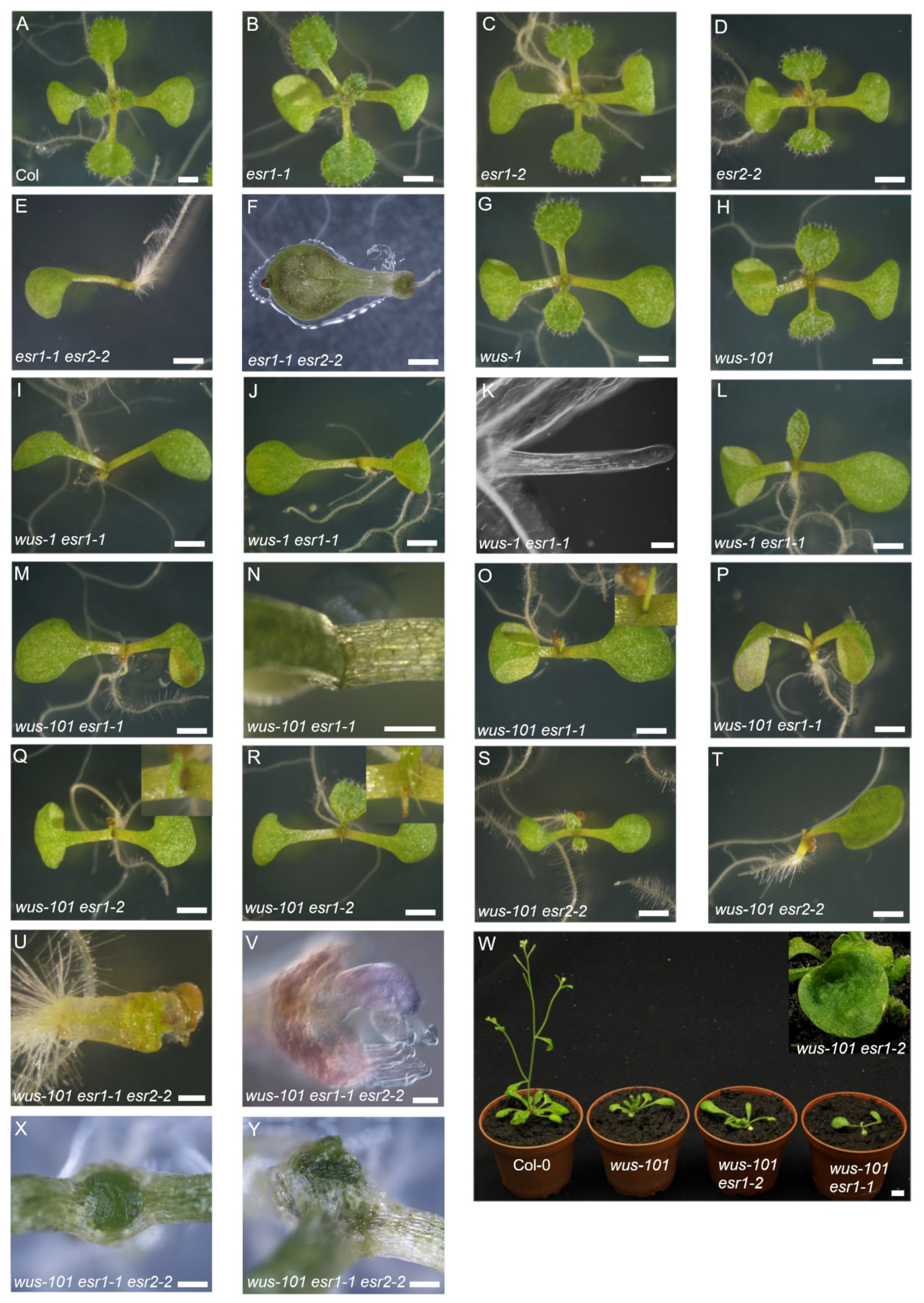
2.1. esr1 Mutations Enhanced Defects in Rosette Leaf Development and Adaxial—Abaxial Polarity in wus Background

By 8 days after germination (d.a.g.), successive lateral organ formation leading to the development of at least four recognizable rosette leaves was observed in the wild type (Figure 1A). In terms of continuous emergence of rosette leaves, esr1-1 was indistinguishable from the wild type (Figure 1B), although, as reported previously, cotyledon phenotypes were observed at low penetrance [13,17,20]. In this study, we identified and characterized a novel esr1 allele, Gabi Kat 369_A3, where T-DNA insertion is located at 7 bp upstream from the stop codon (Supplementary Figure S1A). The allele is termed esr1-2 hereafter (Figure 1C). Endogenous full-length ESR1 transcripts containing 3′-UTR were absent; however, ESR1 transcripts lacking 3’-UTR accumulated in esr1-2 (Supplementary Figure S1B). Although it is not clear how the truncated ESR1 transcripts are efficiently translated, esr1-2 is likely to be a weaker allele than esr1-1 because the penetrance of the cotyledon phenotypes was lower than that of esr1-1 and no gain-of-function phenotypes caused by ESR1 overexpression were observed in esr1-2 under our growth conditions (data not shown). The development of rosette leaves in esr2-2 was indistinguishable from that of the wild type (Figure 1D), although the cotyledon phenotypes appeared at low penetrance [13,17]. Phenotypes observed in esr1-1 esr2-2 were pleiotropic, ranging from the formation of a single cotyledon with delayed emergence of rosette leaves (Figure 1E) to the lack of a hypocotyl with a shorter root, as reported for the monopteros (mp) mutant (Figure 1F). We confirmed retarded rosette leaf emergence in the two independent wus alleles, wus-1 (Figure 1G) and wus-101 (Figure 1H). The original wus-1 in Ler accession was introgressed into Col-0 (see Materials and Methods). The WUS transcript was undetectable in wus-101 [24]. To gain insight into the physiological relevance of the ESR genes in the WUS-independent post-embryonic lateral organ development, esr1-1 was introduced into the two independent wus alleles. Consequently, wus-1 esr1-1 seedlings exhibited a variety of phenotypes; substantially delayed emergence of rosette leaves (Figure 1I), the formation of a radial structure (Figure 1J–K), and moderate delay in leaf emergence (Figure 1L). Under our growth conditions, no radial structure was found in the wus single mutant alleles. We could confirm all the above-mentioned phenotypes in wus-101 esr1-1 seedlings (Figure 1M–P). Hence, the wus-101 allele was used for the genetic crosses. Even on 14 d.a.g. approximately 55% of the wus-101 esr1-1 seedlings did not develop recognizable rosette leaves (Figure 1N and Table 1). We observed weaker enhancement of lateral organ phenotypes in wus-101 esr1-2, resulting in the formation of a radial structure at a lower frequency than wus-101 esr1-1 (Figure 1Q) and intermediate rate of rosette leaf emergence between wus-101 and wus-101 esr1-1 (Figure 1R and Table 1). By 10 d.a.g., 97.9% of the wus-101 esr1-2 seedlings were capable of developing at least one rosette leaf or radial structure (Table 1). The contribution of ESR2 in the WUS-independent rosette leaf development was incomparable with that of ESR1 because the esr2-2 mutation subtly enhanced the wus phenotype up to 10 d.a.g. (Figure 1S-T and Table 1). No radial structure was observed in wus-101 esr2-2 under our growth conditions (Table 1). In the wus-101 esr1-1 esr2-2 triple mutant, in addition to phenotypes found in wus-101 esr1-1, around 21% of the seedlings did not produce fully developed and differentiated cotyledon and rosette leaves (Figure 1U–V), and immaturely died later. By 30 d.a.g., the soil-grown wus-101 esr1-1 adult plant only developed a pair of fully expanded rosette leaves, whereas the wus-101 esr1-2 plant developed rosette leaves more frequently compared to the wus-101 esr1-1 plant (Figure 1W). Approximately 32% of wus-101 esr1-2 formed at least one lotus-like rosette leaf (Figure 1W inset). In the case of wus-101 esr1-1 esr2-2 triple mutants, 34.7% of them failed to develop rosette leaves. Instead, a mass of undifferentiated and disorganized cells accumulated in the shoot apex (Figure 1X) or in the ectopic meristem that emerged beneath the original SAM (Figure 1Y).

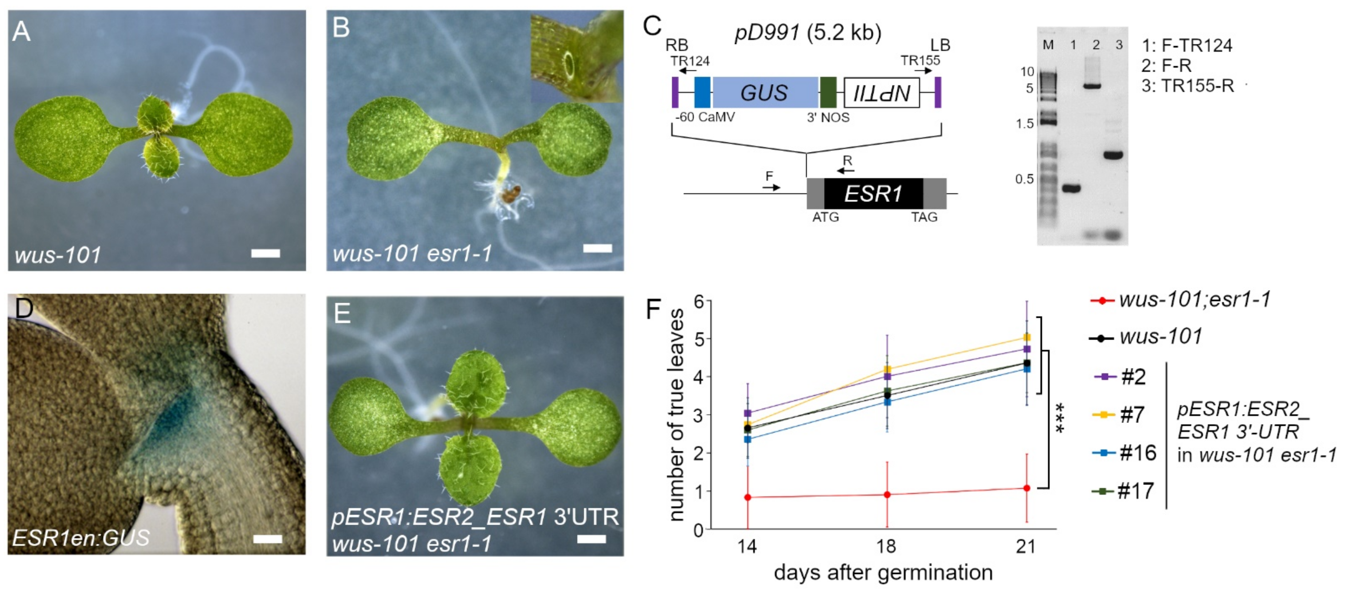
2.2. wus-101 esr1-1 Phenocopied wus-101 rev-5

The consistently observed defects in rosette leaf development of seedlings with the different wus and esr1 allele combinations (Figure 1I–R) bear a striking similarity to those of the wus-1 rev-6 double mutant in Ler [8]. Besides, the REV protein reportedly interacts with ESR1 [23] and ESR2 [17], although the interaction between REV and ESR2 remains a matter of debate [25]. To study the genetic interaction between REV and two ESR genes, rev-5 in Col-0 accession was used for this purpose [8]. In the case of successive emergence of rosette leaves, the rev-5 seedling was indistinguishable from the wild type (Figure 2A and Figure 3C). Consistent with the previous results [8], we were able to confirm substantial enhancement of the wus phenotypes by the rev-5 mutation in the corresponding wus-101 rev-5 (Figure 2C). wus-101 rev-5 double mutant seedlings formed a radial structure more frequently than wus-101 esr1-1 (Figure 2D,I), whereas mutations of its close homologs, phb and phv, did not (Figure 2E,F). A novel T-DNA insertion allele of PHB, SALK_008924C, in which a single T-DNA is inserted into exon 7 (2045 bp downstream from the ATG codon) (Supplementary Figure S2), was employed for crossing with wus-101. This novel phb allele is termed phb-101 hereafter. When rev-5 and esr1-1 mutations were combined in wus-101, the resulting wus-101 rev-5 esr1-1 triple mutant phenotypes appeared to be enhanced in an additive manner (in comparison to the respective double mutants). Unlike the esr1-1 mutation, esr2-2 in wus-101 rev-5 affected in a developmental stage-dependent manner. Until 10 d.a.g., wus-101 rev-5 esr2-2 seedlings failed to develop rosette leaves more frequently than wus-101 rev-5 (Figure 2H), whereas such an enhanced phenotype was mitigated by 17 d.a.g. (Figure 2I).

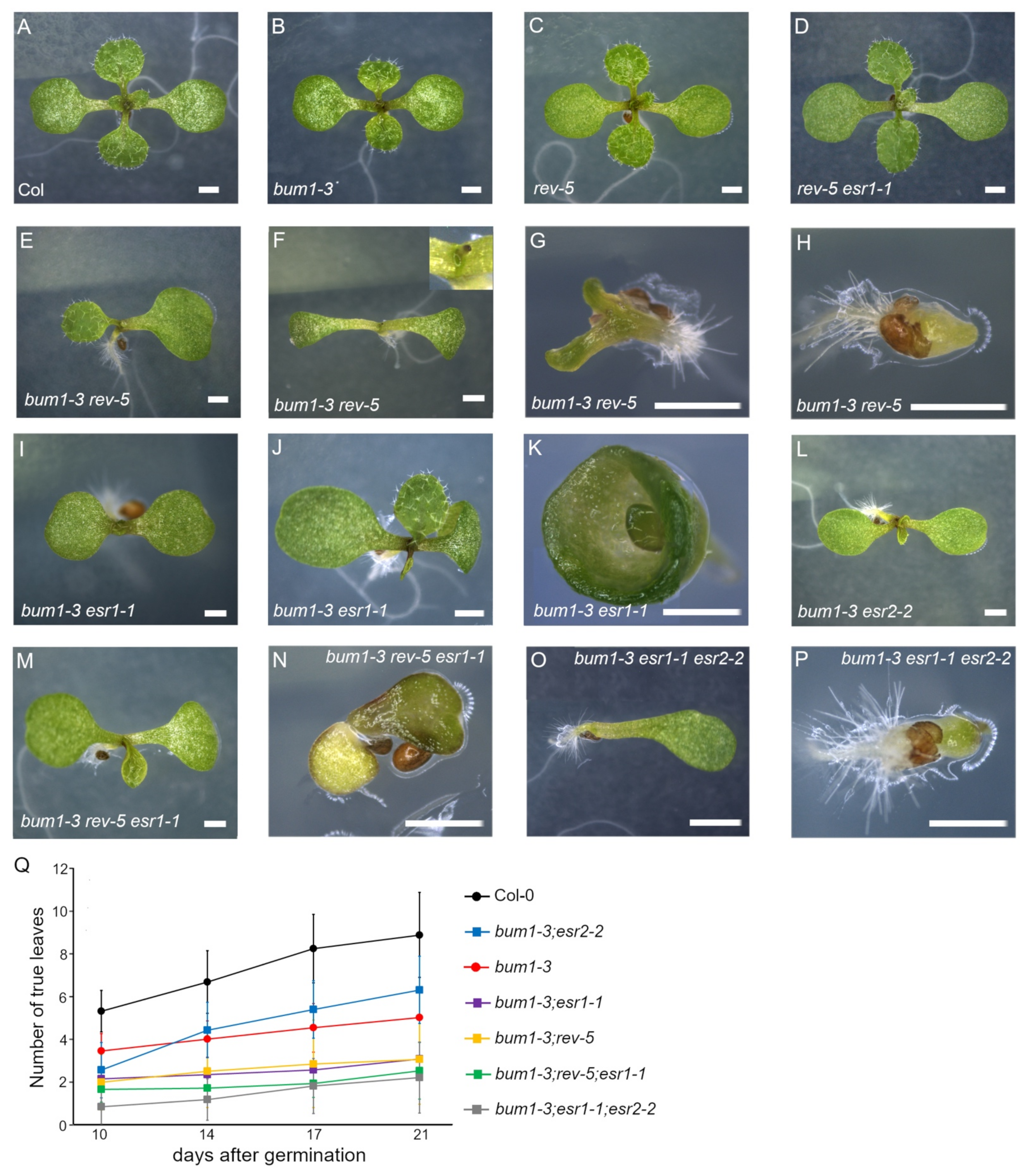
2.3. esr1-1 and esr2-2 Antagonistically Regulate Rosette Leaf Development in bum1-3 in the Later Vegetative Phase

Previously, the rev-6 mutation has been shown to enhance stm-2 phenotypes both in intact plants and in tissue culture [8]. We sought for the role of two ESR genes in the successive development of rosette leaves in the stm background. A weak allele of SHOOTMERISTEMLESS/BUMBERSHOOT1 (BUM1) in the Col-0 accession, bum1-3, was used [26]. Similar to wus seedlings (Figure 1G,H), bum1-3 exhibited a discontinuous rosette leaf emergence (compare Figure 3A,B). Similar to the wild type, both rev-5 and rev-5 esr1-1 seedlings were capable of developing true leaves continuously (Figure 3C,D). As shown in the previous study [8], we observed consistent phenotypes in rosette leaves of bum1-3 rev-5 double mutant seedlings: pronounced delay of rosette leaf emergence (Figure 3E), formation of a radial structure (Figure 3F), aberrant cotyledon in size and shape with delayed emergence of rosette leaves (Figure 3G), and the formation of a pin structure (Figure 3H). Similarly, bum1-3 esr1-1 had delayed emergence of rosette leaves (Figure 3I,J). At 3.34% frequency, the cotyledon was completely fused (Figure 3K). Note that on 17 d.a.g., a rosette leaf developed from the shoot apex, suggesting that the SAM still retained its activity to develop a rosette leaf although the emergence was substantially delayed. Under our growth conditions, we did not find bum1-3 esr1-1 seedlings forming a radial structure. Or the penetrance is too low to discover in bum1-3 esr1-1. The emergence of rosette leaves in bum1-3 esr2-2 seedlings was affected in a developmental stage-dependent manner. In the case of continuous rosette leaf emergence, the esr2-2 mutation enhanced the bum1-3 phenotype until 10 d.a.g. (Figure 3L). Later on, bum1-3 esr2-2 seedlings developed true leaves more effectively than the bum1-3 single mutant seedlings (p < 0.001, n > 50; Figure 3Q), showing that the esr2-2 mutation suppressed the bum1-3 phenotype in the later vegetative phase. On the other hand, introducing either rev-5 or esr2-2 into the bum1-3 esr1-1 background weakly enhanced aberrant lateral organ phenotypes (Figure 3M–P). The number of developed rosette leaves of bum1-3 rev-5 was indistinguishable from that of bum1-3 esr1-1 (p > 0.1, n > 60), suggesting that, consistent with results obtained from the wus-101 background, ESR1 plays a role in successive rosette leaf emergence in the same manner as REV does, presumably by forming a protein complex to modulate gene expression in the STM/BUM-independent pathway. bum1-3 rev-5 esr1-1 seedlings showed a wide range of phenotypes; from relatively milder enhancement (Figure 3M) to aberrant development (Figure 3N). The number of developed rosette leaves on 21 d.a.g. in bum1-3 rev-5 or bum1-3 esr1-1 double mutant seedlings was 2.92 ± 1.91 or 3.15 ± 1.34, respectively, whereas in the bum1-3 rev-5 esr1-1 triple mutant it was 2.51 ± 1.33 leaves. Since both bum1-3 rev-5 and bum1-3 esr1-1 double mutants exhibited a severe phenotype, the triple mutant did not statistically differ from the respective double mutants (p > 0.1 in both cases, n > 50; Figure 3Q).

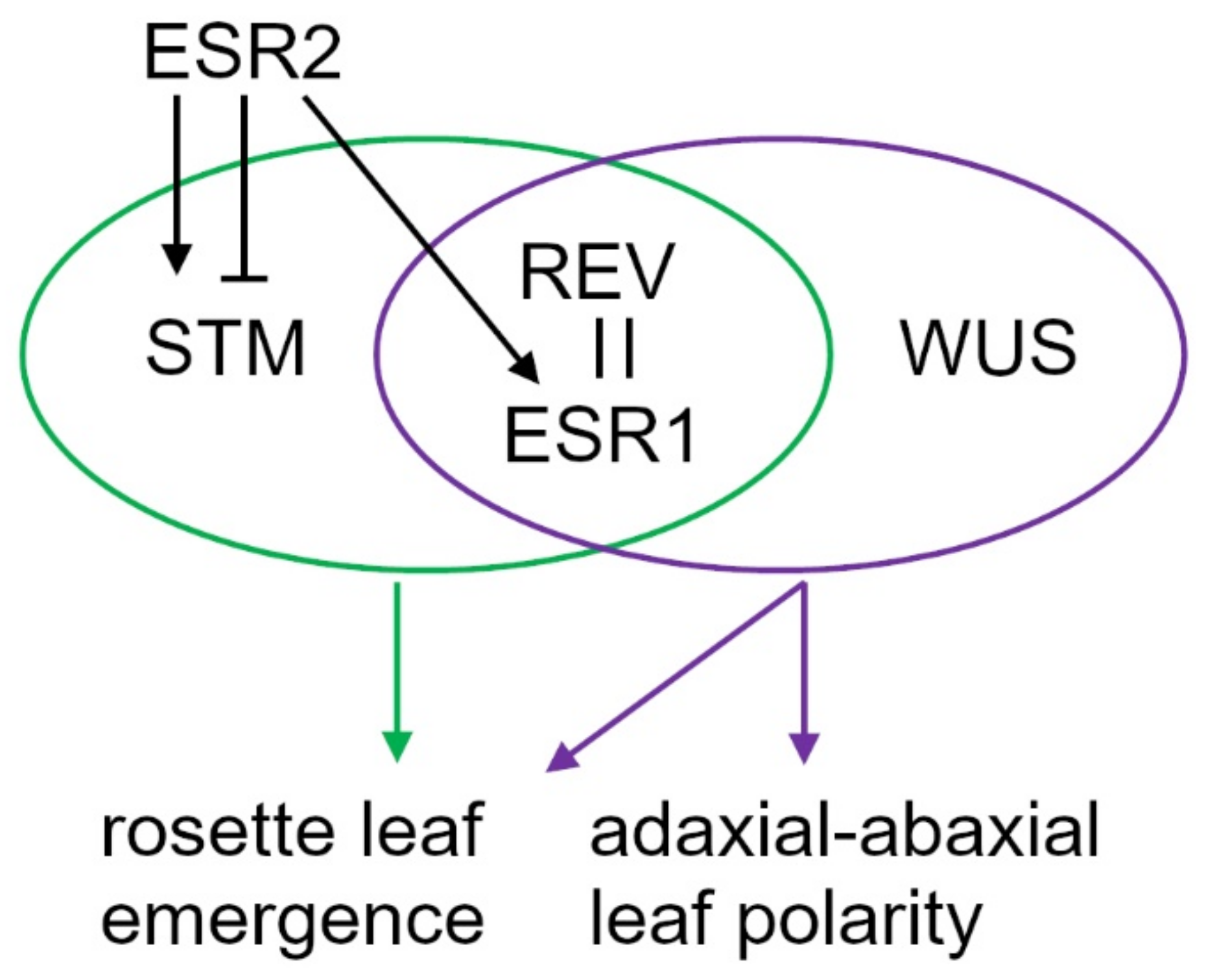
2.4. Distinct Expression Pattern of ESR Genes Defines their Unique Roles

In both the wus (Figure 4A) and bum mutant backgrounds, rev-5 and esr1-1 similarly enhanced defects in rosette leaf development (Figure 4B), whereas esr2-2 had an opposite effect in the later vegetative phase (beyond 10 d.a.g.). Nevertheless, ESR1 and ESR2 are the closest homologs and cause similar phenotypes when overexpressed: cytokinin-independent shoot regeneration in the tissue culture and the accumulation of undifferentiated cells [11,13]. They also share the same downstream target genes [13,21]. These findings suggest that, in terms of regulating downstream gene expression, they are comparable with each other. To tackle this discrepancy, we hypothesize that the distinct expression pattern of the two ESR genes is responsible for such contradicting results. To corroborate the spatial and temporal ESR1 expression, we have identified a GUS enhancer trap line, termed ESR1en:GUS, whereby the reporter is driven under the influence of an endogenous ESR1 locus. In this line, a single copy of pD911 T-DNA that contains a -60 Cauliflower mosaic virus minimal promoter fused to the uidA (GUS) reporter gene [27] is inserted at 73 bp upstream from the ATG codon that corresponds to the putative transcription start site. The right border is oriented toward the ESR1 promoter (Figure 4C). Using this line, we confirmed the consistent expression pattern of ESR1 in the upper layers in the CZ and PZ of the SAM (Figure 4D), as reported previously [17]. On the other hand, the expression of ESR2 is predominantly enriched in the founder cells of leaf primordia in the early vegetative phase [13,28]. The expression pattern of ESR1, ESR2, WUS, STM, and REV in the vegetative shoot has been reported previously [13,29,30,31,32] and their protein distribution is schematically represented (Supplementary Figure S3). The binary vector harboring the ESR1 promoter-driving ESR2 coding sequence containing the 2.86 kb ESR1 downstream region (with ESR1 3′-UTR included) was introduced into the wus-101 +/− esr1-1 −/− genotype. In the T3 generation, four independent lines homozygous for the transgene, termed pESR1:ESR2_ESR1 3′-UTR, in the wus-101 esr1-1 double mutant background, developed rosette leaves in the same manner as the wus-101 single mutant does, demonstrating that loss of ESR1 functions can be replenished by ESR2 driven by the ESR1 regulatory sequence and that the two ESR genes have redundant functions (Figure 4F).

3. Discussion

In this study, we employed the esr1-1/drn-2 and esr2-2 alleles because, unlike drn-1 drnl-2 (null allele combination), esr1-1 esr2-2 double mutant plants still produce a small number of viable seeds, which enabled us to examine genetic interactions with wus or bum and to examine the corresponding triple mutant rosette leaf phenotypes. In the case of lateral organ formation phenotypes on 8 d.a.g., wus-101 appears to exhibit a stronger phenotype than that of wus-1, indicating that wus-101 is a null allele (Table 1). It is intriguing that the lateral organ phenotypes observed in the wus esr1 double mutant combinations bear a striking resemblance to those in wus-1 rev-6 [8], albeit the fact that single esr1 mutant alleles examined so far do not exhibit phenotypes found in rev single mutants. The same holds true in the case of the bum1-3 mutant background that the number of developed rosette leaves of bum1-3 rev-5 is indistinguishable from that of bum1-3 esr1-1 (Figure 3Q). These results support the notion that the ESR1 and REV proteins physically interact with each other to control axillary meristem formation [23]. In fact, the overlapping expression of ESR1 and REV in leaf primordia was shown [23]. It is noteworthy that, unlike REV expression confined within the adaxial region of developing leaves [7], the expression pattern of ESR1 in young leaf primordia is broader [23]. Although it is not clear yet how the esr1 mutation operates to establish adaxial–abaxial polarity in the wus mutant background, we repeatedly observed the adaxial–abaxial polarity defects in rosette leaves of various esr1 wus double mutant backgrounds (Figure 1J,K,O and Table 1) at a lower frequency than in wus-101 rev-5 (Figure 2I). Besides, lotus-like rosette leaves are more frequently found in wus-101 esr1-2, a hypomorphic esr1 allele we introduced in this study, than in wu1-101 esr1-1 (Figure 1W), implying that lotus-like rosette leaf is formed due to the milder adaxial–abaxial polarity defects. The same structure was reported previously in 12.5% of as2-101 single and 23.5% of rev-6 as2-101 double mutant plants [33]. In the same work, the authors also found a needle-like leaf, which resembles what we call a radial structure (Figure 2D), among as2-101 rev-6, as2-101 phb-6, and as2-101 phv-5 double mutants [33]. It is noteworthy to mention that defects in the adaxial–abaxial polarity observed in wus-1 rev-6 and as2-101 rev-6 are Ler accession and that, in the case of the as1 and as2 backgrounds, erecta (er) mutation facilitates leaf polarity defects [34,35]. It is interesting to examine the genetic interaction between ESR1 and ER in the future.
The fact that radial structure formation was more frequently observed in wus-101 esr1-1 than in wus-101 esr1-2, a weak esr1 allele, suggests that, in concert with REV by physical protein–protein interaction, WUS-independent rosette leaf emergence is modulated in an ESR1 dosage-dependent manner (Figure 5). Recently, Xu and colleagues found the remarkably enriched expression of WUS and ESR1/DRN during the regeneration period in mesophyll protoplast regeneration culture and that both of which are required for somatic cell regeneration [36]. The interplay between WUS and ESR1 is implicated and our present genetic results are in agreement with them. It appears that ESR1 genetically interacts with other factors because the previous work showed the aberrant development of rosette leaves in pcn (popcorn) drn-1 double mutant [37].
Although ESR1 and ESR2 have redundant functions and exhibit similar cotyledon phenotypes [13,17,20], ESR1, in concert with WUS and STM, appears to play more important roles in lateral organ emergence and the establishment of adaxial–abaxial polarity.
Our result that the wus-101 esr1-1 double mutant transformed with the construct harboring the ESR1 promoter-driving ESR2 is indistinguishable from wus-101 (Figure 4F) corroborates that the two ESR proteins have redundant functions and are fungible. Reciprocally, the compromised shoot regeneration phenotype of the esr2-2 root explants in the tissue culture system was rescued by the ESR2 promoter-driving ESR1 [20]. These results suggest that the two ESR genes respond differently to internal and external cues. Yet, it is intricate to interpret the fact that the esr2 mutation partially rescued the inconsistent rosette leaf emergence in bum1-3 (Figure 3Q) and in wus-101 rev-5 (compare wus-101 rev-5 with wus-101 rev-5 esr2-2 on Day 17 in Figure 2I) in the later vegetative phase. Unexpectedly, wus-101 esr1-1 esr2-2 triple mutants accumulated numerous undifferentiated cells at the shoot apex (Figure 1X), and, as a consequence, no rosette leaves were differentiated. Monitoring the SAM marker gene expression in the triple mutant shoot apex is anticipated in the future study.

4. Materials and Methods

4.1. Plant Material and Growth Condition

Arabidopsis thaliana accession Columbia-0 (Col-0) was used as the wild type. The seeds described below were obtained from the European Nottingham Arabidopsis Stock Centre (NASC)): esr1-1/drn-2 (N121728) [17,20] wus-101 (N483520) [24], wus-1 (N15) [38], phv (N862830) [17], phb-101 (N654985), esr1-2 (N321463), and bum1-3 (N3781). Homozygous seeds of esr2-2 were kindly obtained from Hiroharu Banno [20]. rev-5 (Col-0 accession) was originally isolated in Luca Comai’s lab and homozygous seeds were kindly obtained through Ida Ruberti [39]. Prior to making higher-order mutants, all mutants employed in this work were backcrossed at least four times and the original wus-1 (Ler accession) was introgressed into Col-0 through repetitive crossing with Col-0 six times. Mutations were genotyped by PCR by a conventional method. For genotyping esr2-2, wus-1, bum1-3, and rev-5, dCAPS markers were developed and the respective PCR products were digested with EcoRV, NcoI, ClaI, or SnaBI, respectively. Primers used for the genotyping are listed in Supplementary Table S1. The GUS enhancer reporter line, ESR1en:GUS, was obtained by PCR-based screening [27] and additional T-DNA insertions present in the original ESR1en:GUS were segregated out by repetitive backcrossing with Col-0 five times. A single pD991 T-DNA insertion in the ESR1 locus was confirmed by kanamycin segregation analysis and Southern blotting. Primers used for the screening and confirming the insertion position of T-DNA are listed in Supplementary Table S1. Seeds were surface-sterilized, sown on MS plant agar medium, and grown at 21 °C in a photoperiod of 16/8 (light/dark) condition at the indicated days. Clearing of seedlings and photographing differential interference contrast (DIC) were carried out as described previously [40]. Seedlings were photographed by a Stereoscopic Zoom Microscope SMZ1000 (Nikon, Tokyo, Japan) operated with NIS Elements software at the indicated time points.

4.2. Construction of Transgene and Transformation

The binary vector, pHLG60, a modified version of pSK34 [13], contains a hygromycin resistant cassette for plant transformation. The ESR1 promoter, its downstream region, and ESR2 coding sequence were PCR-amplified from Col-0 genomic DNA as a template by using Phusion® High-Fidelity DNA Polymerase (NEB, Massachusetts, USA). The primers used are listed in Supplementary Table S1. The resultant PCR products were digested with AscI and BamHI (ESR1 promoter), BamHI and SpeI (ESR2 ORF), and SpeI and NotI (ESR1 downstream region) and sequentially cloned into pHLG60 at the corresponding restriction endonuclease recognition sites. The resulting construct was introduced into Agrobacterium tumefaciens strain GV3101, which was used to transform the wus-101 +/- esr1-1 -/- genetic background by the floral dip method. Harvested seeds were plated on MS agar medium containing 18 mg/L hygromycin B for the transgene and 2 mg/L sulfadiazine sodium salt for wus-101 selections. Four independent T3 lines containing a single insertion for the transgene in the wus-101 +/- esr1-1 -/- background were chosen for the analysis.

4.3. Semi-Quantitative RT-PCR Analysis

Conditions for RNA extraction, first-strand cDNA synthesis, PCR, and agarose gel electrophoresis were previously described [13]. Primers for detecting ESR1 transcripts in esr1-2 are listed in Supplementary Table S1 and those for TUBULIN3 were described [13]. Cycles used for detection of ESR1 or TUB3 are 31 or 18 cycles, respectively.

Supplementary Materials

The following are available online at https://www.mdpi.com/article/10.3390/ijms221910621/s1.

Author Contributions

M.K., D.Z., I.K., M.A. and Y.I. carried out genotyping and making the crosses; M.K., D.Z. and I.K. performed the Agrobacterium-mediated floral dip transformation; M.K. and Y.I. constructed the binary vector; D.Z. performed RNA extraction and semi-quantitative RT-PCR; Y.I. conceived ideas and examined the mutant phenotypes. All authors contributed to writing the manuscript. All authors have read and agreed to the published version of the manuscript.

Funding

This work was supported by the Grant Agency in the Czech Republic (GACR17-23702S and GACR 18-23972Y), the European Regional Development Fund (ERDF) Project (No. CZ.02.1.01/0.0/0.0/16_019/0000827), and the IROAST research unit on plant stem cells and regeneration.

Informed Consent Statement

Not applicable.

Acknowledgments

We thank the European Arabidopsis Stock Centre (NASC), Hiroharu Banno, Luca Comai, and Ida Ruberti for providing the esr2-2 and rev-5 mutant seeds, and Isaac Sugraňes and Iva Hradilova for technical assistance.

Conflicts of Interest

The authors declare no conflict of interest.

References

  1. Brand, U.; Fletcher, J.C.; Hobe, M.; Meyerowitz, E.M.; Simon, R. Dependence of stem cell fate in Arabidopsis on a feedback loop regulated by CLV3 activity. Science 2000, 289, 617–619. [Google Scholar] [CrossRef]
  2. Schoof, H.; Lenhard, M.; Haecker, A.; Mayer, K.F.X.; Jürgens, G.; Laux, T. The stem cell population of Arabidopsis shoot meristems is maintained by a regulatory loop between the CLAVATA and WUSCHEL genes. Cell 2000, 100, 635–644. [Google Scholar] [CrossRef] [Green Version]
  3. Laux, T.; Mayer, K.F.X.; Berger, J.; Jürgens, G. The WUSCHEL gene is required for shoot and floral meristem integrity in Arabidopsis. Development 1996, 122, 87–96. [Google Scholar] [CrossRef] [PubMed]
  4. Lee, C.; Clark, S.E. A WUSCHEL-independent stem cell specification pathway is repressed by PHB, PHV and CNA in arabidopsis. PLoS ONE 2015, 10, 1–19. [Google Scholar] [CrossRef] [Green Version]
  5. Huang, W.; Pitorre, D.; Poretska, O.; Marizzi, C.; Winter, N.; Poppenberger, B.; Sieberer, T. Altered meristem programSuppresses ectopic stem cell niche formation in the shoot apical meristem in a largely cytokinin-independent manner. Plant Physiol. 2015, 167, 1471–1486. [Google Scholar] [CrossRef] [Green Version]
  6. Jung, J.H.; Park, C.M. MIR166/165 genes exhibit dynamic expression patterns in regulating shoot apical meristem and floral development in Arabidopsis. Planta 2007, 225, 1327–1338. [Google Scholar] [CrossRef] [PubMed]
  7. Williams, L.; Grigg, S.P.; Xie, M.; Christensen, S.; Fletcher, J.C. Regulation of Arabidopsis shoot apical meristem and lateral organ formation by microRNA miR166g and its AtHD-ZIP target genes. Development 2005, 132, 3657–3668. [Google Scholar] [CrossRef] [PubMed] [Green Version]
  8. Otsuga, D.; DeGuzman, B.; Prigge, M.J.; Drews, G.N.; Clark, S.E. REVOLUTA regulates meristem initiation at lateral positions. Plant J. 2001, 25, 223–236. [Google Scholar] [CrossRef] [PubMed]
  9. Emery, J.F.; Floyd, S.K.; Alvarez, J.; Eshed, Y.; Hawker, N.P.; Izhaki, A.; Baum, S.F.; Bowman, J.L. Radial Patterning of Arabidopsis Shoots by Class III HD-ZIP and KANADI Genes. Curr. Biol. 2003, 13, 1768–1774. [Google Scholar] [CrossRef] [Green Version]
  10. Prigge, M.J.; Otsuga, D.; Alonso, J.M.; Ecker, J.R.; Drews, G.N.; Clark, S.E. Class III homeodomain-leucine zipper gene family members have overlapping, antagonistic, and distinct roles in Arabidopsis development. Plant Cell 2005, 17, 61–76. [Google Scholar] [CrossRef] [Green Version]
  11. Banno, H.; Ikeda, Y.; Niu, Q.W.; Chua, N.H. Overexpression of Arabidopsis ESR1 induces initiation of Shoot Regeneration. Plant Cell 2001, 13, 2609–2618. [Google Scholar] [CrossRef]
  12. Kirch, T.; Simon, R.; Grünewald, M.; Werr, W. The Dornröschen/enhancer of shoot regeneration1: Gene of Arabidopsis acts in the control of meristem cell fate and lateral organ development. Plant Cell 2003, 15, 694–705. [Google Scholar] [CrossRef] [Green Version]
  13. Ikeda, Y.; Banno, H.; Niu, Q.-W.; Howell, S.H.; Chua, N.-H. The enhancer of shoot regeneration 2 gene in Arabidopsis regulates cup-shaped cotyledon 1 at the transcriptional level and controls cotyledon development. Plant Cell Physiol. 2006, 47, 1443–1456. [Google Scholar] [CrossRef] [Green Version]
  14. Ward, J.M.; Smith, A.M.; Shah, P.K.; Galanti, S.E.; Yi, H.; Demianski, A.J.; Van Der Graaff, E.; Keller, B.; Neff, M.M. A new role for the Arabidopis AP2 transcription factor, leafy petiole, in gibberellin-induced germination is revealed by the misexpression of a homologous gene SOB2/DRN-LIKE. Plant Cell 2006, 18, 29–39. [Google Scholar] [CrossRef] [Green Version]
  15. Marsch-Martinez, N.; Greco, R.; Becker, J.D.; Dixit, S.; Bergervoet, J.H.W.; Karaba, A.; De Folter, S.; Pereira, A. BOLITA, an Arabidopsis AP2/ERF-like transcription factor that affects cell expansion and proliferation/differentiation pathways. Plant Mol. Biol. 2006, 62, 825–843. [Google Scholar] [CrossRef] [PubMed] [Green Version]
  16. Capua, Y.; Eshed, Y. Coordination of auxin-triggered leaf initiation by tomato LEAFLESS. Proc. Natl. Acad. Sci. USA 2017, 114, 3246–3251. [Google Scholar] [CrossRef] [PubMed] [Green Version]
  17. Chandler, J.W.; Cole, M.; Flier, A.; Grewe, B.; Werr, W. The AP2 transcription factors DORNRÖSCHEN and Dornröschen-like redundantly control Arabidopsis embryo patterning via interaction with PHAVOLUTA. Development 2007, 134, 1653–1662. [Google Scholar] [CrossRef] [PubMed] [Green Version]
  18. Durán-Medina, Y.; Serwatowska, J.; Reyes-Olalde, J.I.; De Folter, S.; Marsch-Martínez, N. The AP2/ERF transcription factor DRNL modulates gynoecium development and affects its response to Cytokinin. Front. Plant Sci. 2017, 8, 1–13. [Google Scholar] [CrossRef] [Green Version]
  19. Nag, A.; Yang, Y.; Jack, T. DORNRÖSCHEN-LIKE, an AP2 gene, is necessary for stamen emergence in Arabidopsis. Plant Mol. Biol. 2007, 65, 219–232. [Google Scholar] [CrossRef]
  20. Matsuo, N.; Makino, M.; Banno, H. Arabidopsis ENHANCER OF SHOOT REGENERATION (ESR)1 and ESR2 regulate in vitro shoot regeneration and their expressions are differentially regulated. Plant Sci. 2011, 181, 39–46. [Google Scholar] [CrossRef]
  21. Matsuo, N.; Mase, H.; Makino, M.; Takahashi, H.; Banno, H. Identification of enhancer of shoot regeneration 1-upregulated genes during in vitro shoot regeneration. Plant Biotechnol. 2009, 26, 385–393. [Google Scholar] [CrossRef] [Green Version]
  22. Tian, C.; Zhang, X.; He, J.; Yu, H.; Wang, Y.; Shi, B.; Han, Y.; Wang, G.; Feng, X.; Zhang, C.; et al. An organ boundary-enriched gene regulatory network uncovers regulatory hierarchies underlying axillary meristem initiation. Mol. Syst. Biol. 2014, 10, 755. [Google Scholar] [CrossRef]
  23. Zhang, C.; Wang, J.; Wenkel, S.; Chandler, J.W.; Werr, W.; Jiao, Y. Spatiotemporal control of axillary meristem formation by interacting transcriptional regulators. Development 2018, 145, dev158352. [Google Scholar] [CrossRef] [PubMed] [Green Version]
  24. Kubalová, I.; Zalabák, D.; Mičúchová, A.; Ikeda, Y. Mutations in tetrapyrrole biosynthesis pathway uncouple nuclear WUSCHEL expression from de novo shoot development in Arabidopsis. Plant Cell Tissue Organ Cult. 2019, 139, 395–401. [Google Scholar] [CrossRef]
  25. Magnani, E.; Barton, M.K. A Per-ARNT-sim-like sensor domain uniquely regulates the activity of the homeodomain leucine zipper transcription factor REVOLUTA in Arabidopsis. Plant Cell 2011, 23, 567–582. [Google Scholar] [CrossRef] [PubMed] [Green Version]
  26. Takano, S.; Niihama, M.; Smith, H.M.S.; Tasaka, M.; Aida, M. Gorgon, a novel missense mutation in the shoot meristemless gene, impairs shoot meristem homeostasis in arabidopsis. Plant Cell Physiol. 2010, 51, 621–634. [Google Scholar] [CrossRef] [Green Version]
  27. Campisi, L.; Yang, Y.; Yi, Y.; Heilig, E.; Herman, B.; Cassista, A.J.; Allen, D.W.; Xiang, H.; Jack, T. Generation of enhancer trap lines in Arabidopsis and characterization of expression patterns in the inflorescence. Plant J. 1999, 17, 699–707. [Google Scholar] [CrossRef]
  28. Glowa, D.; Comelli, P.; Chandler, J.W.; Werr, W. Clonal sector analysis and cell ablation confirm a function for DORNROESCHEN-LIKE in founder cells and the vasculature in Arabidopsis. Planta 2021, 253, 1–16. [Google Scholar] [CrossRef]
  29. Caggiano, M.P.; Yu, X.; Bhatia, N.; Larsson, A.; Ram, H.; Ohno, C.K.; Sappl, P.; Meyerowitz, E.M.; Jönsson, H.; Heisler, M.G. Cell type boundaries organize plant development. Elife 2017, 6, 1–32. [Google Scholar] [CrossRef]
  30. Su, Y.H.; Zhou, C.; Li, Y.J.; Yu, Y.; Tang, L.P.; Zhang, W.J.; Yao, W.J.; Huang, R.; Laux, T.; Zhang, X.S. Integration of pluripotency pathways regulates stem cell maintenance in the arabidopsis shoot meristem. Proc. Natl. Acad. Sci. USA 2020, 117, 22561–22571. [Google Scholar] [CrossRef]
  31. Heisler, M.G.; Ohno, C.; Das, P.; Sieber, P.; Reddy, G.V.; Long, J.A.; Meyerowitz, E.M. Patterns of auxin transport and gene expression during primordium development revealed by live imaging of the Arabidopsis inflorescence meristem. Curr. Biol. 2005, 15, 1899–1911. [Google Scholar] [CrossRef] [PubMed] [Green Version]
  32. Luo, L.; Zeng, J.; Wu, H.; Tian, Z.; Zhao, Z. A Molecular Framework for Auxin-Controlled Homeostasis of Shoot Stem Cells in Arabidopsis. Mol. Plant 2018, 11, 899–913. [Google Scholar] [CrossRef] [PubMed] [Green Version]
  33. Fu, Y.; Xu, L.; Xu, B.; Yang, L.; Ling, Q.; Wang, H.; Huang, H. Genetic interactions between leaf polarity-controlling genes and ASYMMETRIC LEAVES1 and 2 in Arabidopsis leaf patterning. Plant Cell Physiol. 2007, 48, 724–735. [Google Scholar] [CrossRef] [PubMed] [Green Version]
  34. Xu, L.; Xu, Y.; Dong, A.; Sun, Y.; Pi, L.; Xu, Y.; Huang, H. Novel as1 and as2 defects in leaf adaxial-abaxial polarity reveal the requirement for ASYMMETRIC LEAVES1 and 2 and ERECTA functions in specifying leaf adaxial identity. Development 2003, 130, 4097–4107. [Google Scholar] [CrossRef] [PubMed] [Green Version]
  35. Qi, Y.; Sun, Y.; Xu, L.; Xu, Y.; Huang, H. ERECTA is required for protection against heat-stress in the AS1/AS2 pathway to regulate adaxial-abaxial leaf polarity in Arabidopsis. Planta 2004, 219, 270–276. [Google Scholar] [CrossRef]
  36. Xu, M.; Du, Q.; Tian, C.; Wang, Y.; Jiao, Y. Stochastic gene expression drives mesophyll protoplast regeneration. Sci. Adv. 2021, 7, eabg8466. [Google Scholar] [CrossRef]
  37. Xiang, D.; Yang, H.; Venglat, P.; Cao, Y.; Wen, R.; Ren, M.; Stone, S.; Wang, E.; Wang, H.; Xiao, W.; et al. POPCORN functions in the auxin pathway to regulate embryonic body plan and meristem organization in Arabidopsis. Plant Cell 2011, 23, 4348–4367. [Google Scholar] [CrossRef] [Green Version]
  38. Mayer, K.F.; Schoof, H.; Haecker, A.; Lenhard, M.; Jurgens, G.; Laux, T. Role of WUSCHEL in regulating stem cell fate in the Arabidopsis shoot meristem. Cell 1998, 95, 805–815. [Google Scholar] [CrossRef] [Green Version]
  39. Turchi, L.; Carabelli, M.; Ruzza, V.; Possenti, M.; Sassi, M.; Peñalosa, A.; Sessa, G.; Salvi, S.; Forte, V.; Morelli, G.; et al. Arabidopsis HD-Zip II transcription factors control apical embryo development and meristem function. Development 2013, 140, 2118–2129. [Google Scholar] [CrossRef] [Green Version]
  40. Aida, M.; Ishida, T.; Fukaki, H.; Fujisawa, H.; Tasaka, M. Genes involved in organ separation in Arabidopsis: An analysis of the cup-shaped cotyledon mutant. Plant Cell 1997, 9, 841–857. [Google Scholar] [CrossRef] [Green Version]
Figure 1. ESR1 controls rosette leaf development in the WUS-independent pathway in a dosage-dependent manner. (A) Eight-day-old Col-0; (B) esr1-1; (C) esr1-2; (D) esr2-2; (E,F) esr1-1 esr2-2; (G) wus-1; (H) wus-101; (IL) wus-1 esr1-1; (K) Normarski image of the plant (J); (MP) wus-101 esr1-1; (N) fourteen-day-old wus-101 esr1-1, note the radial structure (inset) in (O); (Q,R) eight-day-old wus-101 esr1-2, note the single radial structure (inset); (S,T) wus-101 esr2-2; (U) wus-101 esr1-1 esr2-2; (V) Nomarski image of the shoot apex of seedling (U); (W) thirty-day-old plants of Col-0, wus-101, wus-101 esr1-2, and wus-101 esr1-1 (from left to right), note the lotus-like leaf of wus-101 esr1-2 (inset); (X,Y) thirty-day-old wus-101 esr1-1 esr2-2 shoot apex. Scale bars = 1 mm (A–E, G–J, L–T,W), 0.5 mm (F,U,X,Y), and 0.2 mm (K,V).
Ijms 22 10621 g001
Figure 2. rev-5 but not phb-101 or phv enhances wus phenotype. (A) Five-day-old Col-0; (B) wus-101; (C,D) wus-101 rev-5, note the radial structure (inset) in (D); (E) wus-101 phb-101; (F) wus-101 phv; (G) wus-101 rev-5 esr1-1; (H) wus-101 rev-5 esr2-2; (I) frequency of phenotypes in the respective mutant background on 10 d.a.g (left) and 17 d.a.g. (right). At least 50 individual plants per genotype were examined with biological triplicates. The first pair of emerged rosette leaves are categorized into a developed leaf (green) or radial structure (pink). Black indicates no development of lateral organs at the indicated time points.
Ijms 22 10621 g002
Figure 3. esr1 and rev additively enhance the bum phenotype. (A) Eight-day-old Col-0; (B) bum1-3; (C) rev-5; (D) rev-5 esr1-1; (EH) bum1-3 rev-5, note the single cotyledon in (E) and radial structure (inset) in (F); (IK) bum1-3 esr1-1; (L) bum1-3 esr2-2; (M,N) bum1-3 rev-5 esr1-1; (O,P) bum1-3 esr1-1 esr2-2; (Q) number of developed rosette leaves in the respective mutant backgrounds at 10, 14, 17, and 21 d. a. g. Data shown are the mean ± SD of biological triplicates (n > 50 per genotype). Scale bars = 1 mm.
Ijms 22 10621 g003
Figure 4. ESR1 promoter-driving ESR2 complements esr1 phenotypes in wus-101 esr1-1. (A) Eight-day-old Col-0. (B) wus-101 esr1-1. (C) Schematic representation of the ESR1 enhancer trap GUS reporter line (left) and the determination of T-DNA insertion position and orientation. A single copy pD99 T-DNA is inserted at 73 bp upstream from the ATG codon facing the right border toward the ESR1 promoter. Note the correct orientation of the uidA (GUS) transgene. Arrows indicate the primers used for screening and verifying the T-DNA insertion. M, marker. Black, gray, blue, right blue, green, purple, and open rectangle indicate ESR1 coding, ESR1 UTR, -60 Cauliflower mosaic virus minimum promoter, uidA (β-GLUCURONIDASE) coding, NOS terminator, right or left border, and NPTII (NEOMYCIN PHOSPHOTRANSFERASE II), respectively. (D) Histological GUS staining of ESR1 enhancer trap line on 2 d.a.g. (E) Eight-day-old wus-101 esr1-1 double mutant seedlings harboring the homozygous pESR1:ESR2_ESR1 3′-UTR transgene. (F) Number of developed rosette leaves. Four independent transgenic lines harboring a transgene (homozygous single insertion) in the wus-101 esr1-1 background and their parent, wus-101 esr1-1, were compared at the indicated time points. wus-101 was included as a positive control. Data shown are the mean ± SD of biological triplicates (n > 50) (p < 0.001; Student t-tests).
Ijms 22 10621 g004
Figure 5. Schematic representation of genetic interactions. The REV and ESR1 protein interaction is confirmed. Together with STM or WUS, both ESR1 and REV participate in the rosette leaf emergence and the establishment of adaxial–abaxial polarity. ESR2 and ESR1 are functionally interchangeable whereas the effect of ESR2 on rosette leaf emergence in the stm/bum mutant is vegetative phase-dependent.
Ijms 22 10621 g005
Table 1. Frequency of variable shoot phenotypes (%) on day 10 after germination a.
wus bRadial
Structure d
Unrecognizable
True Leaves
mp-Likepin-Like
Shoot
wus-1 (n = 160)1000000
wus-1;esr1-1 (n = 54)68.5 c14.814.8 e1.90
wus-101 (n = 196)1000000
wus-101;esr1-1 (n = 88)35.2 c9.155.7 e00
wus-101;esr1-2 (n = 38)78.9 c21.1000
wus-101;esr2-2 (n = 55)97.902.100
esr1-1;esr2-2e (n = 47)00026.10
wus-101;esr1-1;esr2-2 (n = 72)9.7 c5.634.7 f38.911.1
a: Cotyledon phenotypes are not counted; b: wus phenotype denotes reduced number of developed true leaves; c: wus phenotypes are moderately enhanced; d: Includes seedling developing at least one radial structure; e: Only two fully expanded leaves are developed by 40 d.a.g; f: No visible true leaves developed by 40 d.a.g.
Publisher’s Note: MDPI stays neutral with regard to jurisdictional claims in published maps and institutional affiliations.

